

Advancements in diagnosing oral potentially malignant disorders: leveraging Vision transformers for multi-class detection
- PMID: 38849649
- PMCID: PMC11161543
- DOI: 10.1007/s00784-024-05762-8
Advancements in diagnosing oral potentially malignant disorders: leveraging Vision transformers for multi-class detection
Abstract
Objectives: Diagnosing oral potentially malignant disorders (OPMD) is critical to prevent oral cancer. This study aims to automatically detect and classify the most common pre-malignant oral lesions, such as leukoplakia and oral lichen planus (OLP), and distinguish them from oral squamous cell carcinomas (OSCC) and healthy oral mucosa on clinical photographs using vision transformers.
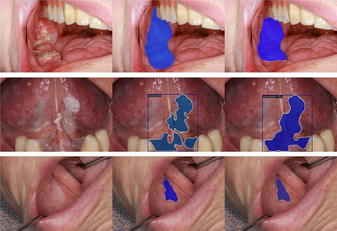
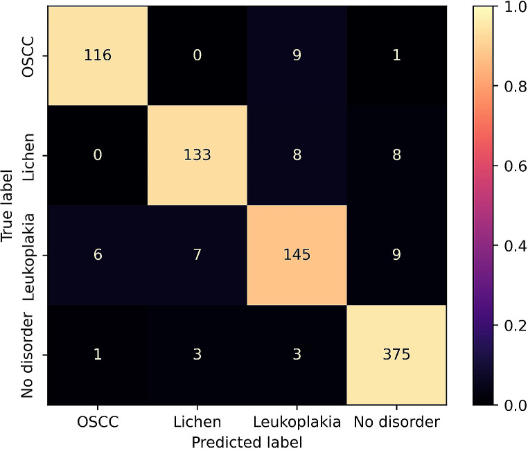
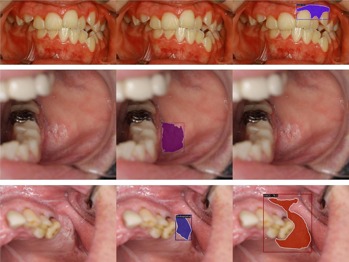
Methods: 4,161 photographs of healthy mucosa, leukoplakia, OLP, and OSCC were included. Findings were annotated pixel-wise and reviewed by three clinicians. The photographs were divided into 3,337 for training and validation and 824 for testing. The training and validation images were further divided into five folds with stratification. A Mask R-CNN with a Swin Transformer was trained five times with cross-validation, and the held-out test split was used to evaluate the model performance. The precision, F1-score, sensitivity, specificity, and accuracy were calculated. The area under the receiver operating characteristics curve (AUC) and the confusion matrix of the most effective model were presented.
Results: The detection of OSCC with the employed model yielded an F1 of 0.852 and AUC of 0.974. The detection of OLP had an F1 of 0.825 and AUC of 0.948. For leukoplakia the F1 was 0.796 and the AUC was 0.938.
Conclusions: OSCC were effectively detected with the employed model, whereas the detection of OLP and leukoplakia was moderately effective.
Clinical relevance: Oral cancer is often detected in advanced stages. The demonstrated technology may support the detection and observation of OPMD to lower the disease burden and identify malignant oral cavity lesions earlier.
Keywords: Artificial Intelligence; Deep learning; Leukoplakia; Malignant transformation; Oral lichen planus; Oral squamous cell carcinoma.
© 2024. The Author(s).
Conflict of interest statement
The authors declare no competing interests.
Figures

References
-
- Lingen MW, Abt E, Agrawal N et al (2017) Evidence-based clinical practice guideline for the evaluation of potentially malignant disorders in the oral cavity a report of the American Dental Association. J Am Dent Assoc 148:712–727e10. 10.1016/j.adaj.2017.07.032 10.1016/j.adaj.2017.07.032 - DOI - PubMed
-
- Meij EHVD, Waal IVD (2003) Lack of clinicopathologic correlation in the diagnosis of oral lichen planus based on the presently available diagnostic criteria and suggestions for modifications. J Oral Pathol Med 32:507–512. 10.1034/j.1600-0714.2003.00125.x 10.1034/j.1600-0714.2003.00125.x - DOI - PubMed
MeSH terms
LinkOut - more resources
Full Text Sources
Medical
