

ESR Essentials: characterisation and staging of adnexal masses with MRI and CT-practice recommendations by ESUR
- PMID: 38849662
- PMCID: PMC11557651
- DOI: 10.1007/s00330-024-10817-1
ESR Essentials: characterisation and staging of adnexal masses with MRI and CT-practice recommendations by ESUR
Abstract
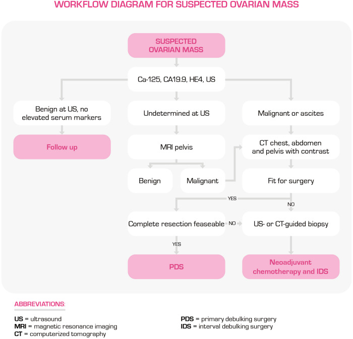
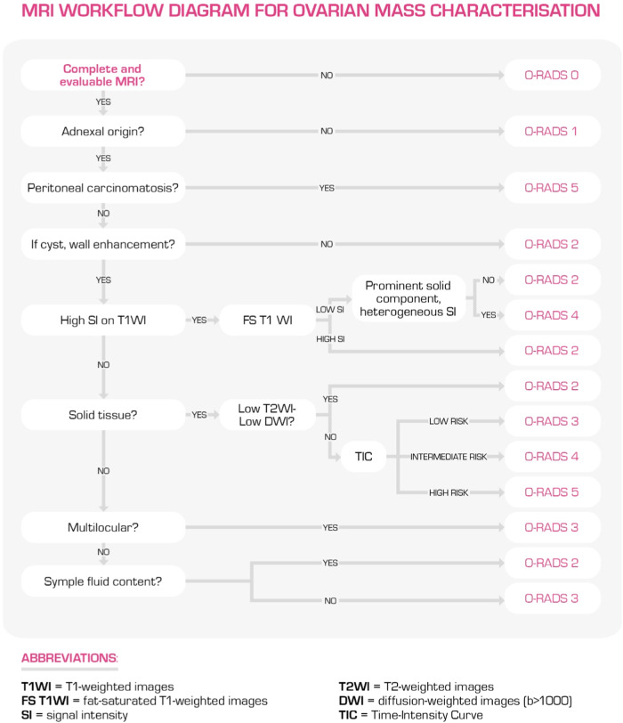
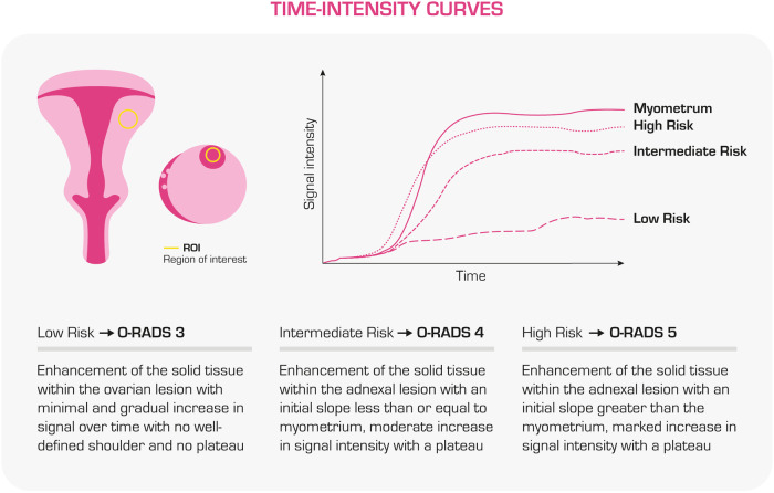
Ovarian masses encompass various conditions, from benign to highly malignant, and imaging plays a vital role in their diagnosis and management. Ultrasound, particularly transvaginal ultrasound, is the foremost diagnostic method for adnexal masses. Magnetic Resonance Imaging (MRI) is advised for more precise characterisation if ultrasound results are inconclusive. The ovarian-adnexal reporting and data system (O-RADS) MRI lexicon and scoring system provides a standardised method for describing, assessing, and categorising the risk of each ovarian mass. Determining a histological differential diagnosis of the mass may influence treatment decision-making and treatment planning. When ultrasound or MRI suggests the possibility of cancer, computed tomography (CT) is the preferred imaging technique for staging. It is essential to outline the extent of the malignancy, guide treatment decisions, and evaluate the feasibility of cytoreductive surgery. This article provides a comprehensive overview of the key imaging processes in evaluating and managing ovarian masses, from initial diagnosis to initial treatment. It also includes pertinent recommendations for properly performing and interpreting various imaging modalities. KEY POINTS: MRI is the modality of choice for indeterminate ovarian masses at ultrasound, and the O-RADS MRI lexicon and score enable unequivocal communication with clinicians. CT is the recommended modality for suspected ovarian masses to tailor treatment and surgery. Multidisciplinary meetings integrate information and help decide the most appropriate treatment for each patient.
Keywords: Magnetic resonance imaging; Ovarian neoplasms; Patient care; Workflow; X-ray computed tomography.
© 2024. The Author(s).
Conflict of interest statement
Figures

References
-
- Taylor EC, Irshaid L, Mathur M (2021) Multimodality imaging approach to ovarian neoplasms with pathologic correlation. Radiographics 41:289–315. 10.1148/rg.2021200086 - PubMed
-
- Andreotti RF, Timmerman D, Strachowski LM et al (2020) O-RADS US risk stratification and management system: a consensus guideline from the ACR ovarian-adnexal reporting and data system committee. Radiology 294:168–185. 10.1148/radiol.2019191150 - PubMed
-
- Thomassin-Naggara I, Belghitti M, Milon A et al (2021) O-RADS MRI score: analysis of misclassified cases in a prospective multicentric European cohort. Eur Radiol 31:9588–9599. 10.1007/s00330-021-08054-x - PubMed
Publication types
MeSH terms
LinkOut - more resources
Full Text Sources
Medical
Miscellaneous
