. 2024 Jun 7;23(1):124.
doi: 10.1186/s12943-024-02016-9.

OLFM4 promotes the progression of intestinal metaplasia through activation of the MYH9/GSK3β/β-catenin pathway

Affiliations

OLFM4 promotes the progression of intestinal metaplasia through activation of the MYH9/GSK3β/β-catenin pathway

Hongfa Wei et al. Mol Cancer. .

Abstract

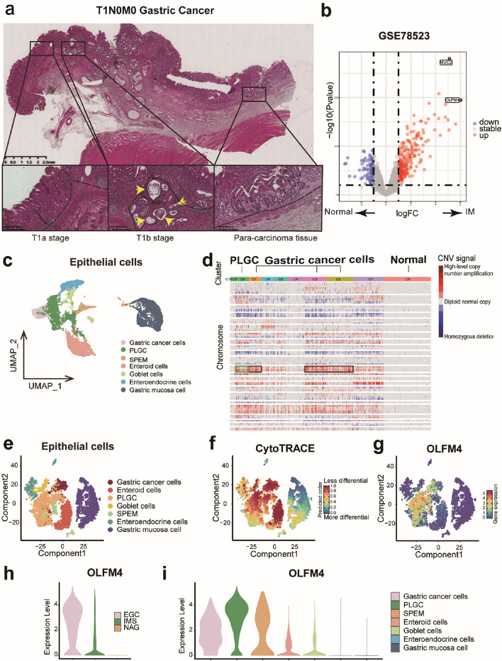
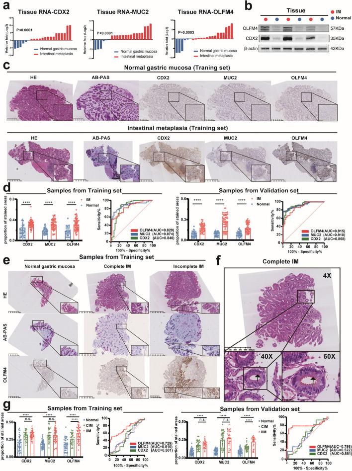
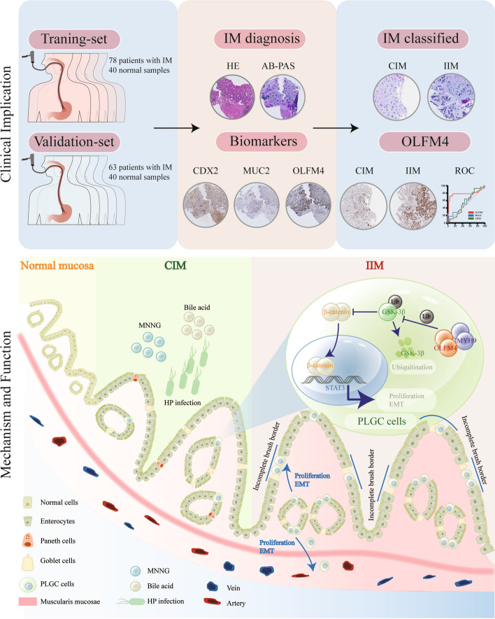
Background: Intestinal metaplasia (IM) is classified into complete intestinal metaplasia (CIM) and incomplete intestinal metaplasia (IIM). Patients diagnosed with IIM face an elevated susceptibility to the development of gastric cancer, underscoring the critical need for early screening measures. In addition to the complexities associated with diagnosis, the exact mechanisms driving the progression of gastric cancer in IIM patients remain poorly understood. OLFM4 is overexpressed in several types of tumors, including colorectal, gastric, pancreatic, and ovarian cancers, and its expression has been associated with tumor progression.

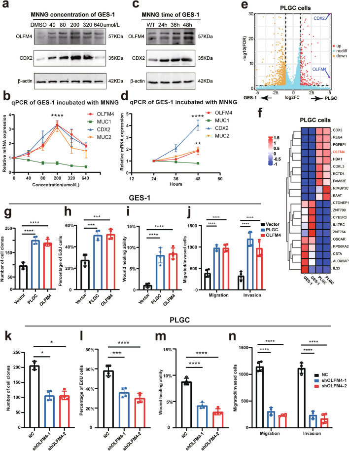
Methods: In this study, we used pathological sections from two clinical centers, biopsies of IM tissues, precancerous lesions of gastric cancer (PLGC) cell models, animal models, and organoids to explore the role of OLFM4 in IIM.

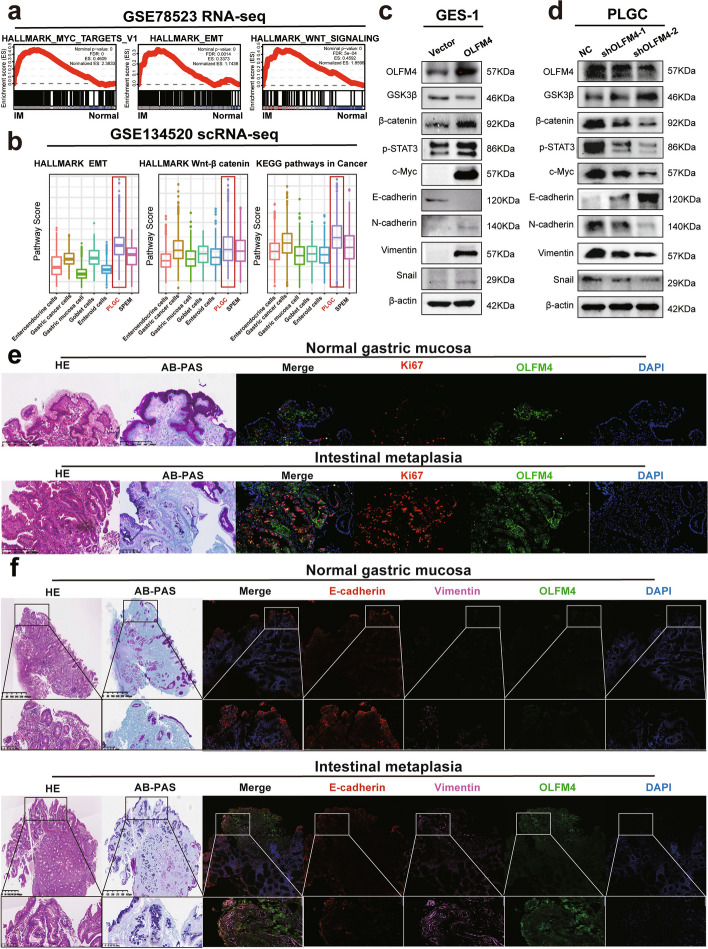
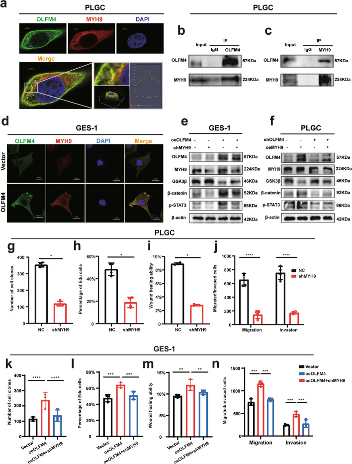
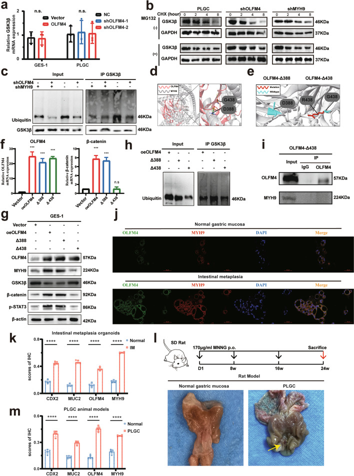
Results: Our results show that OLFM4 expression is highly increased in IIM, with superior diagnostic accuracy of IIM when compared to CDX2 and MUC2. OLFM4, along with MYH9, was overexpressed in IM organoids and PLGC animal models. Furthermore, OLFM4, in combination with Myosin heavy chain 9 (MYH9), accelerated the ubiquitination of GSK3β and resulted in increased β-catenin levels through the Wnt signaling pathway, promoting the proliferation and invasion abilities of PLGC cells.

Conclusions: OLFM4 represents a novel biomarker for IIM and could be utilized as an important auxiliary means to delimit the key population for early gastric cancer screening. Finally, our study identifies cell signaling pathways involved in the progression of IM.


Conflict of interest statement

The authors declare no competing interests.

Figures

Fig. 1
Fig. 1
OLFM4 was the remarkable DEG in PLGC cells: a HE staining of pathological sections in T1N0M0 stage gastric cancer: Intestinal metaplasia lesions co-occurring with gastric cancer infiltration throughout the mucosal layer (left), infiltration that broke through the basement membrane and invaded the submucosa (middle), and normal adjacent glandular epithelium with accompanying intestinal metaplastic lesions (right). Key indicators: The Black dashed line represented the basement membrane, and the yellow arrow highlighted gastric cancer invading the basement membrane with intestinal metaplasia. b Volcano plot from GSE78523 highlighting the top two DEGs, OLFM4 and MUC2, in intestinal metaplasia tissues. c Cluster analysis of epithelial cells in GSE134520. d Analysis of CNV in epithelial cell subgroups using inferCNV. eg Assessment of the differentiation of epithelial cell subgroups with Cytotrace. h-i Evaluation of OLFM4 expression in tissues and cell subgroups. IM: Intestinal metaplasia, EGC: Early gastric cancer, IMS: Sever intestinal metaplasia, NAG: non-atrophic gastritis, SPEM: Spasmolytic polypeptide expressing metaplasia, PLGC: Precancerous lesions of gastric carcinoma
Fig. 2
Fig. 2
OLFM4 was the biomarker of IIM tissues: a, b qPCR and Western Blot were used to assess CDX2, MUC2, and OLFM4 expression in normal gastric mucosa and intestinal metaplasia tissues. c HE staining and AB-PAS staining confirmed intestinal metaplasia diagnosis and the immunohistochemical status of CDX2, MUC2, and OLFM4 was assessed in the Training set. d Statistical analysis of CDX2, MUC2, and OLFM4 immunohistochemical scores and diagnostic efficacy for intestinal metaplasia in both the Training set and the Validation set. e HE staining and AB-PAS staining confirmed intestinal metaplasia subtypes, while OLFM4 immunohistochemistry was used in the Training set. f HE staining of intestinal metaplasia demonstrated red-stained Paneth cells (black arrow) and an intact brush border (red dashed line), confirming CIM. (g) Statistical analysis of CDX2, MUC2, and OLFM4 immunohistochemical scores and diagnostic efficacy for IIM in both the Training set and Validation set. Statistics were expressed as mean ± SD. *p < 0.05, **p < 0.01, ***p < 0.001, ****p < 0.0001. N: Normal gastric mucosa; IM:Intestinal metaplasia
Fig. 3
Fig. 3
OLFM4 was a biomarker of PLGC cells: a-d qPCR and Western Blot were used to assess the markers of intestinal metaplasia in the GES-1 cells incubated with different concentrations or different times of MNNG. e-f Transcriptome sequencing of GES-1 cells induced with 200 μmol/L MNNG for 48 h had been conducted, the volcano plot and the heatmap were drawn to show the DEGs. g-j Statistical analysis of repeated experimental data for cell cloning, EdU, wound healing, and transwell assays in PLGC cells and GES-1 cells overexpressing OLFM4 (oeOLFM4). k-n Statistical analysis of repeated experimental data for cell cloning, EdU, wound healing, and transwell assays after OLFM4 knockdown in PLGC cells (shOLFM4). Statistics were expressed as mean ± SD. *p < 0.05, **p < 0.01, ***p < 0.001, ****p < 0.0001
Fig. 4
Fig. 4
PLGC cells with high OLFM4 expression displayed activation of the Wnt signaling pathway: a The GSEA of the GSE78523 RNA-seq dataset in Hallmark genes and KEGG was analyzed. b The Addmodulescore analysis of the scRNA-seq dataset GSE134520 was analyzed to exhibit the functions enriched in the PLGC cells with high OLFM4 expression. c-d Western Blotting confirmed that the relation between the expression of OLFM4 and the expression of molecules related to Wnt/β-catenin signaling pathway, EMT transition, and c-Myc. e-f HE staining and AB-PAS staining were conducted to diagnose intestinal metaplasia, while the confocal immunofluorescence was performed to show the expression of OLFM4, Ki67, E-cadherin and Vimentin
Fig. 5
Fig. 5
OLFM4 interaction with MYH9 stimulated activation of the Wnt pathway: a The figures demonstrated the co-localization of OLFM4 (green) and MYH9 (red) in fixed and immunostained cells. Cells were imaged using confocal microscopy, and the overlay image (yellow) indicated regions of co-localization, where both proteins were present in close proximity. Nuclei were stained with DAPI (blue) to provide cellular landmarks. The scale bar representeds 5 μm. b-c In the forward Co-IP, an antibody specific to OLFM4 was used to immunoprecipitate the protein complex from cell lysates. Western Blot analysis subsequently revealed the presence of MYH9 in the immunoprecipitated complex, indicating that MYH9 was binding with OLFM4. Conversely, in the reverse Co-IP, an antibody specific to MYH9 was employed to immunoprecipitate the associated proteins. Western Blot analysis confirmed the presence of OLFM4 in the immunoprecipitated complex. d After overexpressing OLFM4 in GES-1 cells, the expression level of MYH9 was evaluated through confocal immunofluorescence analysis. The scale bar representd 10 μm. e-f Western Blot analysis demonstrated the effects of overexpressing OLFM4 followed by knocking down MYH9, or overexpressing MYH9 followed by knocking down OLFM4 on the expression levels of Wnt pathway signals. g-n Statistical analysis of repeated experimental data for cell cloning, EdU, wound healing, and transwell assays after MYH9 knockdown in PLGC cells (shMYH9) or in oeOLFM4 cells. Statistics were expressed as mean ± SD. *p < 0.05, **p < 0.01, ***p < 0.001, ****p < 0.0001
Fig. 6
Fig. 6
OLFM4 interacted with MYH9 to facilitate the ubiquitination of GSK3β: a qPCR assays demonstrated that relative GSK3β mRNA levels after GES-1 cells overexpression of OLFM4 or PLGC cells knockdown of OLFM4. b The CHX assay was performed by incubating the cells with CHX for different periods of time with or without MG132. Cycloheximide, CHX, an intracellular protein synthesis inhibitor. MG132, a proteasome inhibitor. c Western Blot experiments presented the effects of knocking down OLFM4 or MYH9 on ubiquitination levels and the expression of GSK3β in the Co-IP assay of PLGC cells. d The Pymol software constructed two crucial protein–protein dockings between OLFM4 and MYH9. e The Missense3D database predicted and constructed two mutations of OLFM4 protein. f-g GES-1 cells were transfected with vector, OLFM4 or OLFM4 mutation plasmids, and then subjected to qPCR and Western Blot for analysis the effect on the Wnt signaling pathway. h GES-1 cells were transfected with OLFM4 mutation plasmids and conducted with Co-IP assay for the ubiquitination level of GSK3β. i GES-1 cells were transfected with G438 mutation plasmids, and the Co-IP assay was executed to the interaction between OLFM4 and MYH9. j Immunofluorescence assays highlighted the elevated expression of OLFM4 and MYH9 in intestinal metaplasia organoids compared to normal gastric mucosa. k Statistical analysis of immunohistochemical scores for CDX2, MUC2, OLFM4, and MYH9 in intestinal metaplasia organoids. l Illustration of the PLGC animal model construction process and the presentation of rat gastric mucosa. m Statistical analysis of immunohistochemical scores of CDX2, MUC2, OLFM4, and MYH9 in the gastric mucosa of PLGC rats. Statistics were expressed as mean ± SD. *p < 0.05, **p < 0.01, ***p < 0.001, ****p < 0.0001
Fig. 7
Fig. 7
The study overview and the mechanism of OLFM4 promoting intestinal metaplasia: OLFM4 is highly expressed in incomplete intestinal metaplasia, and interacts with MYH9 to promote the progression of intestinal metaplasia via GSK3β/β-catenin signaling pathway

References

    1. Sung H, et al. Global Cancer Statistics 2020: GLOBOCAN Estimates of Incidence and Mortality Worldwide for 36 Cancers in 185 Countries. CA Cancer J Clin. 2021; 71: 209–2491. 10.3322/caac.21660. - PubMed
    1. Leung WK, et al. Screening for gastric cancer in Asia: current evidence and practice. Lancet Oncol. 2008;9:279–287. doi: 10.1016/s1470-2045(08)70072-x. - DOI - PubMed
    1. Wei H, et al. Gastric cancer clinical characteristics and their altered trends in South China: An epidemiological study with 2,800 cases spanning 26 years. 2023; 13. 10.3389/fonc.2023.976854. - PMC - PubMed
    1. Wu J, et al. CHD4 promotes acquired chemoresistance and tumor progression by activating the MEK/ERK axis. Drug resistance updates : reviews and commentaries in antimicrobial and anticancer chemotherapy. 2023;66:100913. doi: 10.1016/j.drup.2022.100913. - DOI - PubMed
    1. Li B, et al. Advances in immunology and immunotherapy for mesenchymal gastrointestinal cancers. Mol Cancer. 2023;22:71. doi: 10.1186/s12943-023-01770-6. - DOI - PMC - PubMed

MeSH terms

LinkOut - more resources