. 2024 Jun 7;22(1):314.
doi: 10.1186/s12964-024-01694-8.

The FABD domain is critical for the oncogenicity of BCR/ABL in chronic myeloid leukaemia

Affiliations

The FABD domain is critical for the oncogenicity of BCR/ABL in chronic myeloid leukaemia

Renren Zheng et al. Cell Commun Signal. .

Abstract

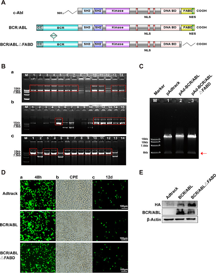
Background: Abnormally expressed BCR/ABL protein serves as the basis for the development of chronic myeloid leukaemia (CML). The F-actin binding domain (FABD), which is a crucial region of the BCR/ABL fusion protein, is also located at the carboxyl end of the c-ABL protein and regulates the kinase activity of c-ABL. However, the precise function of this domain in BCR/ABL remains uncertain.

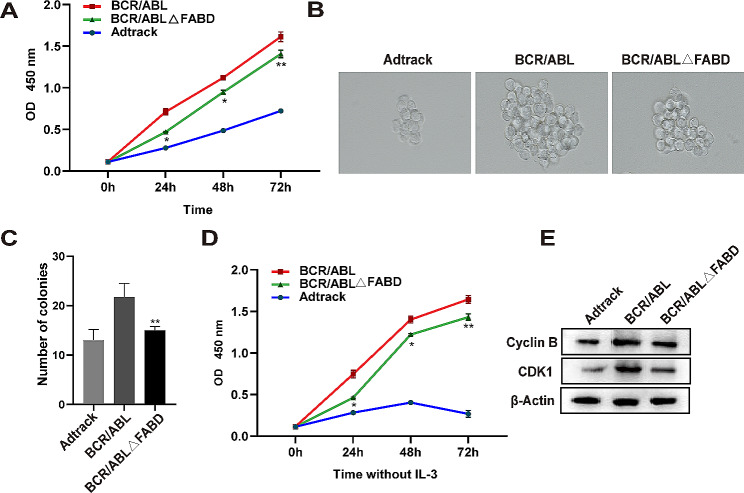
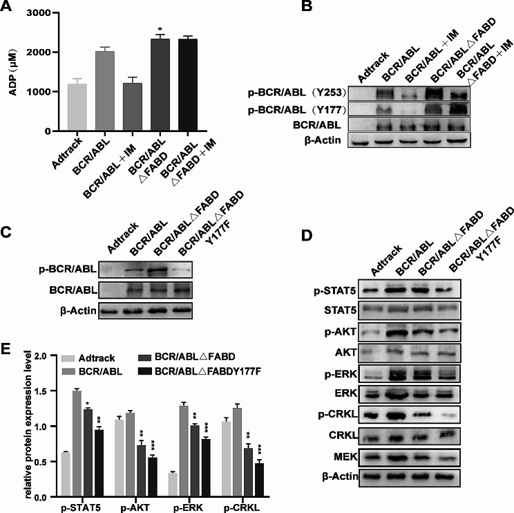
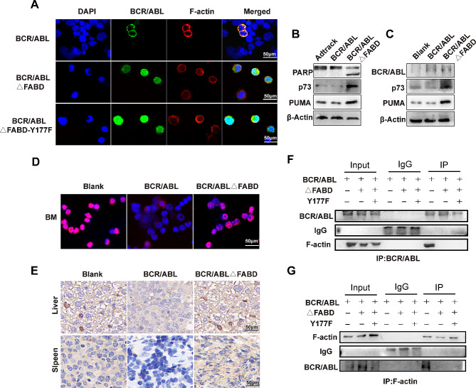
Methods: The FABD-deficient adenovirus vectors Ad-BCR/ABL△FABD, wild-type Ad-BCR/ABL and the control vector Adtrack were constructed, and 32D cells were infected with these adenoviruses separately. The effects of FABD deletion on the proliferation and apoptosis of 32D cells were evaluated by a CCK-8 assay, colony formation assay, flow cytometry and DAPI staining. The levels of phosphorylated BCR/ABL, p73, and their downstream signalling molecules were detected by western blot. The intracellular localization and interaction of BCR/ABL with the cytoskeleton-related protein F-actin were identified by immunofluorescence and co-IP. The effect of FABD deletion on BCR/ABL carcinogenesis in vivo was explored in CML-like mouse models. The degree of leukaemic cell infiltration was observed by Wright‒Giemsa staining and haematoxylin and eosin (HE) staining.

Results: We report that the loss of FABD weakened the proliferation-promoting ability of BCR/ABL, accompanied by the downregulation of BCR/ABL downstream signals. Moreover, the deletion of FABD resulted in a change in the localization of BCR/ABL from the cytoplasm to the nucleus, accompanied by an increase in cell apoptosis due to the upregulation of p73 and its downstream proapoptotic factors. Furthermore, we discovered that the absence of FABD alleviated leukaemic cell infiltration induced by BCR/ABL in mice.

Conclusions: These findings reveal that the deletion of FABD diminished the carcinogenic potential of BCR/ABL both in vitro and in vivo. This study provides further insight into the function of the FABD domain in BCR/ABL.

Keywords: BCR/ABL; Chronic myeloid leukaemia; F-actin binding domain; Oncogenicity; p73.


Conflict of interest statement

The authors declare no competing interests.

Figures

Fig. 1
Fig. 1
The expression of the BCR/ABL and BCR/ABLΔFABD proteins was successfully achieved using adenoviral vectors. (A) Schematic diagrams of c-ABL, BCR/ABL and BCR/ABLΔFABD. (B) The shuttle plasmids pAdtrack-CMV (a), pAdtrack-BCR/ABL (b) and pAdtrack-BCR/ABLΔFABD (c) were recombined with p-AdEasy-1 in BJ5183 bacteria. Agarose gel electrophoresis was then performed to screen the proper recombinant plasmids. The successful recombinant plasmids exhibited a greater molecular weight. The red rectangles in each agarose gel highlight typical successful recombinant plasmids. (C) Identification of recombinant adenoviral plasmids by Pac I enzyme digestion. M represents the DL15000 DNA marker; Lanes 1, 2 and 3 represent the recombinant adenoviral plasmids pAdtrack, pAd-BCR/ABL and pAd-BCR/ABL△FABD digested by PacI respectively. The correct recombinant adenovirus plasmids could be digested by the PacI restriction endonuclease to liberate both inverted terminal repeats (LITR and RITR), resulting in the release of a larger 30 kb fragment and a smaller 4.5 kb or 3 kb fragment (marked with a red arrow) [28]. (D) Packaging of adenoviruses in AD293 cells. AD293 cells were transfected with the respective recombinant adenoviral plasmids. (a) Then, the fluorescence intensity was detected at 48 h after transfection. (b) Cytopathic effects (CPEs) were observed under an inverted microscope 12 days later. (c) The fluorescence intensity decreased after 12 days of transfection. (E) 32D cells were infected with Ad-Adtrack, Ad-BCR/ABL, or Ad-BCR/ABLΔFABD adenoviruses for 48 h. The expression of BCR/ABL or BCR/ABLΔFABD was then detected by western blot analysis with an HA antibody or a c-ABL antibody. The HA tag was incorporated into the N-terminal region of BCR/ABL and BCR/ABLΔFABD
Fig. 2
Fig. 2
Deletion of FABD attenuates the ability of BCR/ABL to promote cell proliferation. (A) 32D cells were infected with Ad-Adtrack, Ad-BCR/ABL, or Ad-BCR/ABLΔFABD adenoviruses for the indicated times, after which cell viability was determined by a CCK-8 assay. (B, C) The effect of FABD deletion on the ability of cells to form colonies was assessed through a colony formation assay. 32D cells were infected with Ad-Adtrack, Ad-BCR/ABL, or Ad-BCR/ABLΔFABD adenoviruses for the indicated times, after which they were plated in semisolid medium. The number and morphology of the colonies were observed 10 days later. Each group was tested in quadruplicate, and the experiment was repeated three times. (D) The influence of FABD deletion on the transformation potential of BCR/ABL was evaluated through an IL-3 withdrawal experiment. 32D cells were infected with Ad-Adtrack, Ad-BCR/ABL, or Ad-BCR/ABLΔFABD adenoviruses and cultured in IL-3-free medium for the indicated times. Then, cell viability was determined by a CCK-8 assay. (E) 32D cells were infected with Ad-Adtrack, Ad-BCR/ABL, or Ad-BCR/ABLΔFABD adenoviruses for 48 h. The protein levels of Cyclin B and CDK1, which are associated with proliferation, were evaluated by western blotting
Fig. 3
Fig. 3
Deletion of FABD reduces the ability of BCR/ABL to antagonize apoptosis. (A) 32D cells were infected with Ad-Adtrack, Ad-BCR/ABL, or Ad-BCR/ABLΔFABD adenoviruses for a period of 48 h. Subsequently, apoptotic cells were identified by flow cytometry. The blank represents 32D cells with no treatment. Each experiment was conducted in triplicate. (B) Statistical analysis of the apoptosis results detected by flow cytometry. (C) 32D cells were infected with Ad-Adtrack, Ad-BCR/ABL, or Ad-BCR/ABLΔFABD adenoviruses for 48 h. Then, the nuclear morphology was observed following DAPI staining. The white arrow indicates typical nucleic fragmentation
Fig. 4
Fig. 4
Deletion of FABD relieves the severity of CML-like disease caused by BCR/ABL in mice. A total of 5 × 106 32D cells infected with Ad-BCR/ABL or Ad-BCR/ABLΔFABD adenoviruses were injected intravenously into each BALB/c mouse. A control group of BALB/c mice was injected with PBS and designated the blank group. There were 5 mice in each group. (A) The number of white blood cells (WBCs) in the peripheral blood was counted and recorded every week. The peak value of the WBC in each mouse was recorded and statistically analysed. (B, C) The weights of the liver and spleen of each mouse were measured at the time of execution and statistically analysed. (D) Representative images of the spleens and livers of mice from each group were shown. (E) Cells from the bone marrow, liver, and spleen of mice were stained with Wright‒Giemsa stain. The infiltration of leukaemic cells in these tissues was observed after staining. (F) Tissues from mouse liver, spleen, and lung were sectioned and stained with HE. The infiltration of leukaemic cells in these tissues was observed after staining. (G) The survival status of each group was recorded and analysed. The survival curves were generated with GraphPad 8.0
Fig. 5
Fig. 5
FABD deletion increases p-Y177 levels. (A) 32D cells were infected with Ad-Adtrack, Ad-BCR/ABL, Ad-BCR/ABL△FABD or Ad-BCR/ABLΔFABD-Y177F adenoviruses for the indicated times. Then, total protein kinase activity was tested by a kinase activity assay according to the kit instructions. (B) The levels of p-BCR/ABL (Y253) and p-BCR/ABL (Y177) in each group were evaluated by western blotting. (C) The tyrosine at the Y177 site in BCR/ABLΔFABD was mutated to phenylalanine. The expression of p-BCR/ABL or BCR/ABL was evaluated by western blotting after infection with the respective adenoviruses. (D) The expression of p-STAT5, p-AKT, p-ERK, p-CRKL and MEK was evaluated by western blotting after infection with the respective adenoviruses. (E) The ratio of p-STAT5, p-AKT, p-ERK or p-CRKL to the corresponding total protein was quantified
Fig. 6
Fig. 6
FABD deletion does not function through p-Y177 elevation. We detected whether Y177F was able to reverse the proliferation inhibition and apoptosis induction caused by BCR/ABLΔFABD. 32D cells were infected with Ad-Adtrack, Ad-BCR/ABL, Ad-BCR/ABL△FABD or Ad-BCR/ABLΔFABD-Y177F adenoviruses and then cultured in medium supplemented with (A) or without (B) IL-3 for the indicated times. The cell viability in each group was determined by a CCK-8 assay. For the colony-forming assay, 32D cells were infected with Ad-BCR/ABL, Ad-BCR/ABLΔFABD, or Ad-BCR/ABLΔFABD-Y177F adenoviruses for the indicated times and then plated in semisolid medium. The morphology (C) and number (D) of colonies were observed 10 days later. Each group was repeated three times. For the apoptosis detection assay, 32D cells were treated as described above, and apoptosis was examined by flow cytometry (E). The results for each group were statistically analysed (F)
Fig. 7
Fig. 7
FABD deletion mediates the translocation of BCR/ABL into the nucleus, where it activates p73 to induce apoptosis. (A) 32D cells were infected with Ad-BCR/ABL, Ad-BCR/ABLΔFABD, or Ad-BCR/ABLΔFABD-Y177F adenoviruses for 48 h. Thereafter, the locations of BCR/ABL and F-actin were examined by immunofluorescence. The cell nuclei were stained with DAPI. BCR/ABL was visualized by a FITC-conjugated specific antibody, while F-actin was detected by a Cy-3-conjugated specific antibody. The scale represents 50 μm. (B) 32D cells were infected with the corresponding adenoviruses for 48 h, after which the expression of PARP, p73 and PUMA was analysed by western blotting. (C) The expression of BCR/ABL, p73 and PUMA in mouse bone marrow cells from the corresponding groups was analysed by western blotting. (D) The localization and expression of p73 in bone marrow cells were detected by immunofluorescence. (E) The expression of p73 in mouse liver and spleen cells from the corresponding groups was detected by immunohistochemistry. (F, G) 32D cells were infected with the corresponding adenoviruses for 48 h, after which the interaction between BCR/ABL and F-actin was analysed by coimmunoprecipitation. ΔFABD represents BCR/ABLΔFABD, and Y177F represents BCR/ABLΔFABD-Y177F
Fig. 8
Fig. 8
A schematic diagram of our study. Wild-type BCR/ABL is located in the cytoplasm through interactions with F-actin and stimulates downstream pathways to promote proliferation and antagonize apoptosis. When the FABD domain is deleted, the interaction between BCR/ABL and F-actin is interrupted. Then, BCR/ABLΔFABD translocates into the nucleus and stimulates p73 and its downstream molecules to induce apoptosis. Moreover, due to the decrease in cytoplasmic BCR/ABL, proliferative stimuli are weakened

References

    1. Cortes J, Pavlovsky C, Saußele S. Chronic myeloid leukaemia. Lancet. 2021;398(10314):1914–26. doi: 10.1016/S0140-6736(21)01204-6. - DOI - PubMed
    1. Braun TP, Eide CA, Druker BJ. Response and resistance to BCR-ABL1-Targeted therapies. Cancer Cell. 2020;37:530–42. doi: 10.1016/j.ccell.2020.03.006. - DOI - PMC - PubMed
    1. Bartram CR, de Klein A, Hagemeijer A, et al. Translocation of c-ab1 oncogene correlates with the presence of a Philadelphia chromosome in chronic myelocytic leukaemia. Nature. 1983;306:277–80. doi: 10.1038/306277a0. - DOI - PubMed
    1. Klil-Drori AJ, Yin H, Azoulay L, et al. Molecular monitoring of therapeutic milestones and clinical outcomes in patients with chronic myeloid leukemia. Cancer. 2019;125:618–25. doi: 10.1002/cncr.31835. - DOI - PubMed
    1. Rousselot P, Coudé MM, Gokbuget N, et al. Dasatinib and low-intensity chemotherapy in elderly patients with Philadelphia chromosome-positive ALL. Blood. 2016;128:774–82. doi: 10.1182/blood-2016-02-700153. - DOI - PMC - PubMed

Publication types

MeSH terms