. 2024 Jul 22;52(13):e56.
doi: 10.1093/nar/gkae472.

iSuRe-HadCre is an essential tool for effective conditional genetics

Affiliations

iSuRe-HadCre is an essential tool for effective conditional genetics

Irene Garcia-Gonzalez et al. Nucleic Acids Res. .

Abstract

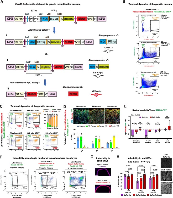
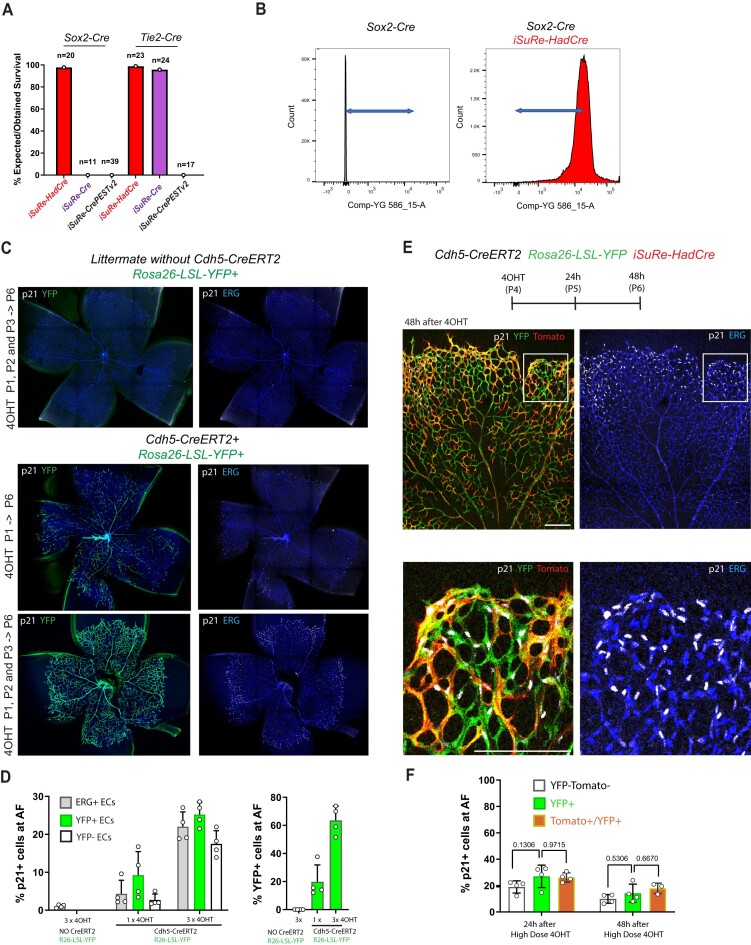
Methods for modifying gene function at high spatiotemporal resolution in mice have revolutionized biomedical research, with Cre-loxP being the most widely used technology. However, the Cre-loxP technology has several drawbacks, including weak activity, leakiness, toxicity, and low reliability of existing Cre-reporters. This is mainly because different genes flanked by loxP sites (floxed) vary widely in their sensitivity to Cre-mediated recombination. Here, we report the generation, validation, and utility of iSuRe-HadCre, a new dual Cre-reporter and deleter mouse line that avoids these drawbacks. iSuRe-HadCre achieves this through a novel inducible dual-recombinase genetic cascade that ensures that cells expressing a fluorescent reporter had only transient Cre activity, that is nonetheless sufficient to effectively delete floxed genes. iSuRe-HadCre worked reliably in all cell types and for the 13 floxed genes tested. This new tool will enable the precise, efficient, and trustworthy analysis of gene function in entire mouse tissues or in single cells.


Figures

Graphical Abstract
Graphical Abstract
Figure 1.
Figure 1.
Caveats of the iSuRe-Cre allele. (A) Simplified schematic of the published iSuRe-Cre allele. After combining it with any other CreERT2 allele, is possible to induce its recombination with tamoxifen and analyse cells with permanent expression of the reporter MbTomato and Cre. (B) FACS analysis of iSuRe-Cre self-leakiness in organs from adult (8 weeks old) and postnatal mice (postnatal day 7). Note that major leakiness occurs only in adult mice. (C) FACS analysis of iSuRe-Cre self-leakiness (MbTomato+ cells) versus non-self-leakiness with the Rosa26-Lox-Stop-Lox-EYFP allele (YFP+ cells). In general, non-self-leakiness is higher than self-leakiness. (D) FACS analysis of iSuRe-Cre self-leakiness (MbTomato+ cells in red bar) versus non-self-leakiness with the Rosa26-Lox-MbTomato-Lox-MbEGFP allele (GFP+ cells in animals with or without iSuRe-Cre). (E) Confocal micrographs of adult mouse tissues, showing inadvertent recombination in a smooth muscle cell (SMC) and EC. (F-H) Images of vascular lesions on snout (F), brain (G) and ear skin (H) of uninduced adult iSuRe-Cre Rosa26-Lox-Stop-Lox-Pik3caH1047R mice. (I) Stereomicroscopy images of leg muscle and heart tissue showing endogenous MbTomato expression due to self-leakiness in iSuRe-Cre mice, higher in animals with 2 copies (ki/ki) of the allele. (J) FACS analysis of the sensitivity of the indicated alleles to CreERT2-dependent recombination. (K) FACS analysis of the frequency of false positives (MbTomato or iSuRe+,YFP), true positives (iSuRe+,YFP+), and false negatives (YFP+, iSuRe) in liver ECs after the administration of different tamoxifen doses to mice carrying the indicated alleles. (L) FACS analysis of the frequency of false and true positives among iSuRe-Cre/Tomato+ cells in postnatal day 7 liver ECs. (M) Expected and obtained survival ratios of animals containing the germline recombined iSuRe-Cre allele (originally on the G4 or C57Bl6x129 background) across generations of breeding with the C57Bl6 strain. (N) Confocal micrographs used to compare the frequency of cells positive for MbTomato-2A-Cre+ (from the iSuRe-Cre allele) and for GFP+ (from the Rosa26-Lox-mT-Lox-mG allele) in retinas of animals in the C57Bl6 background. The drop in the frequency of MbTomato-2A-Cre+ cells from P6 to P28 indicates Cre toxicity in ECs during the development of retinal vessels. (O) Representative confocal micrograph showing that when animals containing the three indicated alleles are in the C57Bl6 background, MbTomato/iSure-Cre+ cells are outcompeted by EYFP+ cells during retinal vascular development. (P) Expected and obtained survival ratios for mice containing the iSuRe-Cre and Tie2-Cre alleles in the mixed C57Bl6 x CD1 genetic background. These mice have similar frequencies of MbTomato+ blood and ECs (iSuRe-Cre + or R26-LSL-TdTomato+), suggesting no toxicity in this case. Confocal micrographs to the right show representative variable recombination and expression in two retinas of the same animal, with no major differences in angiogenesis (ECs are isolectinB4+). Data are presented as mean values ± s.d. For statistics, see Source Data File 1. Scale bars in E, 50μm; in N 200 μm; and in H, O and P, 500 μm.
Figure 2.
Figure 2.
Characterization of the iSuRe-CrePESTv1 allele. (A) Crispr-Cas9 genetic targeting of the preexisting iSuRe-Cre allele, present in Chromosome 17, with the indicated donor DNA to generate the new iSuRe-CrePESTv1 allele shown below. (B) Genetic cascade and expression outcomes after induction of the iSuRe-CrePESTv1 allele with CreERT2 + tamoxifen. After the first recombination event induced by the activation of CreERT2 with tamoxifen, cells will express MbTomato, CreERT2 and FlpO. In a second step, MbTomato+ cells with high FlpO activity will undergo genetic fusion of the PEST domain to Cre and co-express equimolar amounts of MbTomato, CrePEST, and H2B-V5. CrePEST activity is higher than CreERT2 but, due to its higher degradation, presumably lower than Cre activity. Other abbreviations and construct elements are detailed in the DNA engineering section in Methods. (C) FACS analysis of MbTomato+ cells in iSuRe-Cre and iSuRe-CrePESTv1 animals reveals significantly less self-leakiness in the latter. (D) FACS analysis and charts showing the frequency of false positives (MbTomato+, YFP) and true positives (MbTomato+, YFP+) among MbTomato+ cells from several adult organs of animals containing the Cdh5-CreERT2, iSuRe-CrePESTv1 and Rosa26-LSL-YFP alleles (yellow bars) or the Cdh5-CreERT2, iSuRe-Cre, and Rosa26-LSL-YFP alleles (magenta bars). Recombination efficiency (true positives frequency) is lower in mice containing the iSuRe-CrePESTv1 allele. (E) P6 retina confocal micrographs showing the efficiency of the recombination cascade (MbTomato activation followed by H2B-V5) of the iSuRe-CrePESTv1 allele (induced with Cdh5-CreERT2 and tamoxifen at P1). Red arrows indicate cells with only MbTomato expression (false positives); yellow arrows, cells with MbTomato and EYFP expression (partial recombination of the iSuRe-CrePESTv1 allele); and white arrows, cells with expression of MbTomato, EYFP, and H2B-V5 (true positives). (F) Chart showing the low efficiency (% of V5+ cells) of the second, FlpO-mediated, iSuRe-CrePESTv1 recombination event in MbTomato+ cells (these were initially induced by CreERT2 at P1). (G) Quantification of MbTomato reporter signal intensity (from confocal images) in cells that have undergone only the first recombination event (H2B-V5) or all recombination events (H2B-V5+). Each dot is one cell intensity. (H) Frequency of cells expressing the different reporters combinations (EYFP, MbTomato, and H2B-V5) among all recombined cells in P6 retinas of mice containing the alleles iSuRe-CrePESTv1, Rosa26-LSL-YFP and Cdh5-CreERT2. Note the very low frequency of MbTomato+, H2B-V5+ cells, but that most of these are YFP+ (true positives). (I) Frequency of false positives (YFP) and true positives (YFP+) among MbTomato cells (green bar) or MbTomato+, H2B-V5 cells (red bars) or MbTomato+, H2B-V5+ cells (orange bars), revealing that only H2B-V5+ cells accurately indicate cells with high Cre activity, thanks to the permanent CrePEST expression. Data are presented as mean values ± s.d. For statistics, see Source Data File 1. Scale bar, 200 μm.
Figure 3.
Figure 3.
Characterization of the Rosa26-iSuRe-CrePESTv2 allele. (A) Schematic of the Rosa26-iSuRe-CrePESTv2 allele. Tamoxifen–induced CreERT2 activity leads to deletion of the stop cassette containing three Sv40 polyA sequence repeats, triggering co-expression of CrePEST and the reporter MbTomato. (B) Confocal microscopy analysis of the sensitivity to CreERT2-induced recombination of the Rosa26-iSuRe-CrePESTv2 and iSuRe-Cre alleles (MbTomato+) relative to the internal control Rosa26-LSL-EYFP allele (each dot represents one retina sample). (C) FACS analysis of the self-leakiness frequency (MbTomato + cells) in adult organs of iSuRe-Cre versus Rosa26-iSuRe-CrePESTv2 animals (each dot represents a measure in one animal). (D) Quantification of confocal micrographs reflecting the relative frequency of MbTomato+ cells (Rosa26-iSuRe-CrePESTv2) and YFP+ cells (Rosa26-LSL-EYFP) 24 and 96 h after tamoxifen induction at P4. Most MbTomato+ cells are already YFP+ (true positives, yellow arrows) at 24 h postinduction. Very few are MbTomato+ and YFP (false positives, red arrow) at 24 h, and almost none later. (E) MbTomato-2A-CrePEST+ cells become 6 times less frequent 4 days after induction, indicating toxicity of CrePEST expression. (F–H) Retina confocal micrographs and corresponding charts showing a significant decrease in angiogenesis and cell proliferation in animals containing the induced Rosa26-iSuRe-CrePESTv2 allele. Each dot in the charts represents one large retina field (in total 15403 cells were quantified in G and 8795 in H). (I, J) Animals containing the Sox2-Cre (recombines all cells) or Tie2-Cre (recombines only ECs and blood) allele in combination with the Rosa26-iSuRe-CrePESTv2 allele are not born. Data are presented as mean values ± s.d. For statistics, see Source Data File 1. Scale bars, 70 μm in D and 120 μm in E–H.
Figure 4.
Figure 4.
Design and validation of the Rosa26-iSuRe-HadCre mouse allele. (A) Construct and sequential genetic cascade after induction of the Rosa26-iSuRe-HadCre allele. After induction of CreERT2 with tamoxifen, the first recombination event results in deletion of either the LoxP or the LoxN cassette, or of both Lox-flanked genetic cassettes. Since the LoxP-flanked DNA cassette is easier to recombine than the LoxN-flanked cassette, in the presence of relatively low levels of CreERT2 induction, LoxP recombination will predominate after a single 4-OHT injection (as illustrated in the figure), and this will trigger very strong expression of CreERT2 after. This strong CreERT2 expression will facilitate/enhance LoxN cassette recombination, especially if a second injection of tamoxifen (4-OHT) is delivered after 24 h. After recombination of both LoxP and LoxN-flanked cassettes (after a single or multiple injections of tamoxifen), there will be strong, equimolar co-expression of Cre and the weaker FlpO recombinase. FlpO will recombine FRT sites, self-deleting the construct and activating expression of the reporter MbTomato in cells that had, but no longer have, high levels of Cre expression. (B) FACS analysis of the temporal dynamics of the genetic cascade upon 4-OHT induction. Littermate pups with the indicated alleles were injected with a single 4-OHT dose at P4, and lung cells were collected at P5, P6 or P7. (C) FACS analysis of the kinetics of recombination upon induction of the Rosa26-iSuRe-HadCre allele in fibroblasts derived from the indicated mice. (D) Confocal analysis of the temporal dynamics of the genetic cascade upon induction of the Rosa26-iSuRe-HadCre allele in retinal vessels after 15 mg/kg dose of 4-OHT. Note that the dose of 4-OHT used is low and will not recombine all the reporters in all cells. (E) Relative inducibility of the iSure-Cre and Rosa26-iSuRe-HadCre alleles in relation to the reference Rosa26-LSL-EYFP allele at different developmental stages and in different organs. (F) FACS analysis of the correlation between the number of tamoxifen doses and the frequency of recombination. Two doses induce higher recombination rates of the Rosa26-iSuRe-HadCre allele in relation to the Rosa26-LSL-EYFP allele. (G) Comparative analysis on the recombination rate in adult aorta SMCs (Myh11-CreERT2+). (H) Comparative analysis of recombination rates at very high doses of tamoxifen in adult animals, some with 2 copies (2×) of the iSuRe-HadCre allele. Each dot in the charts represents the mean value obtained in one animal. Data are presented as mean values ± s.d. For statistics, see Source Data File 1. Scale bars, 200 μm.
Figure 5.
Figure 5.
Rosa26- iSuRe-HadCre is bright, non-leaky and generates a high rate of true positives. (A) Comparative FACS analysis of reporter (MbTomato) intensity in recombined cells at different stages and in different organs. (B) Comparative FACS analysis of self-leakiness in iSure-Cre and iSuRe-HadCre mice organs. (C) Representative FACS plots showing no leakiness in iSuRe-HadCre mice organs. (D) Comparative FACs analysis of the rate of true positives (YFP+) and false positives (YFP) at embryonic stages in different mouse lines when combined with the compatible reference Rosa26-LSL-YFP allele. (E) Comparative confocal analysis of the rate of true and false positives in postnatal retinas from different mouse lines when combined with the compatible reference Rosa26-LSL-YFP allele. (F) Comparative FACS analysis of the rate of true and false positives in adult liver and lung cells; recombination in these quiescent cells is generally more difficult to achieve than in embryonic or postnatal cells. (G, H) Analysis by FACS of the frequencies of YFP+ (blue laser) and MbTomato+ (yellow laser) CD31CD45 cells (all organ cells except CD45+ blood and CD31+ ECs) extracted from the indicated mice six days after receiving a single injection of 4-OHT. Note the difference in the frequency of false positives between the iSuRe-HadCre and the Rosa26-LSL-tdTomato line. Data are presented as mean values ± s.d. For statistics, see Source Data File 1. Scale bars, 200 μm.
Figure 6.
Figure 6.
Rosa26- iSuRe-HadCre increases the efficiency and reliability of conditional genetics. (A–C) Representative confocal micrographs of P6 retinal vessels from control mice (A) and Notch1 loss-of-function (LOF) mice (B) with high recombination rates of the R26-LSL-EYFP allele; vessels were stained with anti-ERG (labels EC nuclei, for object segmentation), isolectinB4 (labels EC surface), and anti-GFP (detects YFP). Quantification of the contribution of EYFP+ ECs (ERG+, isolectinB4+) to arteries (indicated with a red A), arterial length, and vascular density reveals no significant differences, indicating poor Notch1 deletion (C). (D–F) Representative confocal micrographs of P6 retinal vessels from control mice (D) and Notch1 LOF mice (E) with high recombination rates of the iSuRe-HadCre allele. Quantification of the contribution of MbTomato + ECs to arteries, arterial length, and vascular density shows a very significant difference, confirming complete deletion of the Notch1 gene in MbTomato + cells in animals containing the iSuRe-HadCre allele. (G) Comparative analysis of the contribution of R26-LSL-EYFP (YFP+) and iSuRe-HadCre (MbTomato+) ECs to retinal arteries (indicated with a red A), versus all capillaries, confirming that only cells expressing the recombined iSuRe-HadCre allele have full deletion of Notch1 and cannot form arteries. (H) scRNAseq analysis of ECs collected from livers and hearts by FACS indicate strong Notch1 deletion in cells expressing the iSuRe-Cre or iSuRe-HadCre alleles. (I) Representative confocal micrograph showing retinal vessels (IsolectinB4+, ERG+) and quantification of the contribution of MbTomato+ ECs to the entire vascular network versus arteries, confirming very efficient deletion of Rbpj in the large majority of MbTomato+ cells. Data are presented as mean values ± s.d. For statistics, see Source Data File 1. Scale bars, 200 μm.
Figure 7.
Figure 7.
iSuRe-HadCre enables effective deletion of Vegfr2 in the entire target tissue or in single cells. (A, B) Representative confocal micrographs of P6 retina vessels of control and Vegfr2 floxed mice stained with anti-ERG (EC nuclei), IsolectinB4 (EC surface) and anti-GFP (detects YFP). Only animals containing the iSuRe-HadCre allele have complete and consistent deletion of the Vegfr2 gene, resulting in a profound decrease in the number of blood vessels (including arteries (A) and veins (V)) and ERG + ECs. (C, D) Single cell mosaic analysis in retinas with low induction of the iSuRe-HadCre allele, showing that gene deletion is very effective in single cells and that single Vegfr2 mutant cells cannot migrate to the angiogenic front (area above the dashed line) and do not form tip cells. (E) Immunostaining for VEGFR2 in iSuRe-HadCre mosaic retinas (note that VEGFR2 is expressed by ECs and many non-ECs in retinas) showing that MbTomato + cells (some indicated by white arrowheads) have deletion of Vegfr2, whereas many MbTomato negative cells (green arrowheads) do not have deletion of Vegfr2. (F) Confocal micrographs of liver cryosections showing the deletion of Vegfr2/Kdr in adult mice (8 weeks) liver ECs (Cdh5 + and ERG + nuclei). VEGFR2 immunostaining confirms the high deletion efficiency in MbTomato + cells of Vegfr2 floxed mutants. Loss of VEGFR2 leads to the loss of liver sinusoids. Mosaic induction reveals that most Vegfr2 mutant ECs survive in the larger portal veins and central veins vessels but not in sinusoidal capillaries. (G) Confocal micrographs of skin vessels of the indicated mice induced at P1 and P2 and collected at P21. Immunostaining with anti-VEGFR2 labels both lymphatic and blood endothelial cells. When recombination is mosaically induced by Prox1-CreERT2, only iSuRe-HadCre+/MbTomato + lymphatic ECs of Vegfr2 floxed mutants loose VEGFR2 expression (see also Supplementary Figure S7). Scale Bars 200μm in A, C, F; 50 μm in E and G.
Figure 8.
Figure 8.
iSuRe-HadCre enables the deletion of multiple floxed genes in the entire target tissue or in single cells. (A) Retina confocal micrographs showing effective deletion of Foxo genes in MbTomato+ ECs (FOXO1 imunostaining and phenotypes characteristic of full Foxos depletion, such as defective EC sprouting and dense vasculature). (B) Confocal micrographs showing that one single injection of 4-OHT at P1 efficiently recombines iSuRe-HadCre in all retina ECs and deletes Dll4, leading to the strong upregulation of the tip cell marker gene Esm1 in most cells, and cell-cycle exit. ERG + ECs become Ki67- at the angiogenic front. (C) One single injection of 4-OHT at P1 efficiently recombines iSuRe-HadCre in all retina ECs and deletes the gene Jagged1, leading to the strong inhibition of angiogenesis. (D) The membrane-tagged Tomato protein from iSuRe-HadCre allows visualization of filopodia by confocal microscopy. Deletion of the indicated genes change the number of filopodia per vessel length. (E, F) Effective deletion of Flt1/Vegfr1 is indicated by the loss of FLT1 protein in MbTomato+ ECs from the indicated mice. Note that Flt1 antibody signal is noisy. Specific signal is perinuclear/cytoplasmic. (G) Retina confocal micrographs of animals with mosaic induction of the iSuRe-HadCre allele showing that Myc and Mycn (both detected with Anti-Myc) are only effectively deleted in cells expressing the iSuRe-HadCre allele (MbTomato + cells, white and orange arrows). (H) Confocal micrographs of mouse aortas showing the effective deletion of Rbpj in MbTomato + of SMCs (Myh11+). White arrows indicate some nuclei/cells with MbTomato expression and lacking RBPJ, and green arrows without MbTomato expression and retaining RBPJ expression. Histogram showing the % of cells expressing RBPJ. Data are presented as mean values ± s.d. For statistics, see Source Data File 1. Scale Bars 200μm, except D, 50 μm and H, 25 μm.
Figure 9.
Figure 9.
Expression of the iSuRe-HadCre allele does not elicit cellular toxicity. (A) Comparative analysis of survival rates of C57Bl6 animals containing the Sox2-Cre (recombines all cells), or Tie2-Cre (recombines only ECs and blood) alleles when combined with the different iSuRe-Cre lines. Only the iSuRe-HadCre is devoid of toxicity. (B) Histogram plot showing intensity of MbTomato signals in all liver cells of adult Sox2-Cre iSuRe-HadCre mice, confirming that the expression of the recombined allele is ubiquituous, permanent and non-toxic. (CD) Confocal analysis of retinas from pups induced at P1, P2 and P3 (or only P1) and collected at P6. Cellular toxicity can be scored by the expression of the replicative stress or cell senescence marker p21 in ERG + ECs. The expression of this marker is much higher in retinas from animals containing the Cdh5-CreERT2 allele, and among these in retinas having a higher rate of recombination of the Rosa26-LSL-YFP allele (injected with 3 times more tamoxifen). (E, F) Short-term (24h and 48h after 4-OHT) analysis of the toxicity marker p21 in angiogenic front (AF) ECs expressing the Rosa26-LSL-YFP (YFP+) or the iSuRe-HadCre (MbTomato+/YFP+) allele reveal no additional cellular toxicity by the transient expression of Cre in the first 24–48 h after 4-OHT. Boxed areas showed at higher magnification below. Data are presented as mean values ± s.d. For statistics, see Source Data File 1. Scale bars, 200 μm.

References

    1. Skarnes W.C., Rosen B., West A.P., Koutsourakis M., Bushell W., Iyer V., Mujica A.O., Thomas M., Harrow J., Cox T. et al. A conditional knockout resource for the genome-wide study of mouse gene function. Nature. 2011; 474:337–342. - PMC - PubMed
    1. Luo L., Ambrozkiewicz M.C., Benseler F., Chen C., Dumontier E., Falkner S., Furlanis E., Gomez A.M., Hoshina N., Huang W.H. et al. Optimizing Nervous System-Specific Gene Targeting with Cre Driver Lines: Prevalence of Germline Recombination and Influencing Factors. Neuron. 2020; 106:37–65. - PMC - PubMed
    1. Murray S.A., Eppig J.T., Smedley D., Simpson E.M., Rosenthal N. Beyond knockouts: cre resources for conditional mutagenesis. Mamm. Genome. 2012; 23:587–599. - PMC - PubMed
    1. Schmidt-Supprian M., Rajewsky K. Vagaries of conditional gene targeting. Nat. Immunol. 2007; 8:665–668. - PubMed
    1. Matthaei K.I. Genetically manipulated mice: A powerful tool with unsuspected caveats. J. Physiol. 2007; 582:481–488. - PMC - PubMed