

The Masquelet induced membrane technique with PRP-FG-nHA/PA66 scaffold can heal a rat large femoral bone defect
- PMID: 38851675
- PMCID: PMC11162015
- DOI: 10.1186/s12891-024-07567-y
The Masquelet induced membrane technique with PRP-FG-nHA/PA66 scaffold can heal a rat large femoral bone defect
Abstract
Background: Masquelet membrane induction technology is one of the treatment strategies for large bone defect (LBD). However, the angiogenesis ability of induced membrane decreases with time and autologous bone grafting is associated with donor site morbidity. This study investigates if the PRP-FG-nHA/PA66 scaffold can be used as a spacer instead of PMMA to improve the angiogenesis ability of induced membrane and reduce the amount of autologous bone graft.
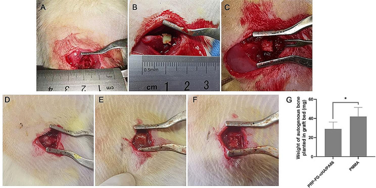
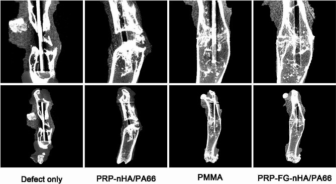
Methods: Platelet rich plasma (PRP) was prepared and PRP-FG-nHA/PA66 scaffold was synthesized and observed. The sustained release of VEGFA and porosity of the scaffold were analyzed. We established a femur LBD model in male SD rats. 55 rats were randomly divided into four groups depending on the spacer filled in the defect area. "Defect only" group (n = 10), "PMMA" group (n = 15), "PRP-nHA/PA66" group (n = 15) and "PRP-FG-nHA/PA66" group (n = 15 ). At 6 weeks, the spacers were removed and the defects were grafted. The induced membrane and bone were collected and stained. The bone formation was detected by micro-CT and the callus union was scored on a three point system.
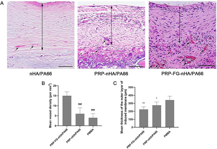
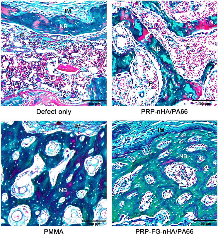
Results: The PRP-FG-nHA/PA66 scaffold was porosity and could maintain a high concentration of VEGFA after 30 days of preparation. The induced membrane in PRP-FG-nHA/PA66 group was thinner than PMMA, but the vessel density was higher.The weight of autogenous bone grafted in PRP-FG-nHA/PA66 group was significantly smaller than that of PMMA group. In PRP-FG-nHA/PA66 group, the bone defect was morphologically repaired.
Conclusion: The study showed that PRP-FG-nHA/PA66 scaffold can significantly reduce the amount of autologous bone graft, and can achieve similar bone defect repair effect as PMMA. Our findings provide some reference and theoretical support for the treatment of large segmental bone defects in humans.
Keywords: Masquelet membrane induction technology; angiogenesis; bone regeneration; large bone defect; platelet rich plasma.
© 2024. The Author(s).
Conflict of interest statement
The authors declare no competing interests.
Figures


Similar articles
-
Engineering the bone reconstruction surgery: the case of the masquelet-induced membrane technique.Eur J Trauma Emerg Surg. 2025 Mar 18;51(1):138. doi: 10.1007/s00068-025-02815-9. Eur J Trauma Emerg Surg. 2025. PMID: 40102268 Free PMC article. Review.
-
The Combination of Platelet Rich Plasma Gel, Human Umbilical Mesenchymal Stem Cells and Nanohydroxyapatite/polyamide 66 Promotes Angiogenesis and Bone Regeneration in Large Bone Defect.Tissue Eng Regen Med. 2022 Dec;19(6):1321-1336. doi: 10.1007/s13770-022-00471-3. Epub 2022 Sep 8. Tissue Eng Regen Med. 2022. PMID: 36074328 Free PMC article.
-
Epidermal growth factor or platelet-rich plasma combined with induced membrane technique in the treatment of segmental femur defects: an experimental study.J Orthop Surg Res. 2020 Dec 11;15(1):601. doi: 10.1186/s13018-020-02142-2. J Orthop Surg Res. 2020. PMID: 33308245 Free PMC article.
-
The Masquelet Technique: Can Disposable Polypropylene Syringes be an Alternative to Standard PMMA Spacers? A Rat Bone Defect Model.Clin Orthop Relat Res. 2021 Dec 1;479(12):2737-2751. doi: 10.1097/CORR.0000000000001939. Clin Orthop Relat Res. 2021. PMID: 34406150 Free PMC article.
-
Mechanisms of the Masquelet technique to promote bone defect repair and its influencing factors.Chin J Traumatol. 2025 May;28(3):157-163. doi: 10.1016/j.cjtee.2024.04.003. Epub 2024 Apr 25. Chin J Traumatol. 2025. PMID: 38734563 Free PMC article. Review.
Cited by
-
Engineering the bone reconstruction surgery: the case of the masquelet-induced membrane technique.Eur J Trauma Emerg Surg. 2025 Mar 18;51(1):138. doi: 10.1007/s00068-025-02815-9. Eur J Trauma Emerg Surg. 2025. PMID: 40102268 Free PMC article. Review.
-
Functionalized Periosteum-Derived Microsphere-Hydrogel with Sequential Release of E7 Short Peptide/miR217 for Large Bone Defect Repairing.Biomater Res. 2025 Jan 7;29:0127. doi: 10.34133/bmr.0127. eCollection 2025. Biomater Res. 2025. PMID: 39780960 Free PMC article.
References
-
- Govaert GA, Ijpma FF, McNally, Gannamani S, Rachakonda KR, Tellakula Y, Takkalapally H, Maryada VR, Gurava Reddy AV. Combining non-vascularized fibula and cancellous graft in the masquelet technique: a promising approach to distal femur compound fracture management with large defects. Injury. 2023;55(2):111233. - PubMed
MeSH terms
Substances
Grants and funding
- LH2022H037/Natural Science Foundation of Heilongjiang Province of China
- LH2022H037/Natural Science Foundation of Heilongjiang Province of China
- LH2022H037/Natural Science Foundation of Heilongjiang Province of China
- LH2022H037/Natural Science Foundation of Heilongjiang Province of China
- LH2022H037/Natural Science Foundation of Heilongjiang Province of China
LinkOut - more resources
Full Text Sources
Research Materials
