Review
. 2024 Jun 8;9(1):155.
doi: 10.1038/s41392-024-01845-w.

Pathology of pain and its implications for therapeutic interventions

Affiliations
Review

Pathology of pain and its implications for therapeutic interventions

Bo Cao et al. Signal Transduct Target Ther. .

Abstract

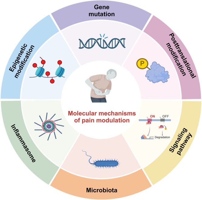
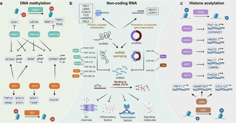
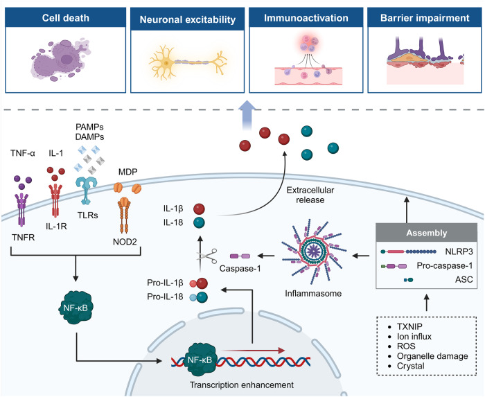
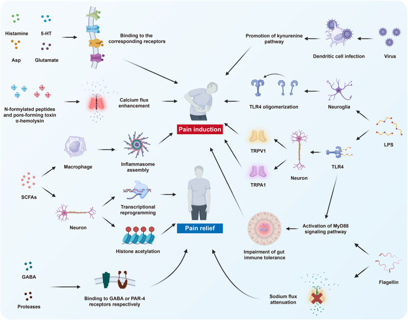
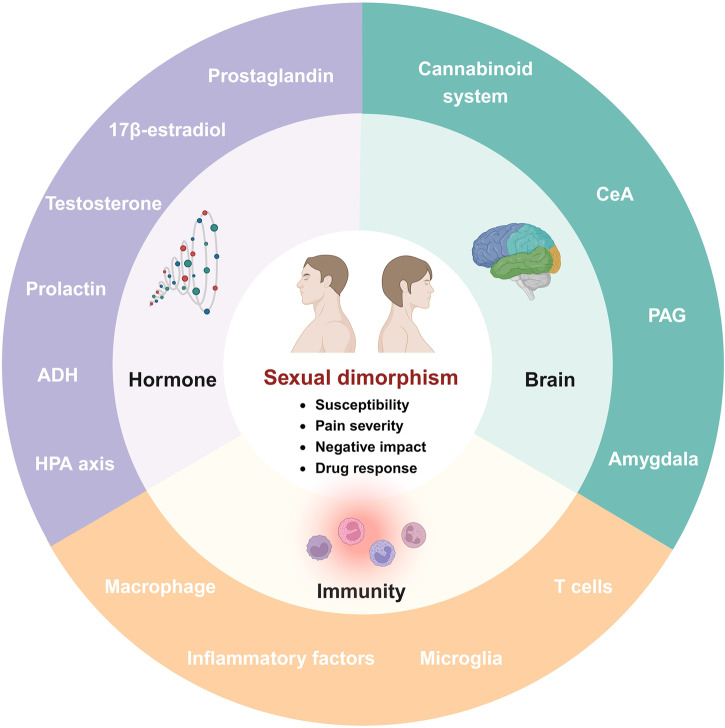
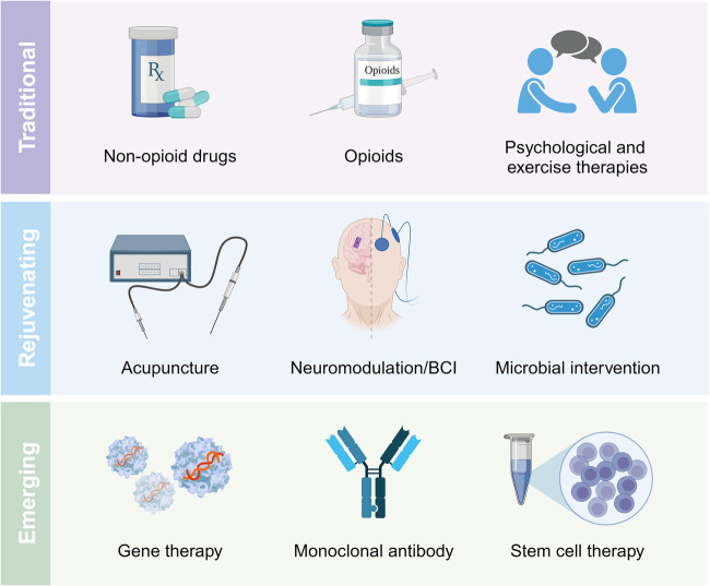
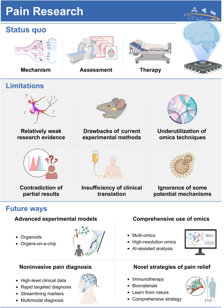
Pain is estimated to affect more than 20% of the global population, imposing incalculable health and economic burdens. Effective pain management is crucial for individuals suffering from pain. However, the current methods for pain assessment and treatment fall short of clinical needs. Benefiting from advances in neuroscience and biotechnology, the neuronal circuits and molecular mechanisms critically involved in pain modulation have been elucidated. These research achievements have incited progress in identifying new diagnostic and therapeutic targets. In this review, we first introduce fundamental knowledge about pain, setting the stage for the subsequent contents. The review next delves into the molecular mechanisms underlying pain disorders, including gene mutation, epigenetic modification, posttranslational modification, inflammasome, signaling pathways and microbiota. To better present a comprehensive view of pain research, two prominent issues, sexual dimorphism and pain comorbidities, are discussed in detail based on current findings. The status quo of pain evaluation and manipulation is summarized. A series of improved and innovative pain management strategies, such as gene therapy, monoclonal antibody, brain-computer interface and microbial intervention, are making strides towards clinical application. We highlight existing limitations and future directions for enhancing the quality of preclinical and clinical research. Efforts to decipher the complexities of pain pathology will be instrumental in translating scientific discoveries into clinical practice, thereby improving pain management from bench to bedside.


Conflict of interest statement

The authors declare no competing interests.

Figures

Fig. 1
Fig. 1
The brief timeline of historic milestones in the field of pain therapy. Morphine was first extracted in 1806, which opened the chapter in fighting with pain using the fruits of modern medicine. Since then, many intervention methods for pain management were discovered and came into clinical application, such as CBT, spinal cord stimulation, monoclonal antibody therapy and gene therapy. The progress in the research on pain mechanisms and interdisciplinary collaboration boosted advances in pain therapy. In recent years, the wide application of high-throughput biotechnologies has further deepened the understanding in pain pathology and has contributed to the development of individualized pain management. Key milestones of pain therapy are chronologically illustrated in the figure. The achievements awarded by the Nobel prizes are marked with the medals
Fig. 2
Fig. 2
Current animal models in pain research. Physical damages, chemical irritants, cancer cell implantation and psychosocial stressors constitute the three primary methods for preparing pain models. Furthermore, composite regimens that combine several of the aforementioned methods have been employed as pain is a multifactorial event. CCI chronic constriction injury, CFA complete Freund’s adjuvant, DSS dextran sulfate sodium, IBD inflammatory bowel disease, IBS irritable bowel syndrome, MS maternal separation, NLB neonatal limited bedding, PTSD posttraumatic stress disorder, SNI spared nerve injury, SNL spinal nerve ligation, TNBS 2,4,6-trinitrobenzene sulfonic acid, WAS water avoidance stress
Fig. 3
Fig. 3
Schematic illustration of pain sensation pathways. The exposure to pain-inducing events changes activity of specific receptors and activates action potential of peripheral nociceptors. The signals are then transmitted from DRG located to the spinal cord via afferent nerves. The nerves are categorized into Aβ, Aδ and C fibers. During neuronal transmission, the presynaptic membrane releases various neurotransmitters into the subsynaptic membrane, inducing potential alterations in the subsequent neuron. The figure shows some representative neurotransmitters in during pain perception. Additionally, neurogliocytes, immune cells and other types of neurons collaboratively modulate pain signals. The DRG, as the relay station, is responsible for ascending transmission to the corresponding sensory cortex, which modulates the ultimate pain sensation. The descending regulatory pathways also play a role in pain modulation. ASIC acid-sensing ion channel, AMPAR α-amino-3-hydroxy-5- methylisoxazole-4-propionate receptor, CGRP calcitonin gene-related peptide, GABA gamma-aminobutyric acid, GPCR G protein-coupled receptor, mGluR metabotropic glutamate receptor, NGF nerve growth factor, NMDAR N-methyl-D-aspartate receptor, P2X3 purinergic receptor 3, TrkA tropomyosin-related kinase A, TRPA1 transient receptor potential ankyrin 1, TRPM8 transient receptor potential melastatin 8, TRPV1 transient receptor potential vanilloid 1, TRPV2 transient receptor potential vanilloid 2, TRPV3 transient receptor potential vanilloid 3, TRPV4 transient receptor potential vanilloid 4, VGCC voltage-gated calcium channel, VGPC voltage-gated potassium channel, VGSC voltage-gated sodium channel
Fig. 4
Fig. 4
The schematic illustration of molecular mechanisms underlying pain modulation. The molecular mechanisms are generally categorized into six aspects, including gene mutation, epigenetic modification, posttranslational modification, inflammasome, signaling pathways and microbiota. They orchestrate pain perception and modulation
Fig. 5
Fig. 5
The mechanisms of epigenetic modification in pain modulation. The mechanisms are categorized into three aspects: DNA methylation, non-coding RNA and histone acetylation. a For DNA methylation, DNMTs and TETs are responsible for DNA methylation and demethylation, respectively. They regulate expression of various genes associated with pain perception. The expression of KCNA2, BDNF and OPRM1 are simultaneously under the control of DNMTs and TETs. b Non-coding RNAs, comprising miRNAs, lncRNAs and circRNAs, play various roles. miRNAs can bind to 3’UTR of mRNAs associated with pain, negatively regulating their expression. Some lncRNAs and circRNAs act as miRNA sponges to counteract the functions of downstream targets. Certain lncRNAs and circRNAs directly interact with proteins to enhance their stabilization, thereby affecting pain sensitivity. Several non-coding RNAs, like lncRNA NEAT1 and circVOPP1, have been shown to stabilize the mRNAs of their parental genes related to pain to promote their expression. c HDACs and HATs collaboratively maintain the balance in histone acetylation. Specific HDACs, including HDAC2, HDAC4, HDAC5, SIRT1 and SIRT3, along with HAT p300, regulate expression of genes involved in pain modulation. Notably, non-coding RNAs regulate expression of enzymes associated with DNA methylation and histone acetylation. The expression of non-coding RNAs are, in turn, regulated by the other two mechanisms. circRNA circular RNA, DNMT DNA methyltransferase, HAT histone acetyltransferase, HDAC histone deacetylase, lncRNA long non-coding RNA, miRNA microRNA, TET ten-eleven-translocation protein, UTR untranslated region
Fig. 6
Fig. 6
The mechanisms underlying NLRP3 inflammasome-medicated hyperalgesia. Certain ligands, including TNF-α, IL-1, PAMPs, DAMPs and MDP, bind to the corresponding receptors, activating NF-κB signaling. The activated NF-κB is transported into the nucleus and promote the expression of pro-IL-1β and pro-IL-18. Various molecular and cellular events regulate the assembly of inflammasomes. Caspase-1, derived from inflammasomes, facilitates the maturation of IL-1β and IL-18. These cytokines are subsequently released, leading to cell death, neuronal hyperexcitability, immune activation and impairment of brain barriers. These processes collectively contribute to hyperalgesia. ASC apoptosis-associated speck-like protein containing a CARD, DAMP damage-associated molecular pattern, IL-1 interleukin-1, IL-1R interleukin-1 receptor, IL-18 interleukin-18, MDP muramyl dipeptide, NF-κB nuclear factor-kappa B, NOD2 nucleotide-binding oligomerization domain 2, PAMP pathogen associated molecular pattern, ROS reactive oxygen species, TLR Toll-like receptor, TNF-α tumor necrosis factor-α, TNFR tumor necrosis factor receptor, TXNIP thioredoxin-interacting protein
Fig. 7
Fig. 7
The direct mechanisms underlying regulation of pain perception by microbiota. The microbiota-derived metabolites, including histamine, 5-HT, Asp and glutamate, bind to their corresponding receptors to enhance pain sensation. In contrast, GABA and proteases can respectively bind to GABA and PAR-4 receptors to alleviate pain. N-formylated peptides and the pore-forming toxin α-hemolysin produced by microbiota, enhance the calcium flux in nociceptors and induce pain perception. SCFAs exhibit dual effects. They interact with macrophages, inducing inflammasome assembly and pain perception. On the other hand, SCFAs promote transcriptional reprogramming and histone acetylation in neurons, reducing pain hypersensitivity phenotypes. Viral infection of dendritic cells facilitates the kynurenine pathway, contributing to pain development. LPS possesses the typical properties of pain induction in a TLR4-dependent manner. TLR4 oligomerization in neuroglia, activation of TRPV1 and TRPA1 in neurons and immune tolerance impairment in intestinal epithelial cells through TLR4/MyD88 signaling pathway are mechanisms underlying LPS-induced pain sensation. Another bacterial component, flagellin, likewise attenuates gut immune tolerance through regulating MyD88 signaling pathway. However, it can decrease sodium flux in nociceptors, inversely alleviating pain perception. 5-HT 5-hydroxytryptamine, Asp aspartic acid, SCFA short-chain fatty acid, γ-aminobutyric acid, PAR-4 protease activated receptor, LPS lipopolysaccharide, TLR4 toll-like receptor 4, TRPV1 transient receptor potential vanilloid 1, TRPA1 transient receptor potential ankyrin 1
Fig. 8
Fig. 8
The mechanisms underlying sexual dimorphism of pain. The sexual dimorphism in pain is characterized as four aspects: susceptibility, pain severity, negative impact of pain and responses to analgesic drug. These differences are attributed to variations in hormones, brain function, and immunity. The specific factors in each mechanism, as identified by existing studies, are displayed in the corresponding outer ring. ADH antidiuretic hormone, CeA central amygdala, HPA hypothalamic-pituitary-adrenal, PAG periaqueductal gray
Fig. 9
Fig. 9
The summary of currently developed therapies for pain management. The nine methods are categorized into three groups, including traditional, rejuvenating, and emerging therapies. BCI brain-computer interface
Fig. 10
Fig. 10
The status quo, limitations and future perspectives of the pain research. Most current preclinical and clinical studies in the pain field focused on its mechanisms, assessment and therapy. However, there are some limitations as follows. i) Current research evidence is relatively weak and the underlying mechanisms remain largely unknown. ii) The in-vitro and in-vivo experimental models cannot thoroughly mimic the clinical conditions of pain. iii) The research value of omics techniques is not fully exploited. iv) There are contradictions in the results from some studies. v) Current progress in clinical translation of pain research achievements is far from clinical requirements. vi) Some potential mechanisms underlying pain modulation should be emphasized, like the regulatory role of fungi. Herein, we propose four future perspectives for pain research, including development of advanced experimental models, comprehensive application of omics, emphasis on noninvasive pain diagnosis and optimization of strategies for pain relief

References

    1. Raja SN, et al. The revised International Association for the Study of Pain definition of pain: concepts, challenges, and compromises. Pain. 2020;161:1976–1982. doi: 10.1097/j.pain.0000000000001939. - DOI - PMC - PubMed
    1. Basbaum AI, Bautista DM, Scherrer G, Julius D. Cellular and molecular mechanisms of pain. Cell. 2009;139:267–284. doi: 10.1016/j.cell.2009.09.028. - DOI - PMC - PubMed
    1. Julius D, Basbaum AI. Molecular mechanisms of nociception. Nature. 2001;413:203–210. doi: 10.1038/35093019. - DOI - PubMed
    1. Yang D, et al. Nociceptor neurons direct goblet cells via a CGRP-RAMP1 axis to drive mucus production and gut barrier protection. Cell. 2022;185:4190–4205.e4125. doi: 10.1016/j.cell.2022.09.024. - DOI - PMC - PubMed
    1. Goodwin G, McMahon SB. The physiological function of different voltage-gated sodium channels in pain. Nat. Rev. Neurosci. 2021;22:263–274. doi: 10.1038/s41583-021-00444-w. - DOI - PubMed

Publication types