

Effect of the COVID-19 pandemic on Vogt-Koyanagi-Harada disease
- PMID: 38851824
- PMCID: PMC11162482
- DOI: 10.1038/s41598-024-63957-1
Effect of the COVID-19 pandemic on Vogt-Koyanagi-Harada disease
Abstract
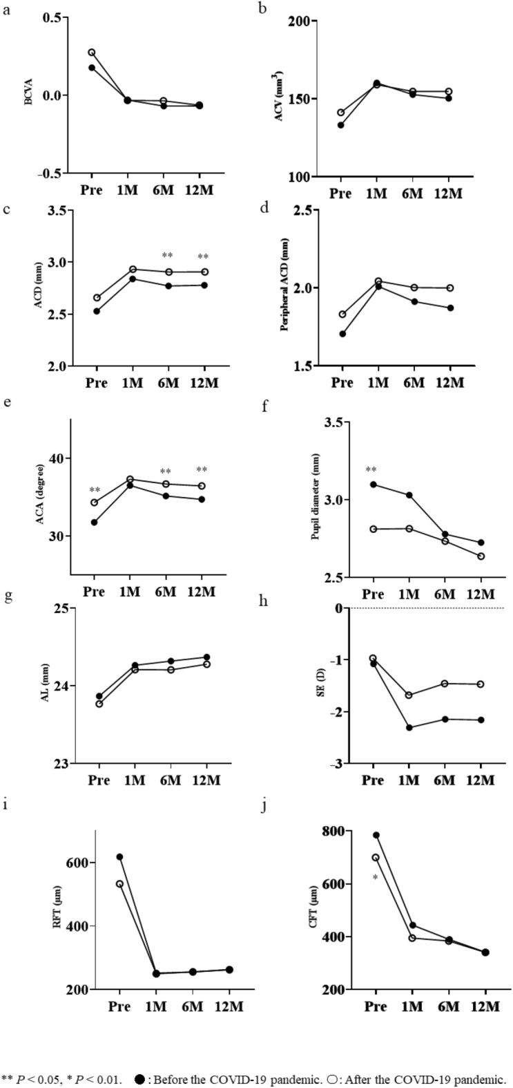
To determine the disease prevalence rate and clinical characteristics of Vogt-Koyanagi-Harada (VKH) disease among new patients before and after the declaration of a state of emergency (April 7, 2020) in Japan. New patients and patients with newly diagnosed VKH disease were categorized into "Before" and "After" groups based on the initial visit. The prevalence rate, sex ratio, and age of patients newly diagnosed with VKH were compared between the groups. Best-corrected visual acuity (BCVA) and recurrence rates were compared among 59 patients observed for > 12 months after receiving pulse steroid therapy. For reference, we also examined the prevalence rate of patients newly diagnosed with acute angle closure (AAC) in the Before and After groups. The prevalence rates of VKH disease among newly diagnosed patients (P < 0.05) or patients with AAC (P < 0.001) were significantly higher in the After group. No significant differences in sex ratio or age of VKH disease were observed in both groups. BCVA and recurrence rates showed no significant differences. The COVID-19 pandemic increased the prevalence of VKH disease among new patients compared with that of AAC. However, the clinical features of VKH disease were unlikely affected by the COVID-19 pandemic.
Keywords: COVID-19; Pandemics; Prevalence; Sex ratio; Visual acuity; Vogt–Koyanagi–Harada disease.
© 2024. The Author(s).
Conflict of interest statement
The authors declare no competing interests.
Figures
Similar articles
-
Predictive factors and adalimumab efficacy in managing chronic recurrence Vogt-Koyanagi-Harada disease.BMC Ophthalmol. 2024 Jun 7;24(1):238. doi: 10.1186/s12886-024-03511-9. BMC Ophthalmol. 2024. PMID: 38849758 Free PMC article.
-
High prevalence of angle-closure glaucoma in Vogt-Koyanagi-Harada disease.Int Ophthalmol. 2022 Dec;42(12):3913-3921. doi: 10.1007/s10792-022-02412-4. Epub 2022 Jul 5. Int Ophthalmol. 2022. PMID: 35789316
-
Comparison of Clinical Features and Visual Outcome between Sympathetic Ophthalmia and Vogt-Koyanagi-Harada Disease in Chinese Patients.Ophthalmology. 2019 Sep;126(9):1297-1305. doi: 10.1016/j.ophtha.2019.03.049. Epub 2019 Apr 6. Ophthalmology. 2019. PMID: 30959067
-
Vogt-Koyanagi-Harada disease after SARS-CoV-2 infection: Case report and literature review.Immun Inflamm Dis. 2024 Apr;12(4):e1250. doi: 10.1002/iid3.1250. Immun Inflamm Dis. 2024. PMID: 38661242 Free PMC article. Review.
-
Vogt-Koyanagi-Harada disease: recurrence rates after initial-onset disease differ according to treatment modality and geographic area.Int Ophthalmol. 2020 Sep;40(9):2423-2433. doi: 10.1007/s10792-020-01417-1. Epub 2020 May 16. Int Ophthalmol. 2020. PMID: 32418076 Review.
Cited by
-
Predictive factors at initial visit for sunset glow fundus in Vogt-Koyanagi-Harada disease.Sci Rep. 2025 Jul 7;15(1):24302. doi: 10.1038/s41598-025-08252-3. Sci Rep. 2025. PMID: 40624077 Free PMC article.
References
MeSH terms
LinkOut - more resources
Full Text Sources
Medical
Miscellaneous
