. 2024 Aug 20;123(16):2422-2430.
doi: 10.1016/j.bpj.2024.06.005. Epub 2024 Jun 8.

Identifying the molecular basis of Laminin N-terminal domain Ca2+ binding using a hybrid approach

Affiliations

Identifying the molecular basis of Laminin N-terminal domain Ca2+ binding using a hybrid approach

Scott Legare et al. Biophys J. .

Abstract

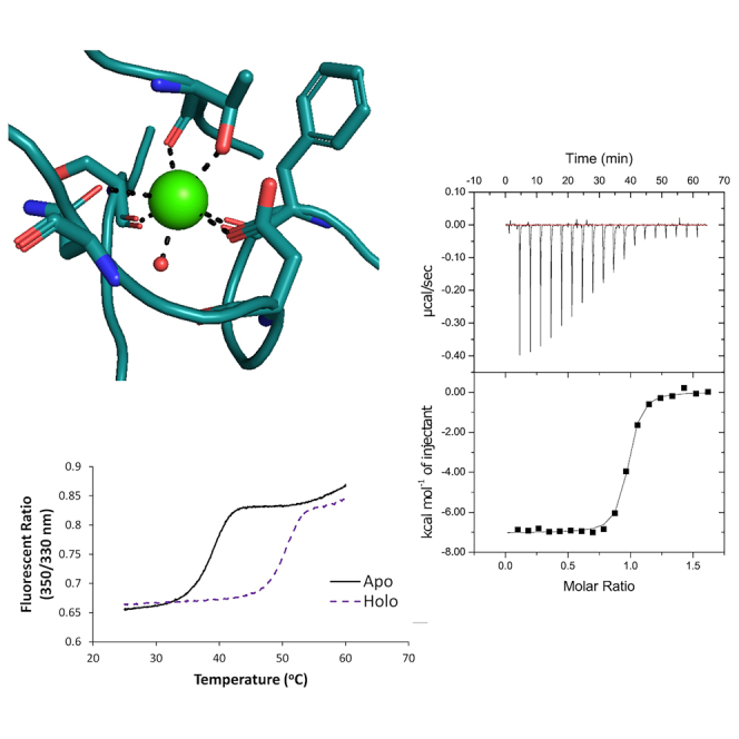
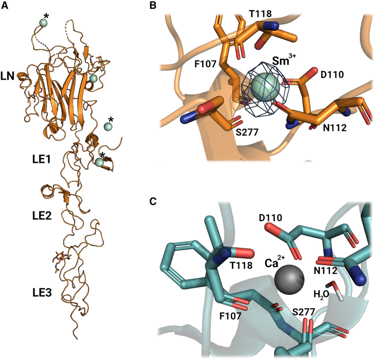
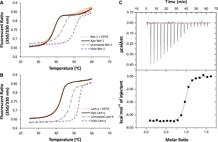
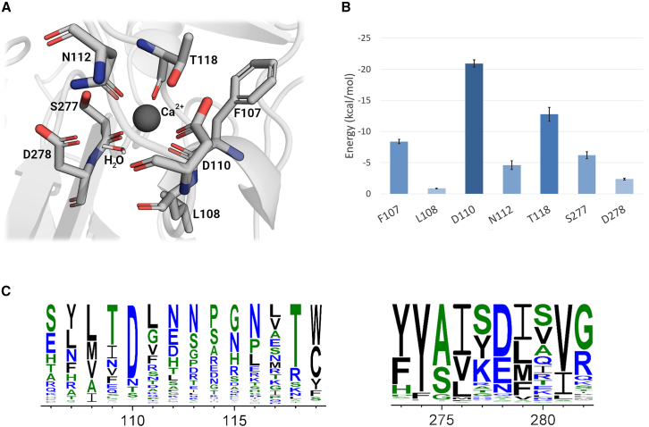
Ca2+ is a highly abundant ion involved in numerous biological processes, particularly in multicellular eukaryotic organisms where it exerts many of these functions through interactions with Ca2+ binding proteins. The laminin N-terminal (LN) domain is found in members of the laminin and netrin protein families where it plays a critical role in the function of these proteins. The LN domain of laminins and netrins is a Ca2+ binding domain and in many cases requires Ca2+ to perform its biological function. Here, we conduct a detailed examination of the molecular basis of the LN domain Ca2+ interaction combining structural, computational, bioinformatics, and biophysical techniques. By combining computational and bioinformatic techniques with x-ray crystallography we explore the molecular basis of the LN domain Ca2+ interaction and identify a conserved sequence present in Ca2+ binding LN domains. These findings enable a sequence-based prediction of LN domain Ca2+ binding ability. We use thermal shift assays and isothermal titration calorimetry to explore the biophysical properties of the LN domain Ca2+ interaction. We show that the netrin-1 LN domain exhibits a high affinity and specificity for Ca2+, which structurally stabilizes the LN domain. This study elucidates the molecular foundation of the LN domain Ca2+ binding interaction and provides a detailed functional characterization of this essential interaction, advancing our understanding of protein-Ca2+ dynamics within the context of the LN domain.


Conflict of interest statement

Declaration of interests The authors declared no competing interests.

Figures

None
Graphical abstract
Figure 1
Figure 1
Structural examination of Net-1 in complex with metal ions. (A) X-ray diffraction structure model of Net-1 in complex with Sm3+ ions. Three Sm3+ ions are coordinated through crystallographic contacts (indicated with asterisk ), while one Sm3+ ion is bound to the Ca2+ binding loop of Net-1 (PDB: 8SNP). (B) Amino acid metal coordination of Sm3+ in the Ca2+ binding loop of Net-1. The ion is coordinated by side chain and backbone functional groups of the residues F107, D110, N112, T118, and S277. The electron density level of the 2Fo-Fc map is set to 0.522 e/Å3 (8 sigma contour level). (C) Ca2+ coordination in the Ca2+ binding loop of Net-1 (PDB: 4OVE) (31) with the indirect coordination of Ca2+ by D278 through a hydrogen bonded water molecule. To see this figure in color, go online.
Figure 2
Figure 2
Characterization of the biophysical properties of the LN domain Ca2+ binding site. Thermal shift assay characterization of Net-1 (A) and Lam-γ (B) in 20 mM HEPES (pH 7.3) and 150 mM NaCl. Melt curve of holo-protein at a physiological extracellular Ca2+ concentration of 1.4 mM shown in a purple short dashed line (Net-1 Tm of 50.6°C, Lam-γ Tm of 52.8°C). Melt curve of Chelex-100-treated apo-protein shown in solid black lines and in the presence of 1.6–50 mM EDTA shown in orange dotted lines (Net-1 Tm of 38.8°C, Lam-γ Tm of 42.6°C), demonstrating Chelex-100 treatment results in the complete removal of Ca2+ to produce apo-protein. Melt curve before treatment with Chelex-100 resin (untreated) shown in a gray long-dashed line (Net-1 Tm of 44.2°C, Lam-γ Tm of 48.2°C). Isothermal calorimetric titration of apo-Net-1 with Ca2+ (C). apo-Net-1 (29.9 μM) in 20 mM HEPES (pH 7.3) and 150 mM NaCl was titrated with 250 μM CaCl2 at 25°C. The binding constant (Ka) is measured to be 1.46 ± 0.20 × 107 M−1 with a stoichiometry of 0.942 ± 0.022. The enthalpy change (ΔH) is −7040 ± 200 cal mol−1 and the entropy change (ΔS) is 9.16 ± 0.96 cal mol−1 K−1. ITC values are reported as the mean ± 1 standard deviation of three experimental replicates. To see this figure in color, go online.
Figure 3
Figure 3
Molecular dynamics simulation analysis of the Ca2+ binding loop of Net-1. (A) Structural model of the Net-1 Ca2+ binding loop bound to Ca2+. Interacting residues and water over the 100 ns molecular dynamics trajectory are shown. (B) MMPBSA residue decomposition analysis of the ligand interaction. D110 is the major contributor to the interaction while L108 is only minimally involved via its carbonyl backbone group. Error bars are based on the standard deviation of the mean energy for each residue. (C) Sequence logo showing conservation of the Ca2+ binding interface for 831 Net-1 sequences from various species. The residue positions that contribute to the Ca2+ binding through their side chain functional groups are highly conserved. To see this figure in color, go online.

Similar articles

References

    1. Carafoli E., Santella L., et al. Brini M. Generation, control, and processing of cellular calcium signals. Crit. Rev. Biochem. Mol. Biol. 2001;36:107–260. - PubMed
    1. Clapham D.E. Calcium Signaling. Cell. 2007;131:1047–1058. - PubMed
    1. Brown E.M., MacLeod R.J. Extracellular calcium sensing and extracellular calcium signaling. Physiol. Rev. 2001;81:239–297. - PubMed
    1. Handschuh G., Candidus S., et al. Becker K.-F. Tumour-associated E-cadherin mutations alter cellular morphology, decrease cellular adhesion and increase cellular motility. Oncogene. 1999;18:4301–4312. - PubMed
    1. Zhang K., Chen J. The regulation of integrin function by divalent cations. Cell Adhes. Migrat. 2012;6:20–29. - PMC - PubMed

Publication types

LinkOut - more resources