. 2024 Aug:74:103194.
doi: 10.1016/j.redox.2024.103194. Epub 2024 May 16.

Histone lactylation-regulated METTL3 promotes ferroptosis via m6A-modification on ACSL4 in sepsis-associated lung injury

Affiliations

Histone lactylation-regulated METTL3 promotes ferroptosis via m6A-modification on ACSL4 in sepsis-associated lung injury

Dan Wu et al. Redox Biol. 2024 Aug.

Erratum in

Abstract

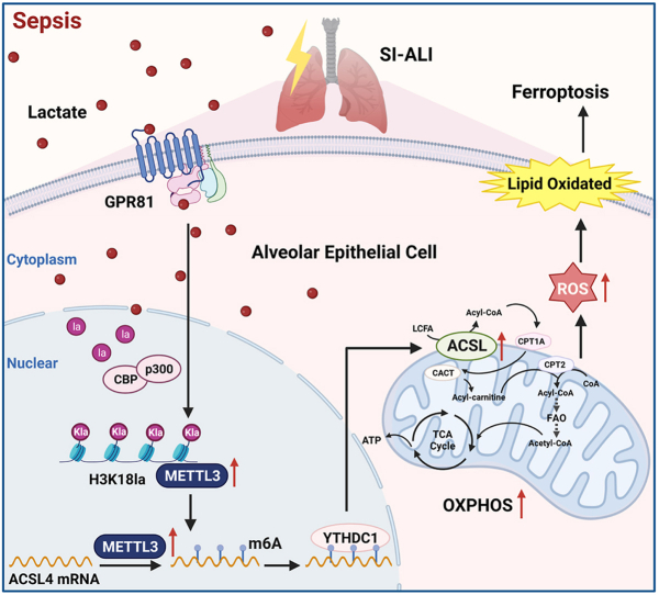
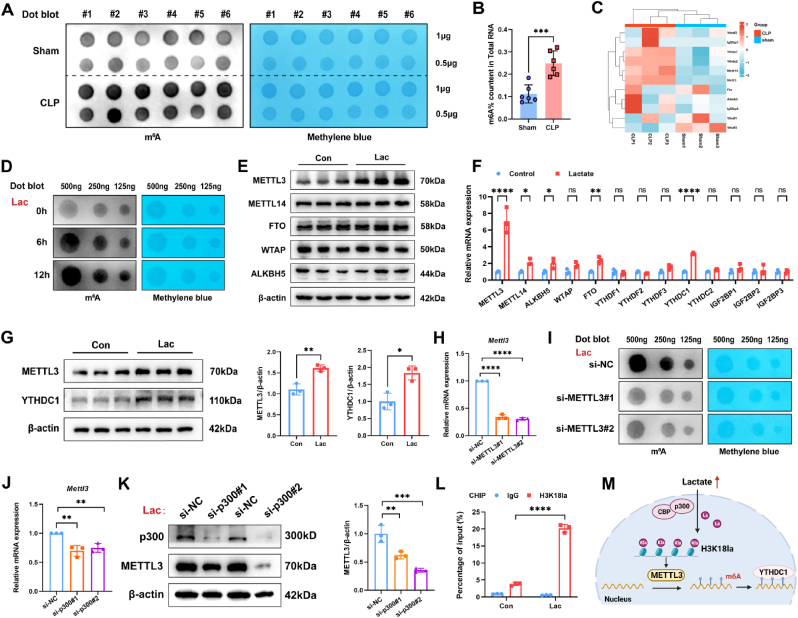
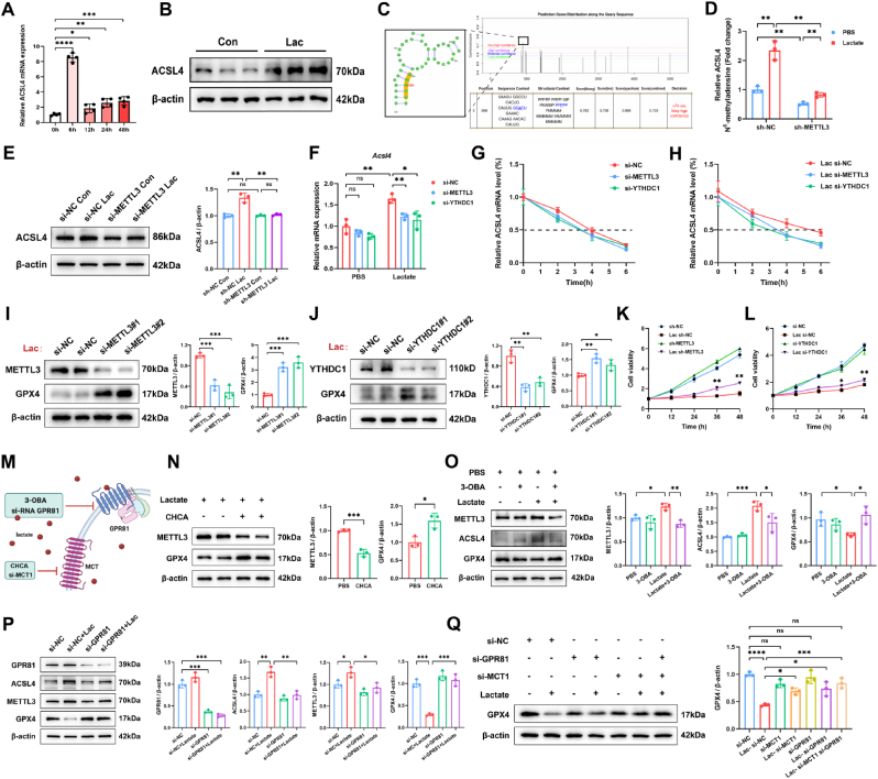
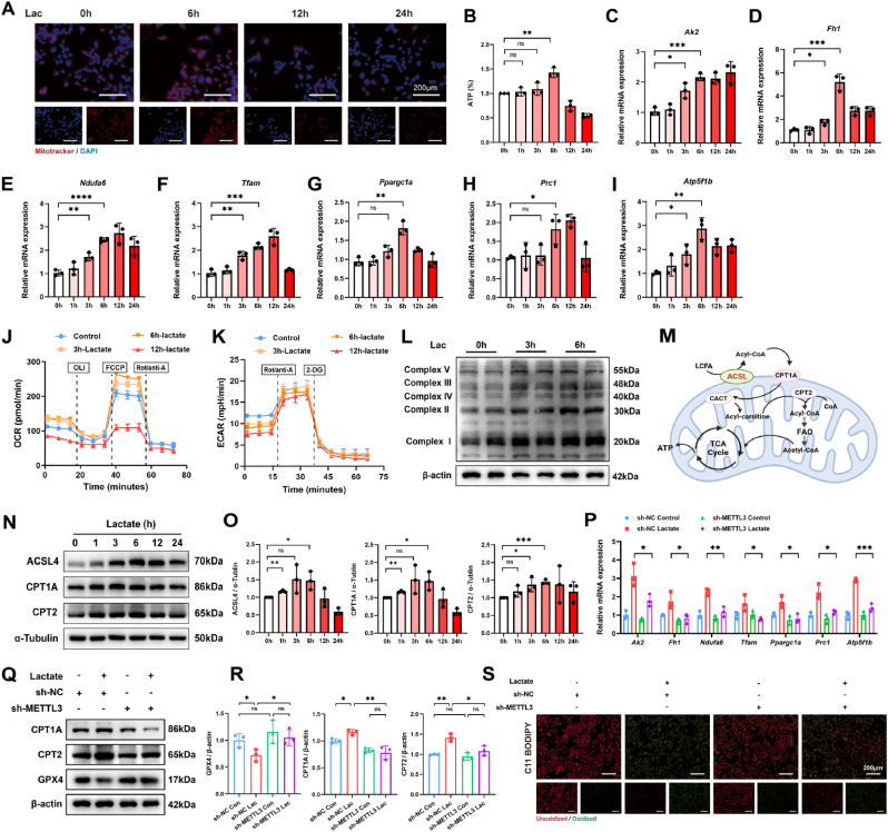
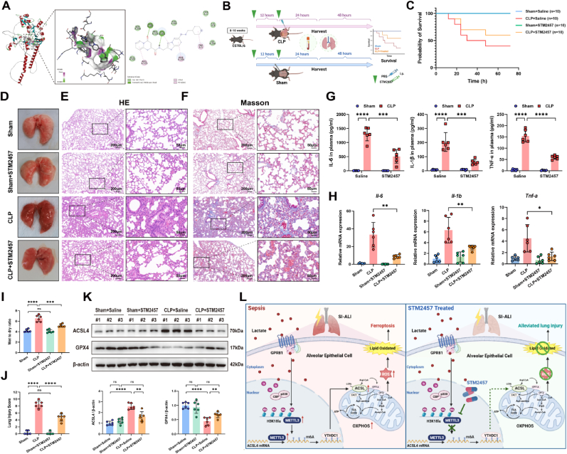
Elevated lactate levels are a significant biomarker of sepsis and are positively associated with sepsis-related mortality. Sepsis-associated lung injury (ALI) is a leading cause of poor prognosis in clinical patients. However, the underlying mechanisms of lactate's involvement in sepsis-associated ALI remain unclear. In this study, we demonstrate that lactate regulates N6-methyladenosine (m6A) modification levels by facilitating p300-mediated H3K18la binding to the METTL3 promoter site. The METTL3-mediated m6A modification is enriched in ACSL4, and its mRNA stability is regulated through a YTHDC1-dependent pathway. Furthermore, short-term lactate stimulation upregulates ACSL4, which promotes mitochondria-associated ferroptosis. Inhibition of METTL3 through knockdown or targeted inhibition effectively suppresses septic hyper-lactate-induced ferroptosis in alveolar epithelial cells and mitigates lung injury in septic mice. Our findings suggest that lactate induces ferroptosis via the GPR81/H3K18la/METTL3/ACSL4 axis in alveolar epithelial cells during sepsis-associated ALI. These results reveal a histone lactylation-driven mechanism inducing ferroptosis through METTL3-mediated m6A modification. Targeting METTL3 represents a promising therapeutic strategy for patients with sepsis-associated ALI.

Keywords: Ferroptosis; Histone lactylation; N6- methyladenosine; Sepsis-associated acute lung injury.


Conflict of interest statement

Declaration of competing interest The authors declare that they have no known competing financial interests or personal relationships that could have appeared to influence the work reported in this paper.

Figures

Image 1
Graphical abstract
Fig. 1
Fig. 1
Inhibition of lactate alleviates sepsis-related lung injury. (A) Concentration of Lactate in plasma of SI-ALI patients and healthy controls (HC). (SI-ALI n = 19, HC n = 19) (B) Correlation between IL-6 level and Lactate concentration in plasma. (C) Correlation between IL-1β level and Lactate concentration in plasma. (D) Correlation between TNF-γ level and Lactate concentration in plasma. (E) Correlation between PaO2/FiO2 level and Lactate concentration in plasma. (F) Representative gross morphology of lung tissues of CLP and Sham mice. (G) Plasma lactate concentration of mice in the sham or CLP group. (n = 6/group) (H) IL-6, IL-1β and TNF-γ levels in plasma. (Sham n = 6, CLP n = 6) (I) Inflammatory factors mRNA levels (IL-6, IL-1β and TNF-γ) in lung tissues. (n = 6/group) (J and K) Schematic diagram of the blocking mechanism of lactate production by 2-DG and Sodium Oxamate in vivo, and the experimental design of drug treatment for CLP model mice. (L and M) Lung injury score and the wet-to-dry ratio of lungs in the sham group, CLP group, or CLP combined with the 2-DG application group. (n = 6/group) (N) Representative HE images of lungs. (Up, scale bar = 200 μm; down, scale bar = 50 μm) (O, P and Q) Plasma IL-6, IL-1β and TNF-γ levels in sham and CLP mice with or without 2-DG administration. (n = 6/group) Data are mean ± SD. *p < 0.05, **p < 0.01, ***p < 0.001, and ****p < 0.001. Statistical significance was determined by one-way ANOVA or two-sided Student's t-test as appropriate.
Fig. 2
Fig. 2
Lactate can induce ferroptosis of the lung in polymicrobial sepsis. (A) Enrichment plot of ferroptosis pathway via GSEA analysis. (B) GPX4 and ACSL4 mRNA level of lung tissues. (C) GPX4 protein expression of lung tissues. (D and E) IHC images and IHC score of GPX4 or ACSL4 in lung tissues. (Scale bar: left = 200, right = 50 μm) (F, G and H) GSH, GSH/GSSG ratio and MDA levels of lung tissues. (I) Ferrous iron level of lung tissues in the sham group, CLP group, or CLP combined with 2-DG or sodium oxamate application group. (J) Representative TEM images of alveolar epithelial cells. (Scale bar = 2 μm) (Animal experiments were validated from 5 independent experiments.) (K and L) Western blotting of ferroptosis marker proteins ACSL4 and GPX4 in lungs. (M) GPX4 protein levels under lactate (10 mM) stimulation for 0, 6, 12, 24, and 48 h. (N) Representative TEM images of MLE12 cells. (Scale bar: left = 1 μm, right = 500 nm) (In vitro experiments were validated from 3 independent experiments.) (O and P) GSH/GSSG ratio and MDA levels of MLE12 cells with or without lactate treatment. (Q) Visualization of MLE12 cells under the corresponding treatment. (Scale bar = 100 μm, n = 3) (R and S) Representative IF images of FeRhNOX-1 and DCFH-DA in MLE12 cells treated with lactate and Fer-1. (For FeRhNOX-1 staining, bar = 50 μm; For DCFH-DA staining, scale bar = 50 μm) For in vivo experiments or tissue samples, n = 6 for each group. For in vitro experiments or cell samples, n = 3 for each group. Data are mean ± SD. *p < 0.05, **p < 0.01, ***p < 0.001, and ****p < 0.001. Statistical significance was determined by one-way ANOVA or two-sided Student's t-test as appropriate.
Fig. 3
Fig. 3
Lactate induces p300-mediated H3K18la increase in SI-ALI. (A) Pan-Kla level of lung tissues in the sham or CLP group. (B) A score of different site Lactylation on Histone H3 and H4 were predicted via CPLM (http://cplm.biocuckoo.cn/index.php). (C) Western blotting analysis of site-specific histone lactylation in the lung tissues of sham and CLP mice. (D) H3K18la site on Histone H3 was predicted on CPLM (http://cplm.biocuckoo.cn/index.php) and Uniprot (https://www.uniprot. org/). (E and F) Representative images of H3K18la co-stained with epithelial cells (Epcam) in lungs. (Scale bar: left, scale bar = 200 μm; right, bar = 20 μm, n = 5, each group) (G) Western blotting analysis of H3K18la and pan-Kla of MLE12 stimulated by lactate. (H) Ep300 and Crebbp mRNA level of lung tissues in sham or CLP mice. (I) p300 protein level in the sham or CLP group. (J) Ep300 mRNA was measured by RT-qPCR after transfection of si-NC or si-p300 in MLE12 cells. (K) Western blotting analysis of p300 and H3K18la in si-NC or si-p300 MLE12 treated with lactate. For in vivo experiments or tissue samples, n = 6 for each group. For in vitro experiments or cell samples, n = 3 for each group. Data are mean ± SD. *p < 0.05, **p < 0.01, ***p < 0.001, and ****p < 0.001. Statistical significance was determined by one-way ANOVA or two-sided Student's t-test as appropriate.
Fig. 4
Fig. 4
Lactate/H3K18la axis upregulates METTL3 and increase m6A modification. (A and B) Dot blot and colourimetric ELISA assay showed m6A abundance of total RNA in lung tissues. (C) Heatmap of m6A-correlated differential mRNA in the sham group and CLP group. (D) Dot blot assay exhibited m6A level of MLE12 after lactate treatment over time. (E) METTL3, METTL4, WTAP, ALKBH5, and FTO protein expression in MLE12 cells after lactate stimulation. (F) Relative mRNA levels of m6A writers (METTL3, METTL4 and WTAP), erasers (ALKBH5 and FTO) and readers (YTHDF1, YTHDF2, YTHDF3, YTHDC1, YTHDC2, IGF2BP1, IGF2BP2, IGF2BP3) in MLE12 cells treated with lactate by RT-qPCR. (G) Western blotting assay measured the METTL3 and YTHDC1 protein levels in MLE12 cells treated with lactate for 12 h. (H) RT-qPCR tested Mettl3 mRNA levels in MLE12 cells transfected with si-NC, si-METTL3#1 or si-METTL3#2. (I) The dot blot showed m6A abundance of total RNA in si-NC, si-METTL3#1, or si-METTL3#2 MLE12 cells. (J) Relative Mettl3 mRNA level measured by RT-qPCR in si-NC, si-p300#1, or si-p300#2 MLE12 cells. (K) Western blotting of p300 and METTL3 in si-NC, si-p300#1, or si-p300#2 MLE12 cells after 12 h-lactate treatment. (L) H3K18la enrichment at Mettl3 promoter in MLE12 cells stimulated by lactate or PBS for 12 h was analyzed by ChIP-qPCR. (M) A scheme depicting the lactate/H3K18la axis that upregulates METTL3 and increases m6A modification. Heatmap data is analyzed from the Sequence Read Archive (SRA) repository (PRJNA836168). For in vivo experiments or tissue samples, n = 6 for each group. For in vitro experiments or cell samples, n = 3 for each group. Data are mean ± SD. *p < 0.05, **p < 0.01, ***p < 0.001, and ****p < 0.001. Statistical significance was determined by one-way ANOVA or two-sided Student's t-test as appropriate.
Fig. 5
Fig. 5
GPR81/METTL3/YTHDC1 axis mediates ACSL4 mRNA stability through m6A modification to regulate lactate-induced ferroptosis. (A) ACSL4 mRNA expression under lactate treatment over time. (B) Protein level of ACSL4 in MLE12 cell after 6 h-lactate administration. (C) The predicted m6A sites of ACSL4 mRNA via SRAMP (http://www.cuilab.cn/sramp/). (D) The relative levels of m6A in ACSL4 were measured by MeRIP-qPCR from MLE12 cells with METTL3 knockdown after lactate administration. (E) The relative mRNA level of ACSL4 after 6 h of lactate treatment in si-NC, si-METTL3, or si-YTHDC1 MLE12 cells. (F) ACSL4 protein level in sh-NC or sh-METTL30 MLE12 cells treated by lactate for 6 h. (G and H) ACSL4 mRNA levels were analyzed by RT-qPCR assay in si-NC, si-METTL3, or si-YTHDC1 MLE12 cells after actinomycin D treatment for 0, 2, 4, and 6 h. (I and J) Western blotting analyses of METTL3, YTHDC1, and GPX4 in MLE12 cells treated with lactate. (K and L) Cell viability of si-METTL3 or si-YTHDC1 MLE12 cell treated with lactate. (M) A schematic graph of Lactate receptors and their specific inhibitors. (N) Western blotting analyses of METTL3 and GPX4 in MLE12 cells pretreated with CHCA after 12 h lactate treatment. (O and P) Western blotting analyses of METTL3, ACSL4, and GPX4 in 12 h-lactate stimulated MLE12 cells pretreated with 3-OBA or transfected with si-GPR81. (Q) GPX4 expression in si-NC, si-MCT1, si-GPR81, or si-MCT1 combined with si-GPR81 MLE12 cells with or without lactate treatment. For in vitro experiments or cell samples, n = 3 for each group. Data are mean ± SD. *p < 0.05, **p < 0.01, ***p < 0.001, and ****p < 0.001. Statistical significance was determined by one-way ANOVA or two-sided Student's t-test as appropriate.
Fig. 6
Fig. 6
METTL3-mediated ACSL4 increase induces ROS accumulation and mitochondria-dependent ferroptosis. (A) Representative images of mito-tracker staining in MLE12 cells treated with lactate over time. (Red, mito-tracker; blue, DAPI. Bar = 200 μm) (B) Relative ATP levels of MLE12 cells accumulated by lactate for 0, 1, 3, 6, 12, and 24 h. (C–I) Relative mitochondria-related mRNA (Ak2, Fh1, Ndufa6, Tfam, Ppargc1a, Prc1, Atp5f1b) in lactate-treated MLE12 cells over time. (J–K) OCR and ECAR analyses of MLE12 treated with lactate for 0, 3, 6, and 12 h by seahorse. (L) Western blotting analyses of mitochondrial complex I–V after 3 h, 6 h-lactate or PBS administration in MLE12 cells. (M) Schematic representation of ACSL-involved intra-mitochondrial FAO metabolism and downstream TCA cycle. (N and O) Western blotting analyses of ACSL4, CPT1A, and CPT2 in MLE12 cells stimulated with lactate over time. (P) Mitochondria-related mRNA of MLE12 cells transfected with sh-NC or sh-METTL3 after PBS or lactate treatment for 6 h. (Q and R) CPT1A, CPT2, and GPX4 protein expression in sh-NC or sh-METTL3 MLE12 cells after lactate treatment. (S) Representative immunofluorescence images of C11 BODIPY staining in sh-NC or sh-METTL3 MLE12 cells after lactate stimulation. (Red indicates unoxidized, and green indicates oxidized. Scale bar = 200 μm). For in vitro experiments or cell samples, n = 3 for each group. Data are mean ± SD. *p < 0.05, **p < 0.01, ***p < 0.001, and ****p < 0.001. Statistical significance was determined by one-way ANOVA or two-sided Student's t-test as appropriate. (For interpretation of the references to colour in this figure legend, the reader is referred to the Web version of this article.)
Fig. 7
Fig. 7
Targeting METTL3 alleviates sepsis-induced lung injury by reducing ferroptosis (A) Schematic illustration of the molecular docking of STM2457 binding to the catalytic domain of METTL3. (B) Experimental design of STM2457 administration before and during CLP-induced SI-ALI. (C) Survival curves of sham or CLP mice co-treated with STM2457 or saline. (D, E and F) Representative images of gross morphology, HE, and Masson staining of lungs. (For HE, and Masson staining, left, scale bar = 200 μm; right, scale bar = 50 μm) (G) IL-6, IL-1β and TNF-γ levels in plasma. (H) IL-6, IL-1β and TNF-γ mRNA levels in lung tissues. (I) Wet-to-dry ratio of lung tissues of mice treated with STM2457 or saline in sham or CLP groups. (J) Lung injury score of mice. (K and L) Protein levels of ACSL4 and GPX4 in lung tissues. (L) Mechanistic diagram of STM2457 reduced lactate-induced ferroptosis in alveolar epithelial cells in SI-ALI. For in vivo experiments or tissue samples, n = 6 for each group. Data are mean ± SD. *p < 0.05, **p < 0.01, ***p < 0.001, and ****p < 0.001. Statistical significance was determined by one-way ANOVA or two-sided Student's t-test as appropriate.

Similar articles

Cited by

References

    1. Singer M., Deutschman C.S., Seymour C.W., et al. The third international consensus definitions for sepsis and septic shock (Sepsis-3) JAMA. 2016;315:801–810. doi: 10.1001/jama.2016.0287. - DOI - PMC - PubMed
    1. Zampieri F.G., Bagshaw S.M., Semler M.W. Fluid therapy for critically ill adults with sepsis: a review. JAMA. 2023;329:1967–1980. doi: 10.1001/jama.2023.7560. - DOI - PubMed
    1. Sinha P., Meyer N.J., Calfee C.S. Biological phenotyping in sepsis and acute respiratory distress syndrome. Annu. Rev. Med. 2023;74:457–471. doi: 10.1146/annurev-med-043021-014005. - DOI - PMC - PubMed
    1. Nolt B., Tu F., Wang X., et al. Lactate and immunosuppression in sepsis. Shock. 2018;49:120–125. doi: 10.1097/SHK.0000000000000958. https://www.ncbi.nlm.nih.gov/pubmed/28767543 - DOI - PMC - PubMed
    1. Iscra F., Gullo A., Biolo G. Bench-to-bedside review: lactate and the lung. Crit. Care. 2002;6:327–329. doi: 10.1186/cc1519. - DOI - PMC - PubMed

MeSH terms