Review
. 2024 Aug:164:57-99.
doi: 10.1016/j.clinph.2024.05.007. Epub 2024 May 23.

Clinical neurophysiology in the treatment of movement disorders: IFCN handbook chapter

Affiliations
Review

Clinical neurophysiology in the treatment of movement disorders: IFCN handbook chapter

Jean-Pascal Lefaucheur et al. Clin Neurophysiol. 2024 Aug.

Abstract

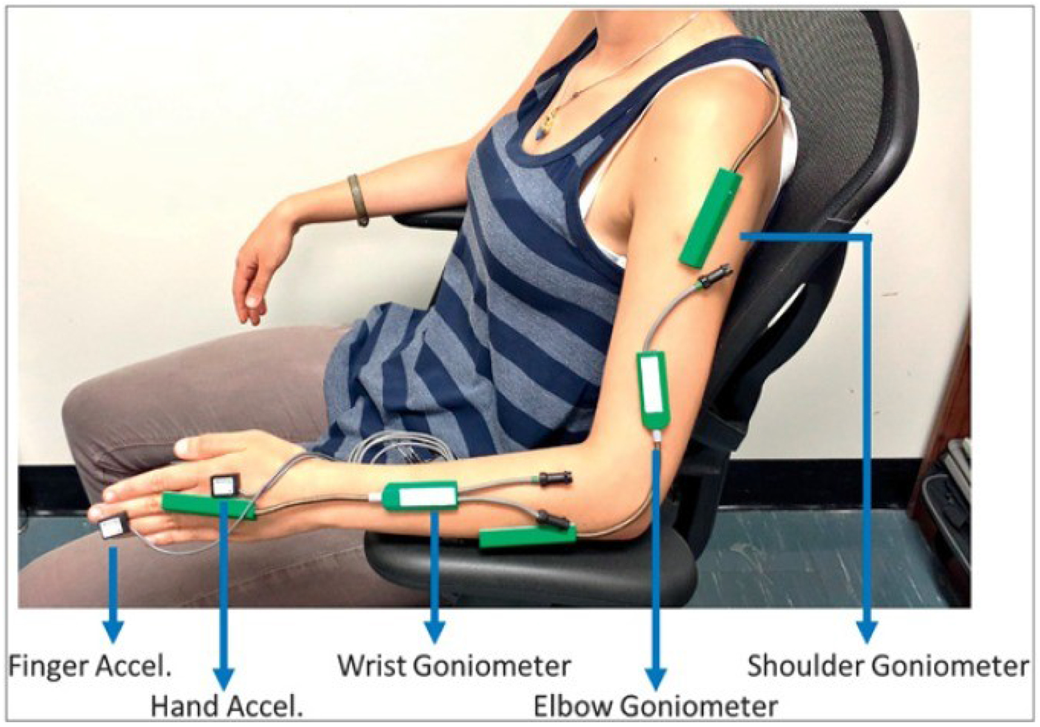
In this review, different aspects of the use of clinical neurophysiology techniques for the treatment of movement disorders are addressed. First of all, these techniques can be used to guide neuromodulation techniques or to perform therapeutic neuromodulation as such. Neuromodulation includes invasive techniques based on the surgical implantation of electrodes and a pulse generator, such as deep brain stimulation (DBS) or spinal cord stimulation (SCS) on the one hand, and non-invasive techniques aimed at modulating or even lesioning neural structures by transcranial application. Movement disorders are one of the main areas of indication for the various neuromodulation techniques. This review focuses on the following techniques: DBS, repetitive transcranial magnetic stimulation (rTMS), low-intensity transcranial electrical stimulation, including transcranial direct current stimulation (tDCS) and transcranial alternating current stimulation (tACS), and focused ultrasound (FUS), including high-intensity magnetic resonance-guided FUS (MRgFUS), and pulsed mode low-intensity transcranial FUS stimulation (TUS). The main clinical conditions in which neuromodulation has proven its efficacy are Parkinson's disease, dystonia, and essential tremor, mainly using DBS or MRgFUS. There is also some evidence for Tourette syndrome (DBS), Huntington's disease (DBS), cerebellar ataxia (tDCS), and axial signs (SCS) and depression (rTMS) in PD. The development of non-invasive transcranial neuromodulation techniques is limited by the short-term clinical impact of these techniques, especially rTMS, in the context of very chronic diseases. However, at-home use (tDCS) or current advances in the design of closed-loop stimulation (tACS) may open new perspectives for the application of these techniques in patients, favored by their easier use and lower rate of adverse effects compared to invasive or lesioning methods. Finally, this review summarizes the evidence for keeping the use of electromyography to optimize the identification of muscles to be treated with botulinum toxin injection, which is indicated and widely performed for the treatment of various movement disorders.

Keywords: Botulinum toxin; Deep brain stimulation; Dystonia; Electromyography essential tremor; Focused ultrasound; Parkinson’s disease; Repetitive transcranial magnetic stimulation; Transcranial alternating current stimulation; Transcranial direct current stimulation.


Figures

Figure 1.
Figure 1.. Literature search results on the therapeutic use of repetitive transcranial magnetic stimulation (rTMS) in movement disorders
rTMS: repetitive transcranial magnetic stimulation.
Figure 2.
Figure 2.. Relationship between publication year and effect size derived from the meta-analyses on the therapeutic use of repetitive transcranial magnetic stimulation (rTMS) in Parkinson’s disease
Summary of meta-analyses on rTMS treatment for motor symptoms of PD. The area of each circle represents sample size (number of patients).
Figure 3.
Figure 3.. Comparison of included studies across the meta-analyses on the therapeutic use of repetitive transcranial magnetic stimulation (rTMS) in Parkinson’s disease
Each row indicates an original study sorted by the publication year, and each column a meta-analysis; the leftmost one shows our original Pubmed search. Filled cells represent that the original study was included in the Pubmed search or the meta-analysis. The double line shows the publication year of each meta-analysis.
Figure 4.
Figure 4.. Literature search results on the therapeutic use of transcranial direct current stimulation (tDCS) in movement disorders
tDCS: transcranial direct current stimulation.
Figure 5.
Figure 5.. The molecular structure of botulinum neurotoxin
HC: heavy chain C-terminal domain, HN: heavy chain N-terminal domain. From Choudhury et al., 2021. Reproduced under creative commons distribution. Courtesy of PMC and Toxins.
Figure 6.
Figure 6.. Steps of botulinum neurotoxin function inside the nerve terminal
Internalization and trafficking, translocation, disulfide bond reduction and proteolysis of SNARE proteins. BoNT: botulinum neurotoxin, HSP: heat shock protein, NT: botulinum neurotoxin, PSG: polysialoganglioside, SNAP: synaptosomal-associated protein, SNARE: SNAP receptor, Stx: syntaxin, SV: synaptic vesicle, Syt: synaptotagmin, Trx: thioredoxin, TrxR: thioredoxin reductase, VAMP: vesicle-associated membrane protein. From Pirazzini et al., 2017. Reproduced under open access article distributed CC BY-NC Attribution 4.0 International license. Courtesy of publisher American Society for Pharmacology and Experimental Therapeutics
Figure 7.
Figure 7.. Position of longus colli muscle
From Selyverstov et al., 2020. Courtesy of PIMD.
Figure 8.
Figure 8.. Guitar player’s dystonia
On the left: 30 seconds into playing. On the right: 60 seconds into playing. From author’s collection.
Figure 9.
Figure 9.. Location of thyroarytenoid muscle
Hyper-adduction of the thyroarytenoid muscle causes adductor laryngeal dystonia (spasmodic dysphonia).
Figure 10.
Figure 10.. Approaching thyroarytenoid muscle for botulinum neurotoxin injection
The thyroarytenoid muscle is injected through cricothyroid space for treatment of adductor laryngeal dystonia. From Yeung et al., 2022.
Figure 11.
Figure 11.. Changes of tremor in 4 patients documented by Archimedes spiral test
Courtesy of Dr. Shivam Mittal.
Figure 12.
Figure 12.. Sensor-based kinematic measurement of tremor
Accel: accelerometer. From Rahimi et al., 2015. Courtesy of Ubiquity Press.
Figure 13.
Figure 13.. Localization of posterior tibial muscle
Patient with Parkinson’s disease and foot inversion dystonia. From author’s collection.
Figure 14.
Figure 14.. Jitter during voluntary activation using concentric needle electrodes
A, Normal jitter. B, Increased jitter. C, Increased jitter with intermittent impulse blocking (arrows). From Sanders et al., 2022.

Similar articles

References

    1. Abe K, Taira T. Focused Ultrasound Treatment, Present and Future. Neurol Med Chir (Tokyo). 2017. Aug 15;57(8):386–91. - PMC - PubMed
    1. Abeliovich A, Hefti F, Sevigny J. Gene Therapy for Parkinson's Disease Associated with GBA1 Mutations. J Parkinsons Dis 2021;11(s2):S183–8. - PMC - PubMed
    1. Abo M, Shigematsu T, Hara H, Matsuda Y, Nimura A, Yamashita Y, et al. Efficacy and Safety of OnabotulinumtoxinA 400 Units in Patients with Post-Stroke Upper Limb Spasticity: Final Report of a Randomized, Double-Blind, Placebo-Controlled Trial with an Open-Label Extension Phase. Toxins (Basel) 2020;12(2):127. - PMC - PubMed
    1. Abrahao A, Meng Y, Llinas M, Huang Y, Hamani C, Mainprize T, et al. First-in-human trial of blood–brain barrier opening in amyotrophic lateral sclerosis using MR-guided focused ultrasound. Nat Commun 2019;10(1):4373. - PMC - PubMed
    1. Adenzato M, Manenti R, Enrici I, Gobbi E, Brambilla M, Alberici A, et al. Transcranial direct current stimulation enhances theory of mind in Parkinson's disease patients with mild cognitive impairment: a randomized, double-blind, sham-controlled study. Transl Neurodegener 2019;8:1. - PMC - PubMed

MeSH terms