. 2024 Aug 10:597:217023.
doi: 10.1016/j.canlet.2024.217023. Epub 2024 Jun 7.

Dual inhibition of the TrkA and JAK2 pathways using entrectinib and pacritinib suppresses the growth and metastasis of HER2-positive and triple-negative breast cancers

Affiliations

Dual inhibition of the TrkA and JAK2 pathways using entrectinib and pacritinib suppresses the growth and metastasis of HER2-positive and triple-negative breast cancers

Angelina T Regua et al. Cancer Lett. .

Abstract

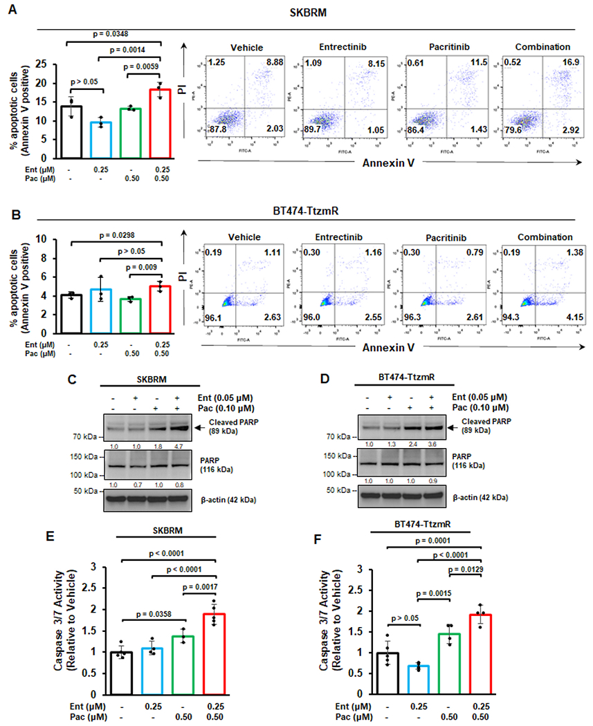
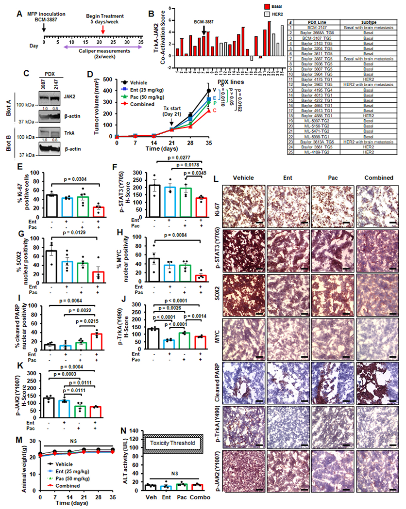
HER2-positive and triple-negative breast cancers (TNBC) are difficult to treat and associated with poor prognosis. Despite showing initial response, HER2-positive breast cancers often acquire resistance to HER2-targeted therapies, and TNBC lack effective therapies. To overcome these clinical challenges, we evaluated the therapeutic utility of co-targeting TrkA and JAK2/STAT3 pathways in these breast cancer subtypes. Here, we report the novel combination of FDA-approved TrkA inhibitors (Entrectinib or Larotrectinib) and JAK2 inhibitors (Pacritinib or Ruxolitinib) synergistically inhibited in vitro growth of HER2-positive breast cancer cells and TNBC cells. The Entrectinib-Pacritinib combination inhibited the breast cancer stem cell subpopulation, reduced expression of stemness genes, SOX2 and MYC, and induced apoptosis. The Entrectinib-Pacritinib combination suppressed orthotopic growth of HER2-positive Trastuzumab-refractory breast cancer xenografts and basal patient-derived xenograft (PDXs), reduced tumoral SOX2 and MYC, and induced apoptosis in both mouse models. The Entrectinib-Pacritinib combination inhibited overall metastatic burden, and brain and bone metastases of intracardially inoculated TNBC cells without toxicity. Together, our results demonstrate for the first time that co-inhibition of TrkA and JAK2 synergistically suppresses breast cancer growth and metastasis, thereby providing preclinical evidence that supports future clinical evaluations.

Keywords: Breast cancer; Breast cancer metastasis; Combined targeted therapy; JAK2; TrkA.


Conflict of interest statement

Declaration of competing interest The authors declare that they have no known competing financial interests or personal relationships that could have appeared to influence the work reported in this paper.

Figures

Fig. 1.
Fig. 1.. TrkA and JAK2 inhibitors synergize to reduce cell viability of HER2-positive breast cancer cells and TNBC in vitro.
(A) Western blot panel of 2 normal mammary epithelial cells and 14 breast cancer cell lines to examine relative expression of TrkA and JAK2. Densitometry values (normalized to β-actin) are displayed below each blot. LumA, luminal A. LumB, luminal B. Representative IC50 values of JAK2is (Pacritinib or Ruxolitinib) and TrkAis (Larotrectinib or Entrectinib) derived from treatment of parental BT474 and its trastuzumab-resistant variant (BT474-TtzmR), TNBC cell line MDA-MB-231, and SKBRM (brain-metastatic variant of HER2-enriched SKBR3). Lower IC50 values for each inhibitor are highlighted in red (B). Representative combination index (CI) values for combinations of TrkA and JAK2 inhibitors. CI < 1 indicates synergy, CI = 1 indicates additive and CI > 1 indicates antagonism. Synergistic CI values are highlighted in red (C). Representative cell viability plots of BT474-TtzmR (D), MDA-MB-231 (E), and SKBRM (F) after treatment with Entrectinib-Pacritinib combination at fixed 1:2 ratio. Experiments were repeated at least three times to derive averages. Data are presented as mean ± SEM. One-way ANOVA with Tukey’s multiple comparison post hoc test was used to compare p values.
Fig. 2.
Fig. 2.. Breast CSCs are inhibited by the combination of TrkA and JAK2 inhibitors.
Mammosphere formation assay of HER2-positive SKBRM (A) or BT474-TtzmR (B) after treatment with vehicle, Entrectinib, Pacritinib, or combination. Representative mammosphere images for each cell line are shown. Scale bar represents 200 μm. CD44high/CD24low flow cytometry of SKBRM (C) and BT474-TtzmR (D) after treatment with vehicle, Entrectinib, Pacritinib, or combination. Representative flow cytometry plots are shown. Western blot analysis of SKBRM (E) and BT474-TtzmR (F) after treatment with vehicle, Entrectinib, Pacritinib, or combination. Densitometry values (normalized to β-actin) are displayed below each blot. ALDH activity in SKBRM (G) or BT474-TtzmR (H) after treatment with vehicle, Entrectinib, Pacritinib, or combination. Data of three to four experimental repeats are presented as mean ± SEM. One-way ANOVA with Tukey’s multiple comparison post hoc test was used to compare p values.
Fig. 3.
Fig. 3.. Co-inhibition of TrkA and JAK2 enhances apoptosis of breast cancer cells.
Annexin V flow cytometry of SKBRM (A) or BT474-TtzmR (B) cells after treatment with vehicle, Entrectinib, Pacritinib, or combination. Cells were counterstained with propidium iodide (PI). Representative flow cytometry plots are displayed for each treatment condition. Western blot analysis of SKBRM (C) or BT474-TtzmR (D) for cleaved and total PARP after single agent or combination therapy treatment. Densitometry values (normalized to β-actin) are displayed below each blot. Caspase 3/7 activity of SKBRM (E) and BT474-TtzmR (F) after single agent or combination therapy treatment. Data of at least three experimental repeats are presented as mean ± SEM. One-way ANOVA with Tukey’s multiple comparison post hoc test was used to compare p values.
Fig. 4.
Fig. 4.. Orthotopic growth of HER2-positive trastuzumab-resistant breast cancer xenografts is suppressed by the Entrectinib-Pacritinib combination therapy.
(A) Scheme for the mammary fat pad (MFP) tumor treatment model. Trastuzumab-refractory BT474 (BT474-TtzmR) cells stably expressing luciferase were inoculated into the right inguinal MFP of female nude mice and assessed bi-weekly for tumor volume via caliper measurement. Once MFP tumors reached an average tumor volume of ~100 mm3, mice were randomized and began receiving vehicle, Entrectinib (25 mg/kg), Pacritinib (50 mg/kg), or combination orally b.i.d. 5 times per week (N = 10–11/group). (B) MFP tumor growth curve for each treatment condition. V: vehicle; E: Entrectinib; P: Pacritinib; C: combination. Tx: treatment. (C) Average ex vivo MFP tumor mass at study endpoint. (D-J) Percent nuclear positivity or H-score quantitation of MFP tumors after IHC staining. For all analyses, at least 3 fields were quantified per tumor section (N = 3–5 tumors/group). (K) Representative IHC images of MFP tumors for each treatment group. All images were captured at 40x magnification. Scale bar, 50 μm. (L) Average animal weights for each treatment group throughout the course of the study. (M) Liver toxicity following systemic therapy administration was measured using ALT activity assay. Data of at least three experimental repeats are presented as mean ± SEM. One-way ANOVA with Tukey’s multiple comparison post hoc test was used to compare p values.
Fig. 5.
Fig. 5.. The Entrectinib-Pacritinib combination therapy suppresses the orthotopic growth of TNBC PDX MFP tumors.
(A) Schema of PDX MFP tumor treatment model. 1-mm3 fragment of BCM-3887 PDX tissue was implanted into the right inguinal mammary fat pad of female nude mice. Once tumors reached an average volume of ~100 mm3, animals were randomized based on tumor volume and began receiving vehicle, Entrectinib only (25 mg/kg), Pacritinib (50 mg/kg), or combination via oral gavage b.i.d., 5 times/week (N = 11/group). BCM-3887 was chosen due to its basal subtype, relatively higher level of JAK2-TrkA pathway co-activation score, and propensity to develop brain metastases (B). (C) Western blot analysis of basal PDX tissues BCM-3887 BCM-2147. Densitrometry values (normalized to β-actin) are displayed below each blot. (D) MFP tumor growth curve for each treatment condition. V: vehicle; E: Entrectinib; P: Pacritinib; C: combination, Tx: treatment. (E-K) Percent nuclear positivity or H-score quantitation of MFP tumors after IHC staining. For all analyses, at least 3 fields were quantified per tumor section (N = 3–5 tumors/group). (L) Representative IHC images of MFP tumors for each treatment group. All images were captured at 40x magnification. Scale bar, 50 μm. (M) Average animal weights throughout the course of the study. (N) Liver toxicity was measured using ALT activity assay. Data are represented as mean ± SEM. One-way ANOVA with Tukey’s multiple comparison post hoc test was used to compare p values.
Fig. 6.
Fig. 6.. Combined treatment with Entrectinib and Pacritinib reduces TNBC metastatic burden in vivo.
(A) Scheme for the intracardiac inoculation treatment model. Luciferase-expressing MDA-MB-231 cells were inoculated into the left ventricle of athymic nude mice and assessed for metastasis burden using bi-weekly bioluminescent imaging (BLI). Mice were randomized and began receiving oral administration (b.i.d.) of vehicle, Entrectinib (25 mg/kg), Pacritinib (50 mg/kg), or combination, 5 days/week (N = 12/group). (B) Average metastatic burden for each treatment group throughout the study as measured by BLI. One-way ANOVA with Tukey’s multiple comparison post-hoc test was used to compute p-values for differences in metastatic burden at study endpoint (Day 25). (C) Representative bioluminescent images of mice 25 days post-inoculation (study endpoint). (D) Representative ex vivo organ images are shown. Ex vivo brain (E), bone (F), and liver (G) bioluminescence was quantified. (H) Average mouse weights throughout the course of the study. (I) ALT assay was performed on serum collected at study endpoint to assess liver toxicity. Data are represented as mean ± SEM. One-way ANOVA with Tukey’s multiple comparison post-hoc test was used to compute p-values.

References

    1. Siegel RL, Miller KD, Fuchs HE, Jemal A, Cancer statistics, CA A Cancer J. Clin 72 (2022) 7–33, 2022. - PubMed
    1. Howlader N, Noone AM, Krapcho M, Miller D, Brest A, Yu M, et al. SEER Cancer Statistics Review, 1975-2016, National Cancer Institute. Bethesda, MD, https://seer.cancer.gov/csr/1975_2016/, based on November 2018 SEER data submission, posted to the SEER web site, April 2019.
    1. Perou CM, Sorlie T, Eisen MB, van de Rijn M, Jeffrey SS, Rees CA, et al., Molecular portraits of human breast tumours, Nature 406 (2000) 747–752. - PubMed
    1. Waks AG, Winer EP, Breast cancer treatment: a review, JAMA 321 (2019) 288–300. - PubMed
    1. Kim YJ, Kim JS, Kim IA, Molecular subtype predicts incidence and prognosis of brain metastasis from breast cancer in SEER database, J. Cancer Res. Clin. Oncol 144 (2018) 1803–1816. - PMC - PubMed

MeSH terms