Review
. 2025 Jun;92(6):1189-1206.
doi: 10.1016/j.jaad.2024.03.056. Epub 2024 Jun 7.

Dermatologic fungal neglected tropical diseases-Part I. Epidemiology and clinical features

Affiliations
Review

Dermatologic fungal neglected tropical diseases-Part I. Epidemiology and clinical features

Kaya L Curtis et al. J Am Acad Dermatol. 2025 Jun.

Abstract

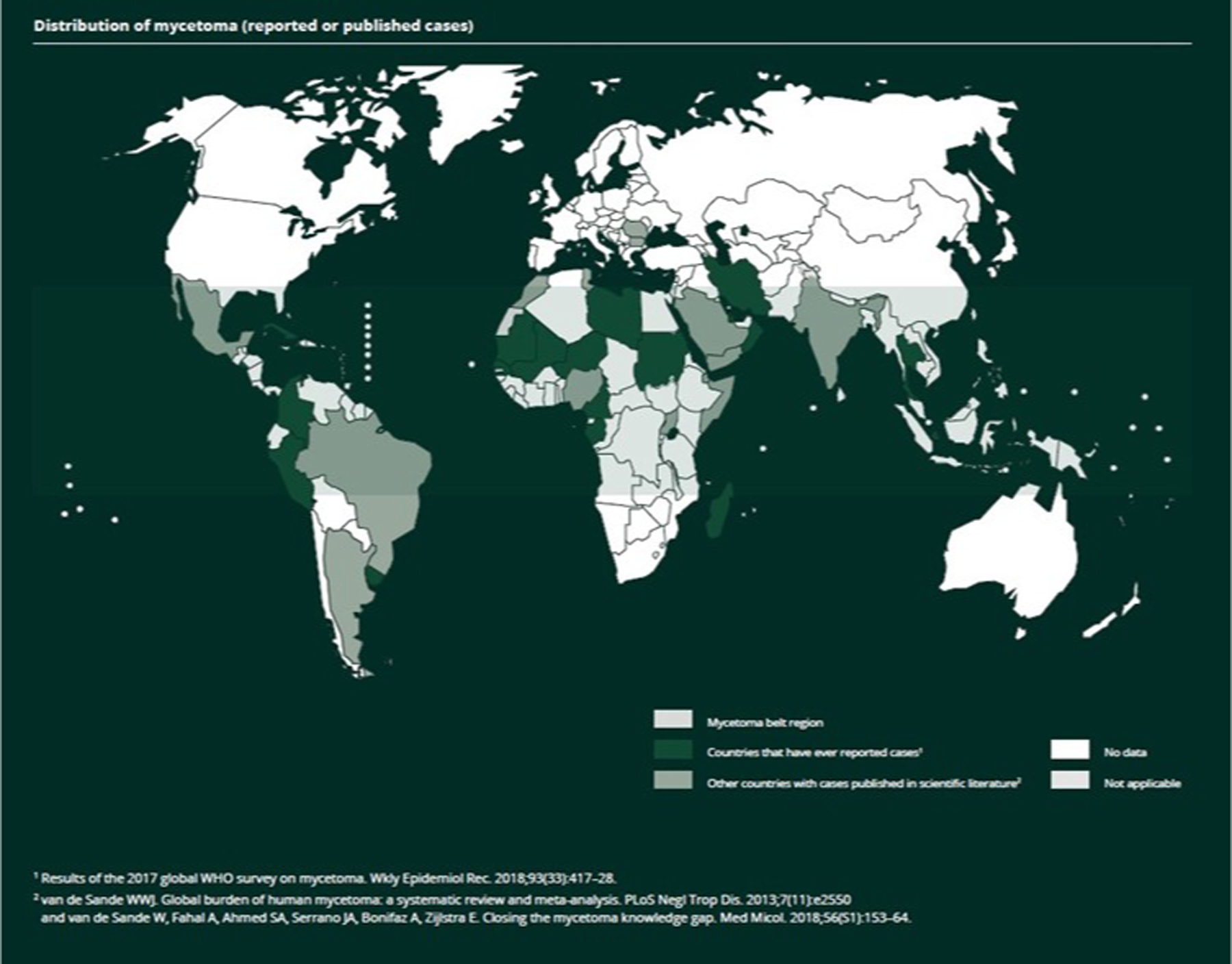
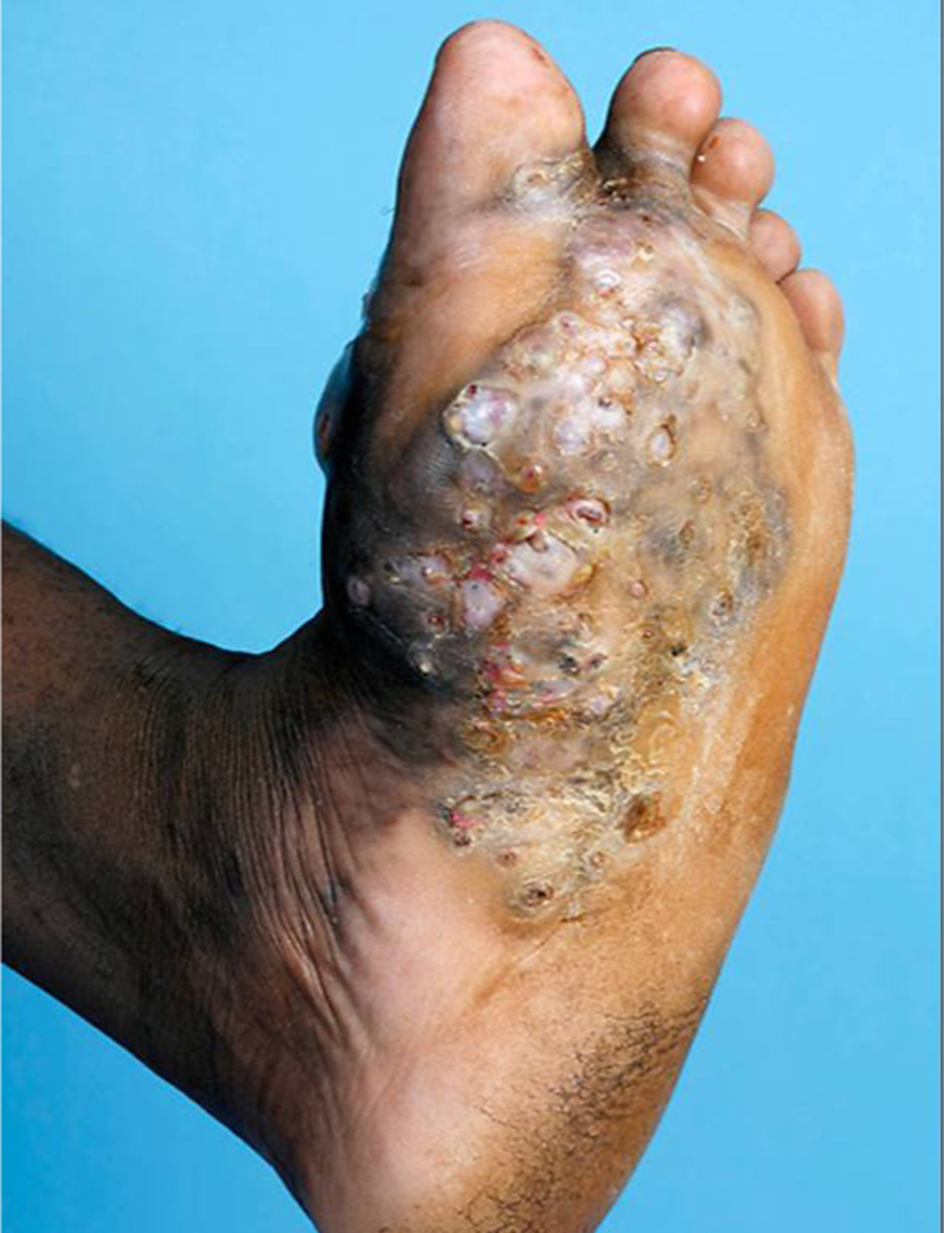
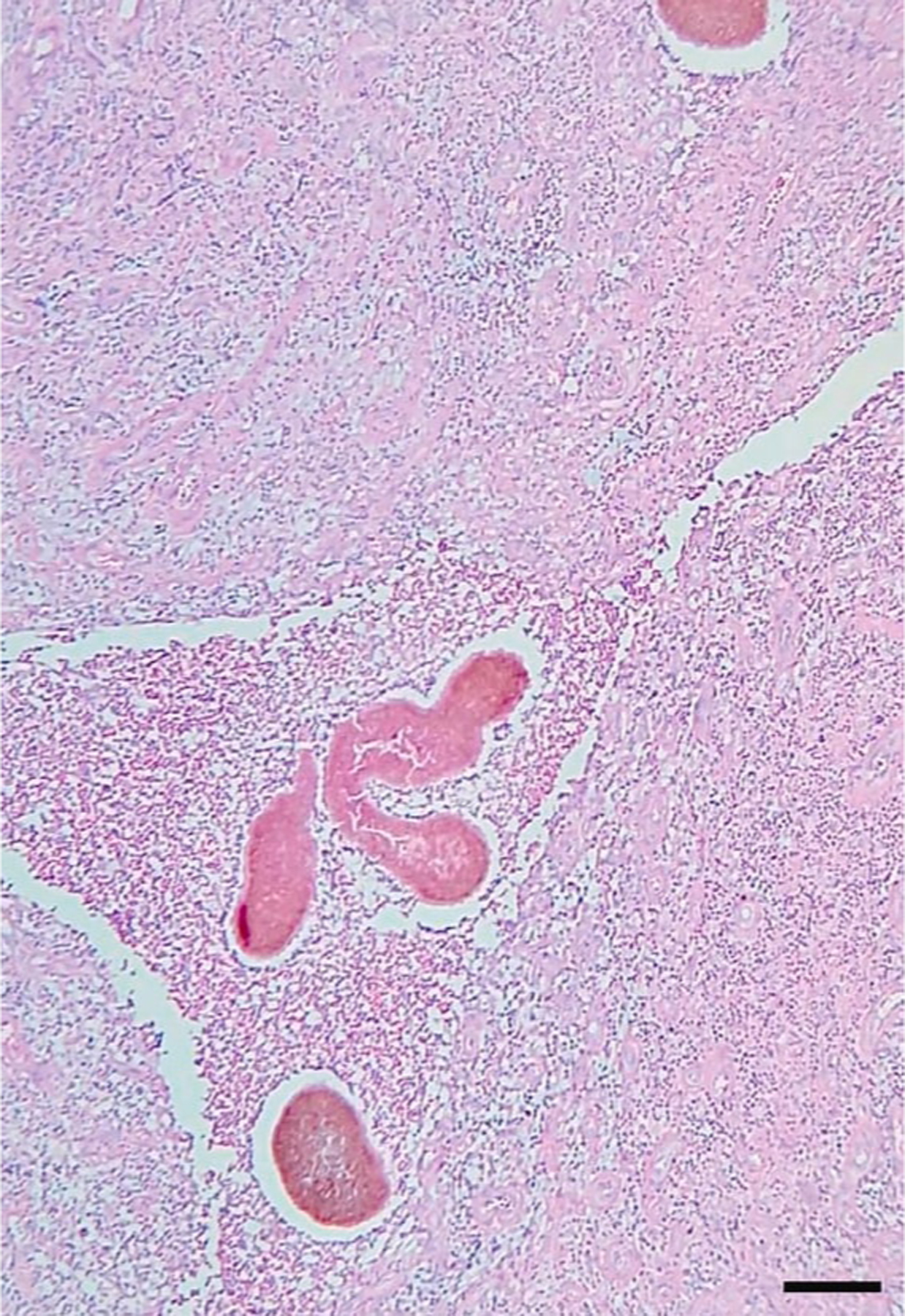
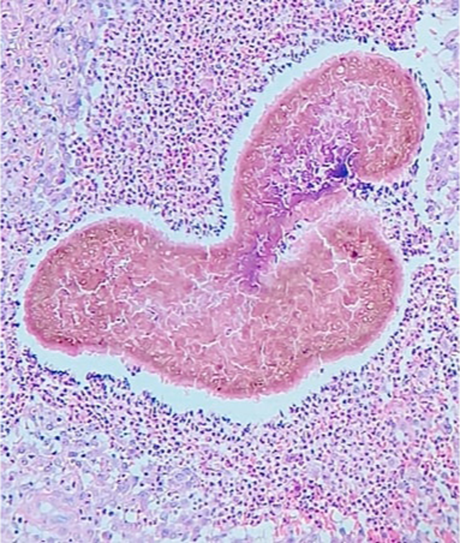
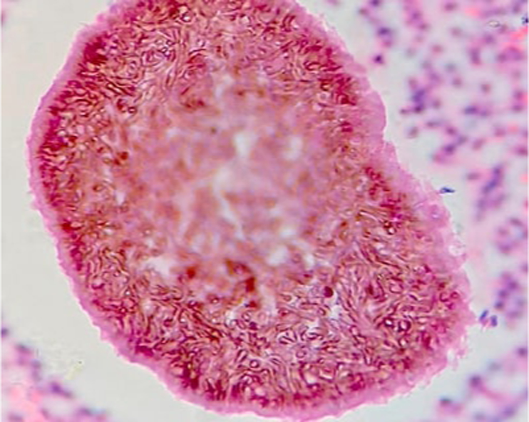
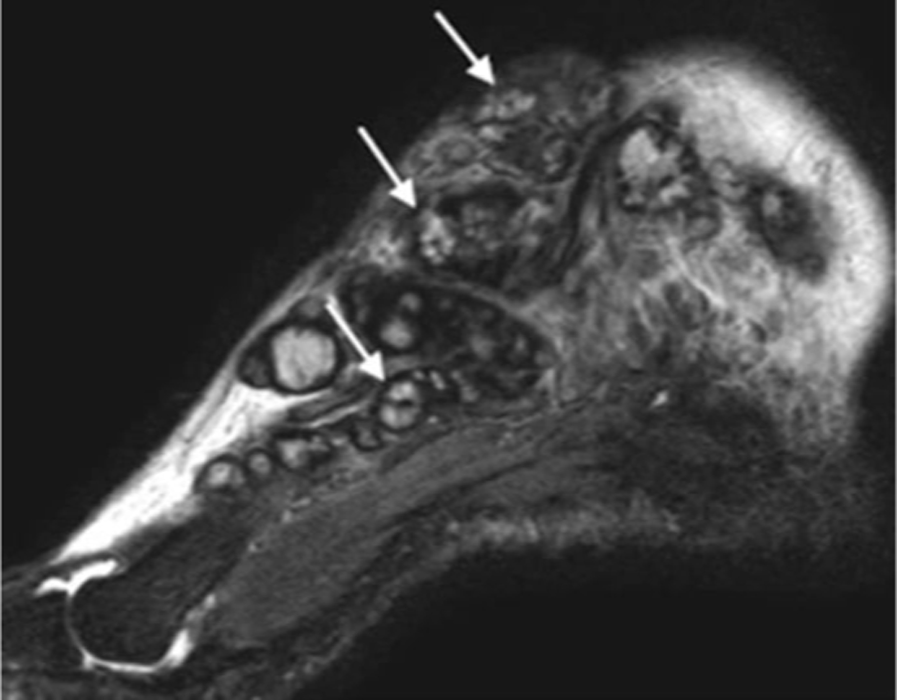
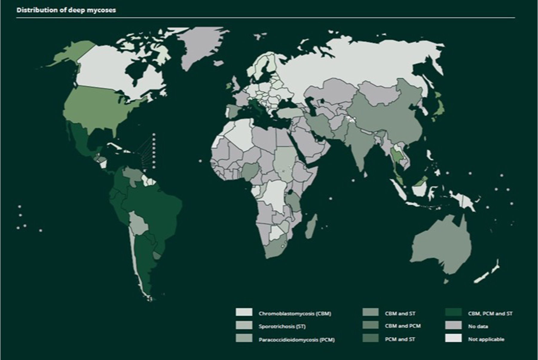
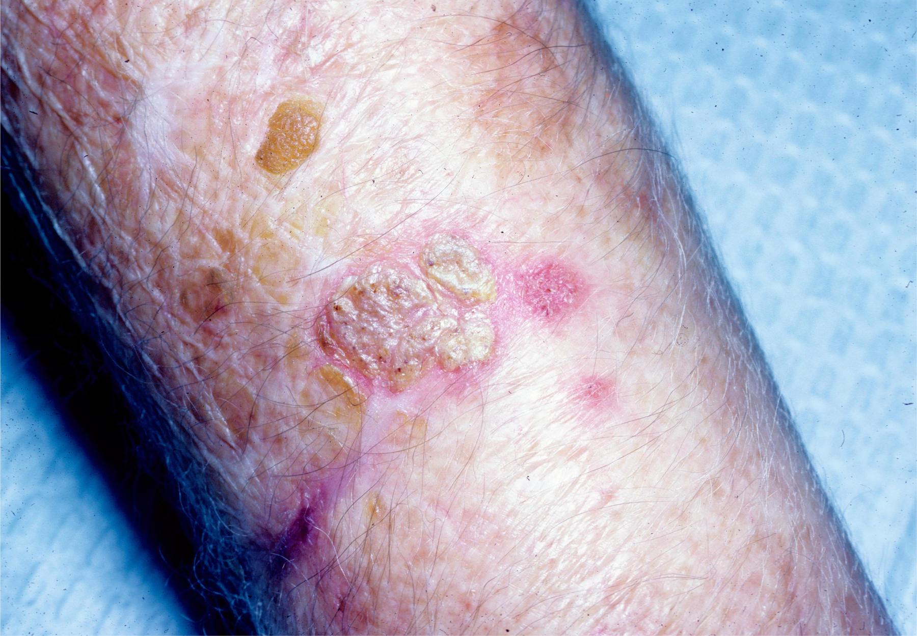
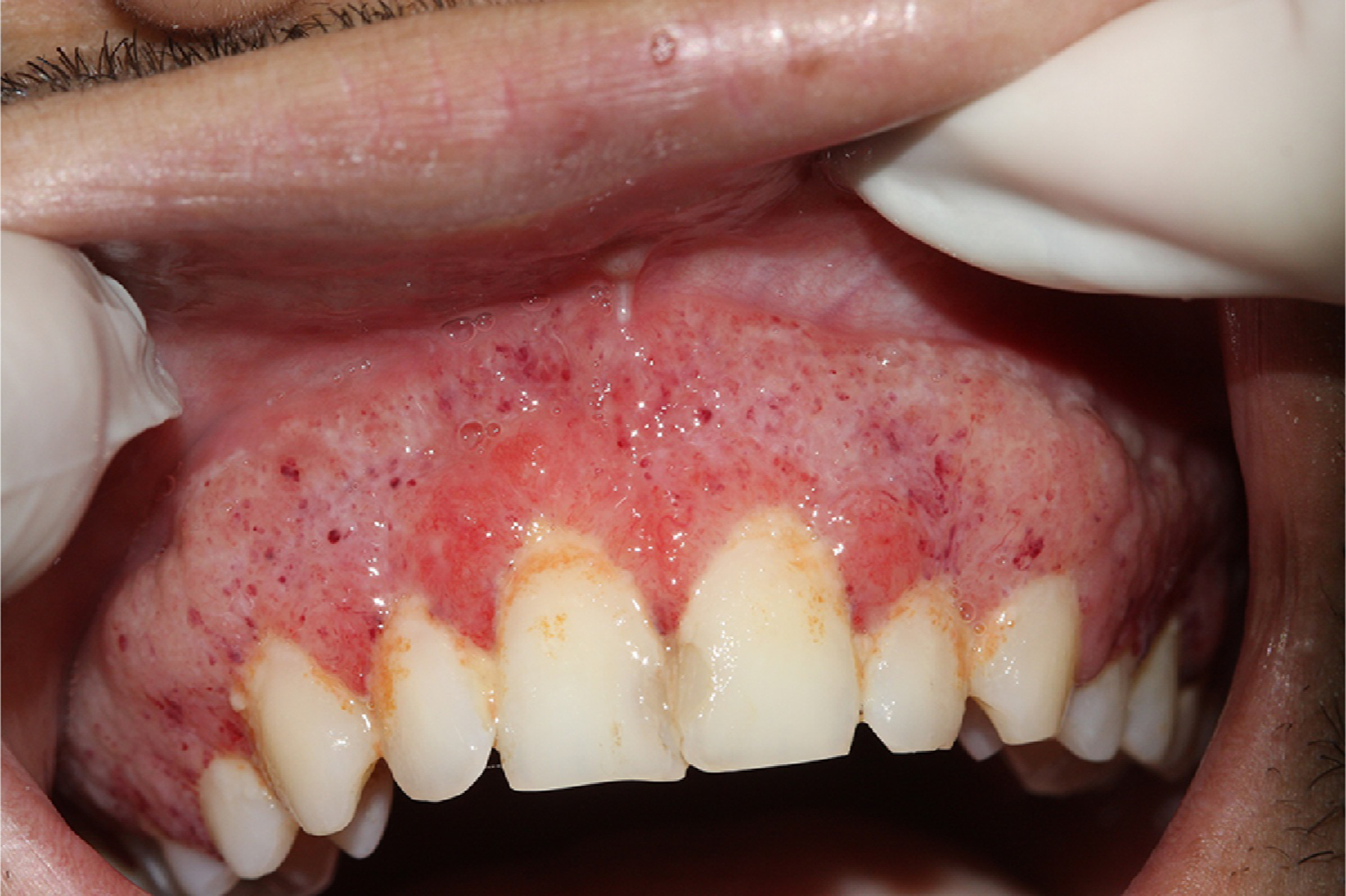
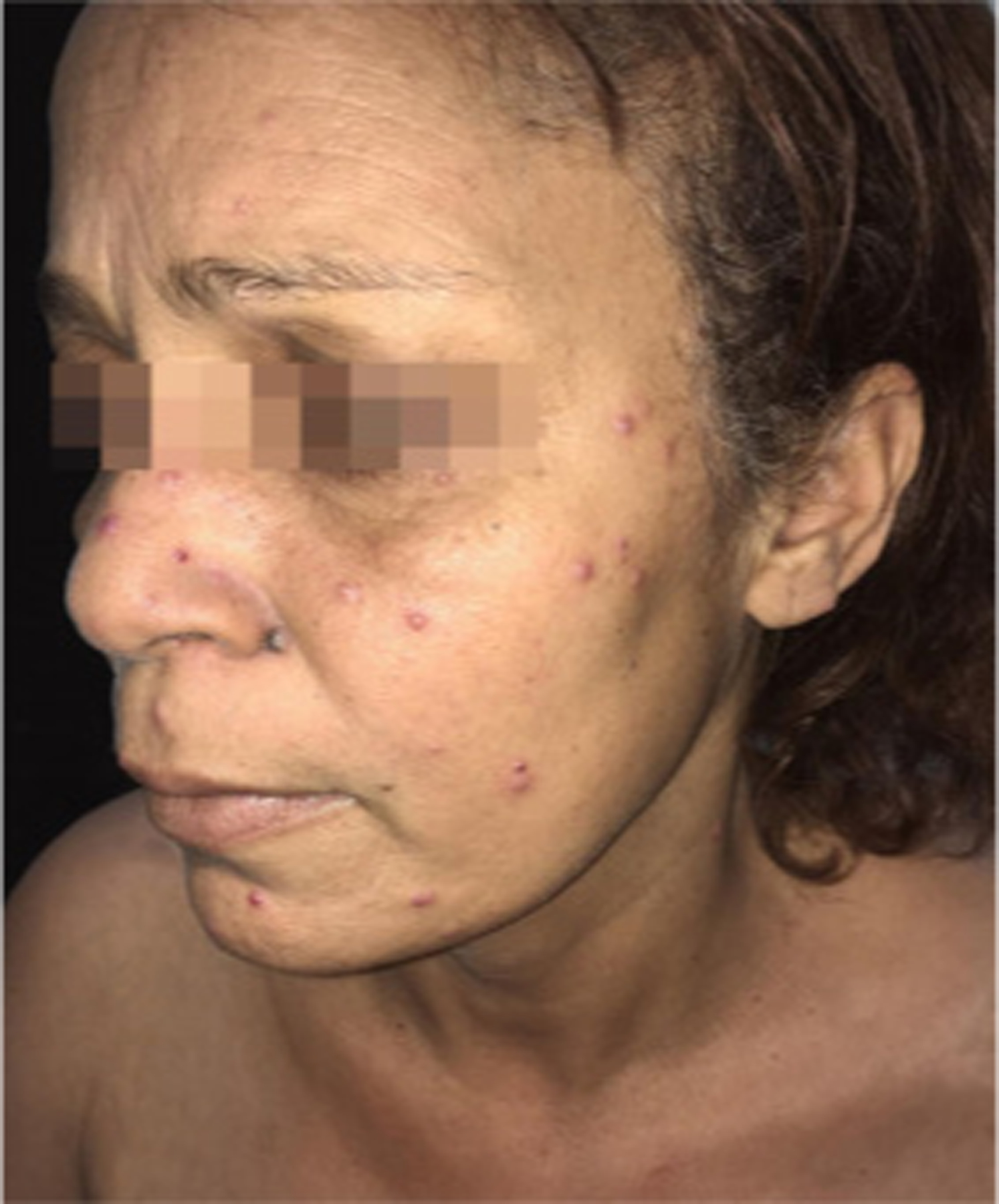
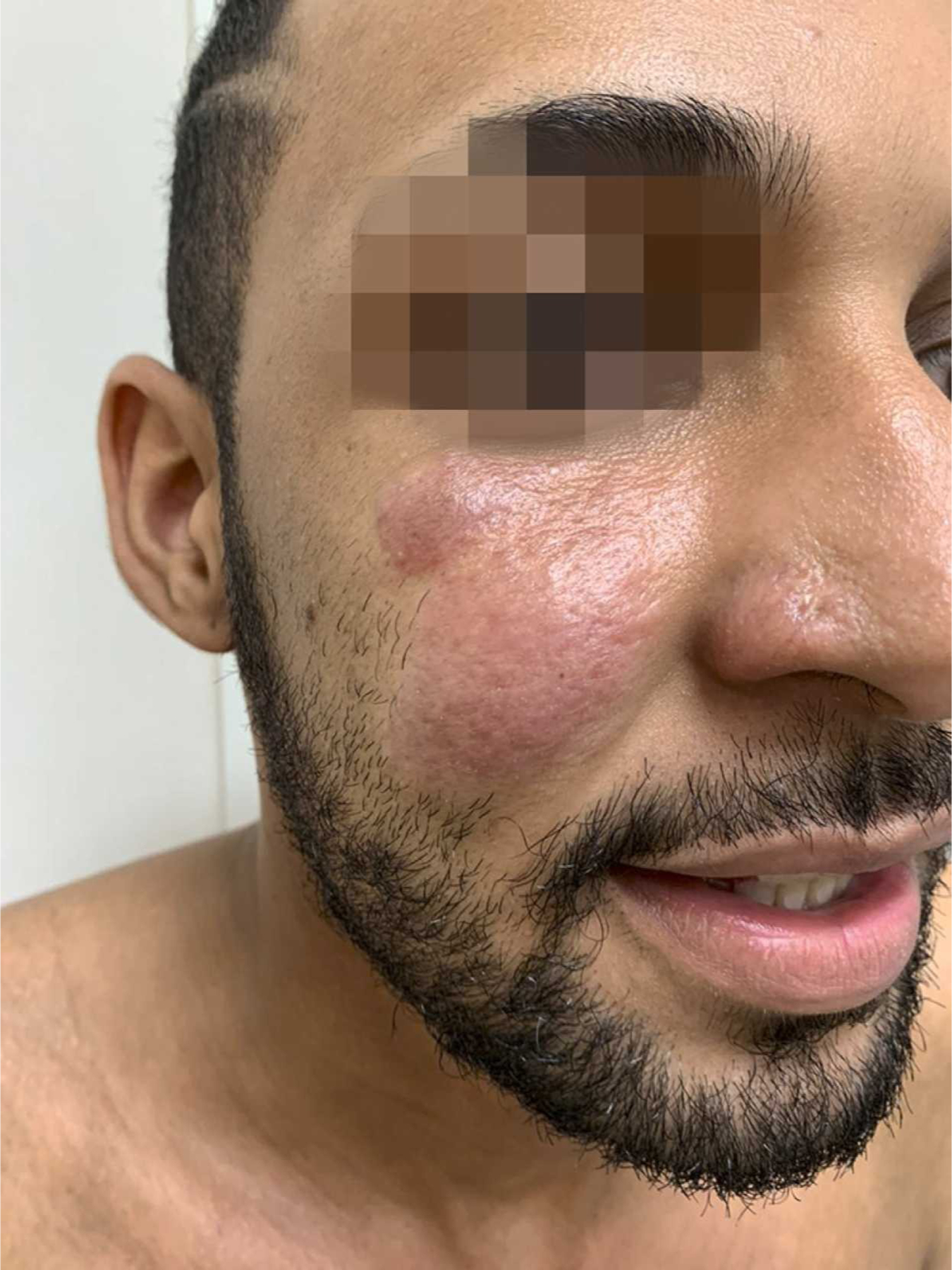
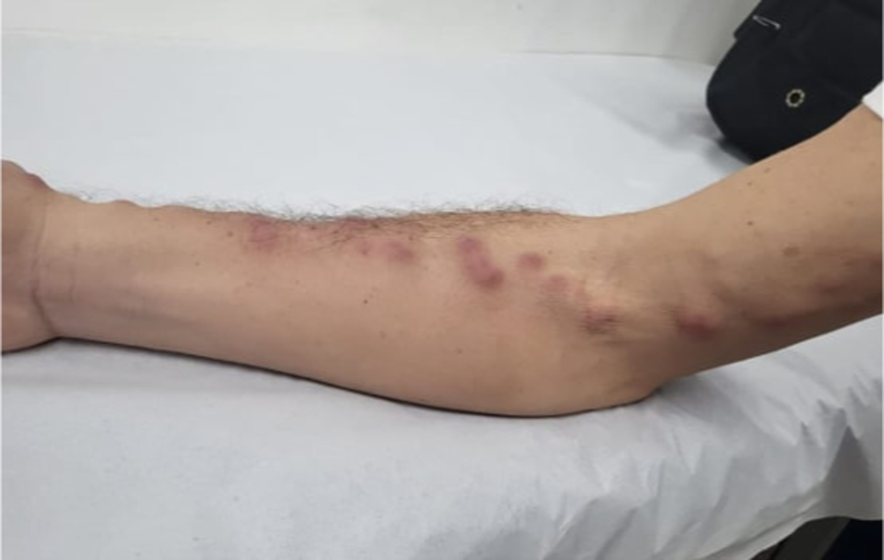
In this part 1 of a 2-part continuing medical education series, the epidemiology, clinical features, and diagnostic methods for fungal skin neglected tropical diseases (NTDs), which include eumycetoma, chromoblastomycosis, paracoccidioidomycosis, sporotrichosis, emergomycosis, talaromycosis, and lobomycosis, are reviewed. These infections, several of which are officially designated as NTDs by the World Health Organization, cause substantial morbidity and stigma worldwide and are receiving increased attention due to the potential for climate change-related geographic expansion. Domestic incidence may be increasing in the setting of global travel and immunosuppression. United States dermatologists may play a central role in early detection and initiation of appropriate treatment, leading to decreased morbidity and mortality.

Keywords: chromoblastomycosis; diagnostics; emergomycosis; endemic mycoses; epidemiology; eumycetoma; fungal infections; implantation mycoses; lobomycosis; neglected tropical diseases; paracoccidioidomycosis; sporotrichosis; systemic mycoses; talaromycosis.


Conflict of interest statement

Conflicts of interest None disclosed.

Figures

Figure 1.
Figure 1.
Distribution of mycetoma (reported or published cases). Reproduced from the World Health Organization (published under Creative Commons CC BY-NC-SA 4.0 DEED license).
Figure 2.
Figure 2.
A massive eumycetoma lesion of the plantar foot with multiple discharging sinuses and black grains, Mexico.
Figure 3.
Figure 3.
Eumycetoma histopathologic examination (skin biopsy) demonstrating A. suppurative granulomatous inflammatory infiltrates with neutrophils, lymphocytes, and histiocytes; B. large multilobed grains with Splendore-Hoeppli phenomenon; C. clusters of radially branched brown hyphae (Hematoxylin and eosin stain, 100x). Reproduced from Arteaga et al. (published under Creative Commons CC-BY-NC-ND license).
Figure 3.
Figure 3.
Eumycetoma histopathologic examination (skin biopsy) demonstrating A. suppurative granulomatous inflammatory infiltrates with neutrophils, lymphocytes, and histiocytes; B. large multilobed grains with Splendore-Hoeppli phenomenon; C. clusters of radially branched brown hyphae (Hematoxylin and eosin stain, 100x). Reproduced from Arteaga et al. (published under Creative Commons CC-BY-NC-ND license).
Figure 4.
Figure 4.
Sagittal MRI show multiple, small, round-to-spherical hyperintense mycetoma lesions separated by peripheral hypointense tissue. Some of the lesions contain a central hypointense dot, resulting in the ‘dot-in-circle’ sign (arrows). Reproduced from Laohawiriyakamol et al. (published under Creative Commons CC-BY license).
Figure 5.
Figure 5.
Distribution of deep mycoses. Reproduced from the World Health Organization (published under Creative Commons CC BY-NC-SA 4.0 DEED license).
Figure 6.
Figure 6.
Small, scale-crust covered lesion on the forearm found to be chromoblastomycosis; submitted for biopsy as Bowen’s disease. Patient from Houston, Texas.
Figure 7.
Figure 7.
Chromoblastomycosis, hyperkeratotic plaque, with warty dry lesions, with moderate severity disease localized to the distal third of the leg. Lesions with small black dots (cayenne pepper appearance).
Figure 8.
Figure 8.
Tumoral mass clinically diagnosed as verrucous squamous cell carcinoma and diagnosed with biopsy as chromoblastomycosis. Patient from Panama.
Figure 9.
Figure 9.
Chromoblastomycosis, clustered muriform cells depicted as round to polyhedral (chestnut-like) cells, visualized on direct microscopy potassium hydroxide exam.
Figure 10.
Figure 10.
Chromoblastomycosis, skin, H&E stain: Clusters of round, thick-walled, brown-pigmented fungal spores (“sclerotic bodies,” “Medlar bodies” or “copper bodies”) within multinucleated giant cells in a focus of subepidermal granulomatous inflammation. Sclerotic bodies are fungal spores that are usually 5–12 microns and divide by internal septation (arrows). Original magnification: x630.
Figure 11.
Figure 11.
Oral lesions of paracoccidioidomycosis in the gingiva and mulberry-like ulcers with hemorrhagic dots. Photograph from a patient treated at University Hospital Cassiano Antonio Moraes, Federal University of Espirito Santo. Reproduced from Dutra et al. (published under Creative Commons CC-BY-NC-ND license).
Figure 12.
Figure 12.
Paracoccidioidomycosis and liver transplantation. Acneiform lesions and erythematous papules, some with an ulcerated center, disseminated on the face. Reproduced from Valentim et al. (published under Creative Commons CC-BY license).
Figure 13.
Figure 13.
Paracoccidioidomycosis, infiltrated, sarcoid-like cutaneous lesions in the right malar region. Reproduced from Cárcano et al. (published under Creative Commons CC-BY-NC-ND license).
Figure 14.
Figure 14.
Paracoccidioidomycosis and liver transplantation, histopathologic examination (skin biopsy) demonstrating multi-budding fungal cells, “ship’s wheel” finding pathognomonic for disease (Grocott-Gomori. Immersion). Reproduced from Valentim et al. (published under Creative Commons CC-BY license).
Figure 15.
Figure 15.
Sporotrichosis, feline dissemination. Erythematous lesions, with subcutaneous nodules, painful on palpation, with lymphatic distribution involving the right arm.
Figure 16.
Figure 16.
Sporotrichosis fixed cutaneous form, verrucous lesion on the dorsum of the hand. Reproduced from Orofino-Costa et al. (published under Creative Commons CC-BY license).
Figure 17.
Figure 17.
Primary chancriform lesion of sporotrichosis after trauma with rose thorn.
Figure 18.
Figure 18.
Sporotrichosis histopathologic examination (skin) demonstrating suppurative granuloma and parasitic fungus. Epithelioid cells on the left, neutrophils and myocytes on the right, round yeast-like (black arrow) and elongated or navicular fungal cells (red arrow) (Periodic acid-Schiff stain, 1000x). Reproduced from Orofino-Costa et al. (published under Creative Commons CC-BY license).
Figure 19.
Figure 19.
Mucocutaneous manifestations of HIV-associated disseminated emergomycosis in a South African patient who presented with a 1-year history of nodular lesions on the face, limbs, and trunk. Papules and plaques with central necrosis. Reproduced from Reddy et al. with permission. Figure courtesy of Dr. Matilda Mphahlele (Division of Dermatology, Chris Hani Baragwanath Academic Hospital, Faculty of Health Sciences, University of the Witwatersrand, Johannesburg, South Africa).
Figure 20.
Figure 20.
Clinical image of a South African patient with HIV-associated disseminated emergomycosis, showing hyperpigmented and violaceous plaques with surface scaling. Reproduced from Reddy et al. with permission.
Figure 21.
Figure 21.
Histopathology of skin biopsies from South African patients with HIV-associated disseminated emergomycosis caused by Es. Africanus. Es. africanus organisms occupying the papillary dermis, as seen under oil emersion (haematoxylin and eosin, original magnification x 1,000). Reproduced from Govender et al. with permission.
Figure 22.
Figure 22.
Talaromycosis physical examination. Multiple cuticolor follicular papules with or without central umbilication, nodules, and acne-like lesions were developed densely on his face, neck, and upper anterior chest. Some of them were covered with reddish brown crusts. Reproduced from Xian et al. (published under Creative Commons CC-BY license).
Figure 23.
Figure 23.
Talaromycosis histopathologic examination (skin biopsy) demonstrating abundant yeast-like organisms in the cytoplasm of histocytes. The organisms were spherical to oval, about 3-μm in diameter and occasional contained septum (Periodic acid-Schiff stain, 40x). Reproduced from Xian et al. (published under Creative Commons CC-BY license).
Figure 24.
Figure 24.
Lobomycosis, classic infiltrative nodules on the right outer ear. Reproduced from Talhari et al. Permission granted for figure reproduction.
Figure 25.
Figure 25.
Large aggregate of keloid-like nodules admixed with sclerotic areas diagnosed as lobomycosis on biopsy. Patient from Panama.
Figure 26.
Figure 26.
Lobomycosis histopathology examination demonstrating multiple isolated and chained parasites (Grocott stain, 40x). Reproduced from Talhari et al. Permission granted for figure reproduction.

Similar articles

Cited by

References

    1. Hotez PJ, Alvarado M, Basanez MG et al. The global burden of disease study 2010: interpretation and implications for the neglected tropical diseases. PLoS Negl Trop Dis 2014; 8:e2865. - PMC - PubMed
    1. Gadre A, Enbiale W, Andersen LK, Coates SJ. The effects of climate change on fungal diseases with cutaneous manifestations: a report from the International Society of Dermatology Climate Change Committee. J Clim Change Health. 2022;6: 100156.
    1. Lafferty KD. The Ecology of Climate Change and Infectious Diseases. Vol. 90, Source: Ecology. 2009. - PubMed
    1. Thompson GR 3rd, Le T, Chindamporn A, Kauffman CA, Alastruey-Izquierdo A, Ampel NM, Andes DR, Armstrong-James D, Ayanlowo O, Baddley JW, Barker BM, Lopes Bezerra L, Buitrago MJ, Chamani-Tabriz L, Chan JFW, Chayakulkeeree M, Cornely OA, Cunwei C, Gangneux JP, Govender NP, Hagen F, Hedayati MT, Hohl TM, Jouvion G, Kenyon C, Kibbler CC, Klimko N, Kong DCM, Krause R, Lee Lee L, Meintjes G, Miceli MH, Rath PM, Spec A, Queiroz-Telles F, Variava E, Verweij PE, Schwartz IS, Pasqualotto AC. Global guideline for the diagnosis and management of the endemic mycoses: an initiative of the European Confederation of Medical Mycology in cooperation with the International Society for Human and Animal Mycology. Lancet Infect Dis 2021. Dec;21(12):e364–e374. - PMC - PubMed
    1. Bonifaz A, Robles-Tenorio A, Tirado-Sánchez A (2022). Climate Change Impact on Chromoblastomycosis. In: Frías-De-León MG, Brunner-Mendoza C, Reyes-Montes M.d.R., Duarte-Escalante E (eds) The Impact of Climate Change on Fungal Diseases. Fungal Biology. Springer, Cham.