. 2024 Jun 9;14(1):13229.
doi: 10.1038/s41598-024-63946-4.

Motor assessment of X-linked dystonia parkinsonism via machine-learning-based analysis of wearable sensor data

Affiliations

Motor assessment of X-linked dystonia parkinsonism via machine-learning-based analysis of wearable sensor data

Federico Parisi et al. Sci Rep. .

Abstract

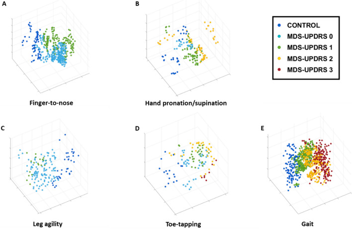
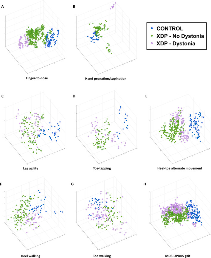
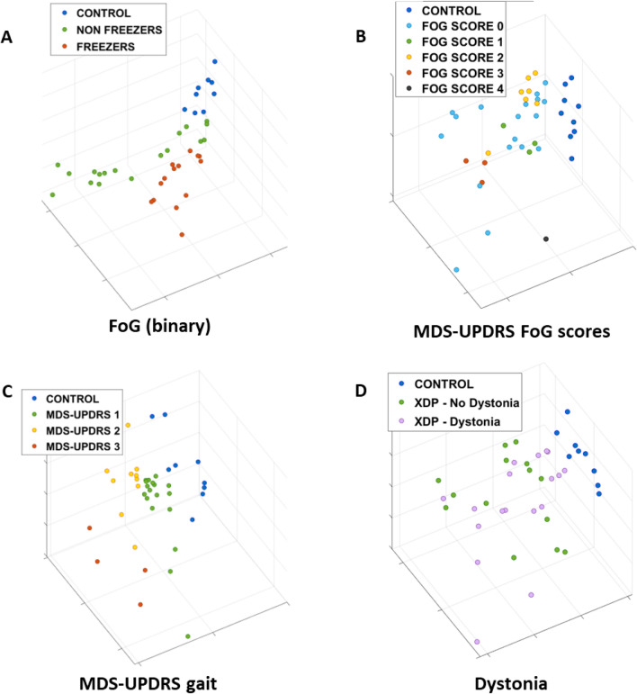
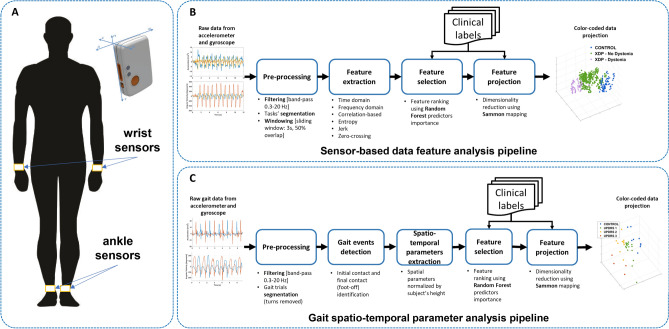
X-linked dystonia parkinsonism (XDP) is a neurogenetic combined movement disorder involving both parkinsonism and dystonia. Complex, overlapping phenotypes result in difficulties in clinical rating scale assessment. We performed wearable sensor-based analyses in XDP participants to quantitatively characterize disease phenomenology as a potential clinical trial endpoint. Wearable sensor data was collected from 10 symptomatic XDP patients and 3 healthy controls during a standardized examination. Disease severity was assessed with the Unified Parkinson's Disease Rating Scale Part 3 (MDS-UPDRS) and Burke-Fahn-Marsden dystonia scale (BFM). We collected sensor data during the performance of specific MDS-UPDRS/BFM upper- and lower-limb motor tasks, and derived data features suitable to estimate clinical scores using machine learning (ML). XDP patients were at varying stages of disease and clinical severity. ML-based algorithms estimated MDS-UPDRS scores (parkinsonism) and dystonia-specific data features with a high degree of accuracy. Gait spatio-temporal parameters had high discriminatory power in differentiating XDP patients with different MDS-UPDRS scores from controls, XDP freezing of gait, and dystonic/non-dystonic gait. These analyses suggest the feasibility of using wearable sensor data for deriving reliable clinical score estimates associated with both parkinsonian and dystonic features in a complex, combined movement disorder and the utility of motion sensors in quantifying clinical examination.

Keywords: Digital health; Dystonia; Dystonia parkinsonism; Machine Learning; Parkinsonism; Wearable sensors.


Conflict of interest statement

Dr. Parisi reports no relevant disclosures. Dr. Corniani reports no relevant disclosures. Dr. Bonato has received grant support from the American Heart Association, the Department of Defense, the Michael J Fox Foundation, the National Institutes of Health (NIH), the National Science Foundation (NSF), and the Peabody Foundation including sub-awards on NIH and NSF SBIR grants from Barrett Technology (Newton MA), BioSensics (Watertown MA) and Veristride (Salt Lake City UT). He has also received grant support from Emerge Diagnostics (Carlsbad CA), MC10 (Lexington MA), Mitsui Chemicals (Tokyo Japan), Pfizer (New York City NY), Shimmer Research (Dublin Ireland), and SynPhNe (Singapore). He has served on the Advisory Board of SwanBio (Boston MA). Dr. Bonato serves in an advisory uncompensated role the Michael J Fox Foundation, the NIH-funded New England Pediatric Device Consortium, and the Walking Tall-PD clinical trial carried out by Neuroscience Research Australia. He also serves in an uncompensated role on the Scientific Advisory Boards of ABLE Human Motion (Barcelona, Spain), FormSense (San Diego CA, USA), Hocoma AG (Zurich, Switzerland), and Trexo (Toronto, Canada). Dr. Acuna reports no relevant disclosures. Dr. Go reports no relevant disclosures. Dr. Sharma receives support from Wiley Publishing for her role as editor-in-chief of Brain and Behavior. has received grant support from the Department of Defense and the National Institutes of Health (NIH). Dr. Stephen has received financial support from Encora Therapeutics, SwanBio Therapeutics, Sanofi-Genzyme, Biogen and Biohaven for the conduct of clinical trials. He has received honoraria from the Movement Disorders Society, American Academy of Neurology and from Oakstone CME. Dr. Stephen’s institution has received research funding from Sanofi-Genzyme for a study of video oculography in late-onset GM2 gangliosidosis.

Figures

Figure 1
Figure 1
Sensor-based data feature projections color-coded by MDS-UPDRS clinical scores. Three-dimensional data feature projections for the (A) finger-to-nose (item 3.16), (B) hand pronation/supination (item 3.6), (C) leg agility (item 3.8), (D) toe-tapping (item 3.7), and (E) gait (item 3.10) motor tasks. The points in each plot correspond to the representation in the reduced dimensionality space of the data features derived from the sensor signals and are color-coded according to the clinical labels (control and MDS-UPDRS scores). For sample numbers for each task, please see Supplementary Table S3.
Figure 2
Figure 2
Sensor-based data feature projections color-coded by presence/absence of dystonia. Three-dimensional data feature projections for the (A) finger-to-nose (item 3.16), (B) hand pronation/supination (item 3.6), (C) leg agility (item 3.8), (D) toe-tapping (item 3.7), and (H) gait (item 3.10) motor tasks, as well as for the non-MDS-UPDRS dystonia provoking maneuvers (E) heel-toe alternate movement, (F) heel walking, and (G) toe walking. The points in each plot correspond to the representation in the reduced dimensionality space of the data features derived from the sensor signals and are color-coded according to the clinical labels (control and XDP with/without dystonia). For sample numbers for each task, please see Supplementary Table S4.
Figure 3
Figure 3
Projections of the gait spatio-temporal parameters color-coded by clinical characteristics. Three-dimensional data feature projections of gait spatio-temporal parameters color-coded by (A) presence/absence of freezing of gait (FoG) on examination, (B) MDS-UPDRS FoG score (Item 3.11), (C) MDS-UPDRS gait score (Item 3.10), and (D) presence/absence of dystonia. The points in each plot correspond to the representation in the reduced dimensionality space of the aggregated statistics (mean, standard deviation, coefficient of variation, and right/left ratio) of the gait parameters extracted from each trial. For sample numbers for each task, please see Supplementary Table S5.
Figure 4
Figure 4
Cadence, stride length, and stride velocity boxplots for different clinical characteristics. Boxplots of cadence, stride length, and stride velocity for presence/absence of freezing of gait (FoG) on clinical examination, MDS-UPDRS FoG scores (Item 3.11), MDS-UPDRS gait scores (Item 3.10), and presence/absence of dystonia on clinical gait examination. The boxplots visually summarize the distribution of data. Each boxplot displays the median (central line), interquartile range (box edges), and overall range excluding outliers (whiskers). Outliers are marked with individual points. Pairwise significant differences were assessed with a mixed regression model and are indicated by a horizontal red line. *** indicates p-value < 0.001, while * indicates p-value < 0.01.
Figure 5
Figure 5
Experimental set-up and data processing pipelines. (A) Two wearable motion sensors were placed on the wrists during the performance of upper-limb tasks and repositioned on the ankles when performing lower-limb tasks. An enlarged view of a motion sensor (Shimmer3 by Shimmer Research Ltd, Dublin, Ireland) and its reference system are also shown. (B) Sensor-based data feature analysis pipeline. The diagram illustrates the processing steps used to derive data feature projections from the raw motion sensor data when participants performed standardized motor tasks. (C) Gait spatio-temporal parameter analysis pipeline. The diagram depicts the steps of the algorithm used to extract gait spatio-temporal parameters from the raw motion sensor signals recorded during gait and visualize them via projections.

Similar articles

Cited by

References

    1. Lee LV, Maranon E, Demaisip C, et al. The natural history of sex-linked recessive dystonia parkinsonism of Panay, Philippines (XDP) Parkinsonism Relat. Disord. 2002;9(1):29–38. doi: 10.1016/s1353-8020(02)00042-1. - DOI - PubMed
    1. Bragg DC, Mangkalaphiban K, Vaine CA, et al. Disease onset in X-linked dystonia-parkinsonism correlates with expansion of a hexameric repeat within an SVA retrotransposon in TAF1. Proc. Natl. Acad. Sci. U. S. A. 2017;114(51):E11020–e11028. doi: 10.1073/pnas.1712526114. - DOI - PMC - PubMed
    1. Lee LV, Kupke KG, Caballar-Gonzaga F, Hebron-Ortiz M, Müller U. The phenotype of the X-linked dystonia-parkinsonism syndrome. An assessment of 42 cases in the Philippines. Medicine (Baltimore) 1991;70(3):179–87. doi: 10.1097/00005792-199105000-00002. - DOI - PubMed
    1. Evidente VG, Advincula J, Esteban R, et al. Phenomenology of “Lubag” or X-linked dystonia-parkinsonism. Mov. Disord. 2002;17(6):1271–1277. doi: 10.1002/mds.10271. - DOI - PubMed
    1. Lee LV, Rivera C, Teleg RA, et al. The unique phenomenology of sex-linked dystonia parkinsonism (XDP, DYT3, “Lubag”) Int. J. Neurosci. 2011;121(Suppl 1):3–11. doi: 10.3109/00207454.2010.526728. - DOI - PubMed

Supplementary concepts