. 2024 Jun 10;9(1):144.
doi: 10.1038/s41392-024-01858-5.

Farnesyltransferase inhibitor lonafarnib suppresses respiratory syncytial virus infection by blocking conformational change of fusion glycoprotein

Affiliations

Farnesyltransferase inhibitor lonafarnib suppresses respiratory syncytial virus infection by blocking conformational change of fusion glycoprotein

Qi Yang et al. Signal Transduct Target Ther. .

Abstract

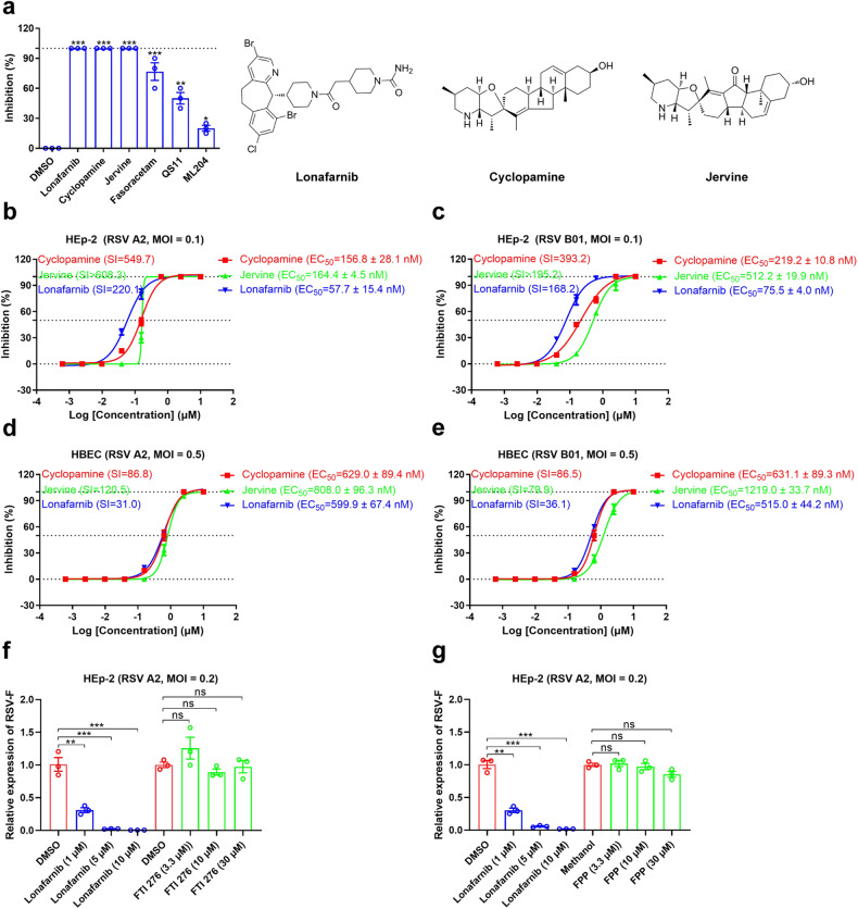
Respiratory syncytial virus (RSV) is the major cause of bronchiolitis and pneumonia in young children and the elderly. There are currently no approved RSV-specific therapeutic small molecules available. Using high-throughput antiviral screening, we identified an oral drug, the prenylation inhibitor lonafarnib, which showed potent inhibition of the RSV fusion process. Lonafarnib exhibited antiviral activity against both the RSV A and B genotypes and showed low cytotoxicity in HEp-2 and human primary bronchial epithelial cells (HBEC). Time-of-addition and pseudovirus assays demonstrated that lonafarnib inhibits RSV entry, but has farnesyltransferase-independent antiviral efficacy. Cryo-electron microscopy revealed that lonafarnib binds to a triple-symmetric pocket within the central cavity of the RSV F metastable pre-fusion conformation. Mutants at the RSV F sites interacting with lonafarnib showed resistance to lonafarnib but remained fully sensitive to the neutralizing monoclonal antibody palivizumab. Furthermore, lonafarnib dose-dependently reduced the replication of RSV in BALB/c mice. Collectively, lonafarnib could be a potential fusion inhibitor for RSV infection.


Conflict of interest statement

The authors declare no competing interests.

Figures

Fig. 1
Fig. 1
Activity of lonafarnib against RSV infection. a Antiviral activity of six compounds (lonafarnib, cyclopamine, jervine, fasoracetam, QS11, and ML204) from high-throughput screening at 5 μM against RSV infection at a multiplicity of infection (MOI) of 0.1 in a cytopathic effect (CPE) inhibition assay with HEp-2 cells. The chemical structures of lonafarnib, cyclopamine, and jervine with antiviral activity are in the top three. For statistical analysis, one-way ANOVA compared with the DMSO group was used. Antiviral activity of cyclopamine, jervine, and lonafarnib against RSV A2 (b) and B01 (c) at an MOI of 0.1 in a CPE inhibition assay with HEp-2 cells. Antiviral activity of cyclopamine, jervine, and lonafarnib against RSV A2 (d) and B01 (e) in a fluorescence focus assay (FFA) with HBEC. be Selectivity index (SI) = CC50/EC50 were calculated for all compounds tested. Treatment with the farnesyltransferase antagonist FTI 276 (f) and agonist FPP (g) did not affect RSV infection. FTI 276 and FPP were tested at concentrations exceeding their EC50. Lonafarnib (10, 5, 1 μM) was used as a control. For statistical analysis, non-parametric Mann-Whitney for comparison was used. The results are representatively shown with three random experiments. The error bars indicate mean ± SEM. *P < 0.05, **P < 0.01, ***P < 0.001; ns, not significant
Fig. 2
Fig. 2
Lonafarnib inhibits the entry process of RSV. a Time-of-addition experiment of lonafarnib. Schematic illustration of the time-of-addition experiment (left). HEp-2 cells were infected with RSV A2 at an MOI of 2, and treated with 3.3 μM lonafarnib pre (−2–0 h), during (0–2 h), and post (2, 4, 6, 8, and 16 h) infection; 0.03% DMSO was added at the same time as in the control. The viral RNA was extracted and measured at 22 h postinfection (right). Statistical significance was assessed by Student’s t-test compared with the DMSO group. b HEp-2 cells were infected with the pseudotypes harboring RSV F protein (RSV-FPP) and treated with lonafarnib (1 or 5 μM), AK0529 (5 μM), palivizumab (10 μg/mL), and D25 (10 μg/mL). At 48 h postinfection, the luciferase activity of RSV-FPP was analyzed. Lonafarnib did not modulate RSV binding and internalization. RSV A2 was incubated with HEp-2 cells pretreated with lonafarnib, AK0529, palivizumab, or D25 mAb (c, d, left), or RSV A2 was incubated with lonafarnib, AK0529, palivizumab or D25 mAb before infection (c, d, right). Cells were collected and the viral RNA was detected. e Lonafarnib inhibits the RSV F protein-induced cell-to-cell fusion process. RSV F protein was transiently expressed in HEK293T cells. Different compounds at 5 μM were diluted and added to the wild-type RSV-F-expressing cells. The cell-to-cell fusion was observed using microscopy on day 2 after compound addition. The results are representatively shown with three random experiments. The error bars indicate mean ± SEM. Statistical significance was assessed by ANOVA for comparison. *P < 0.05, **P < 0.01, ***P < 0.001; ns, not significant
Fig. 3
Fig. 3
Cryo-EM structure of pre-fusion F in complex with lonafarnib. a Cryo-EM map of lonafarnib bound to RSV F-D25 Fab complex and colored according to the subunit. Slate blue, RSV F promoter a; Salmon, RSV F promoter b; Gray, RSV F promoter c; Wheat and thistle, D25 Fab; Lonafarnib, yellow, cyan, and purple. b Orthogonal views of lonafarnib bound to the RSV F-D25 Fab complex. c Density map and constructed model of lonafarnib near the lonafarnib pocket. The density map is shown as mesh and the threshold level is 0.06. d Close-up view of the hydrophobic interactions in the lonafarnib-binding site. e Close-up view of the hydrophilic interactions of one lonafarnib molecule. Hydrogen bonds are indicated with dashed lines. f 2D ligand-interaction diagram of lonafarnib binding to pre-fusion RSV F. g The relative percentage of RSV F remaining in the pre-fusion conformation after a 55 °C heat shock for 15 min was performed with an increasing concentration of AK0529, lonafarnib, or palivizumab. n = 3 biological replicates. h Differential scanning fluorimetry (DSF) for the stability of lonafarnib and AK0529 to DS-Cav1. The black, blue, and red lines represent the melting temperature (Tm) value fitted curve for DS-Cav1 incubated with DMSO, AK0529, or lonafarnib, respectively. The results are representatively shown with three random experiments
Fig. 4
Fig. 4
HDX-MS analysis and structural mapping of lonafarnib binding to the fusion protein of RSV. Differential HDX-MS analysis of RSV pre-fusion F trimer and monomer ± lonafarnib is shown as the change in deuterium uptake mapped onto the cryo-EM structure (PDB: 8KG5). Deuterium uptake plots for peptides affected by ligand binding in the absence (gray) or presence (green) of lonafarnib. The data represent mean ± SEM of three experimental replicates
Fig. 5
Fig. 5
Impact of RSV F mutations on cell-to-cell fusion activity and lonafarnib binding. a, HEK293T cells were transfected with wild-type F, F137L, F140L, M396A, T397A, S398A, D486N, D486A, E487A, F488L, or D489A plasmids for 36 h. The cells were fixed followed by staining, and imaging using confocal microscopy. The pcDNA3.1 plasmid was used as a vector control. b Relative fusion activity for RSV F mutations (such as F137L, F140L, M396A, T397A, S398A, D486N, D486A, E487A, F488L, or D489A) was normalized to wild-type F. Statistical significance was assessed by ANOVA for comparison. c RSV F constructs carrying M396A, S398A, D486A, F488L, or D489A mutations were tested for their ability to induce the cell-to-cell fusion process during lonafarnib (5 μM) addition to the cells. The pcDNA3.1 plasmid was used as a vector control. d Quantification of the area of the syncytia was analyzed using Image J. For statistical analysis, non-parametric Mann-Whitney for comparison was used. The results are representatively shown with three random experiments. The error bars indicate mean ± SEM. **P < 0.01, ***P < 0.001; ns, not significant
Fig. 6
Fig. 6
In vivo efficacy of lonafarnib against RSV infection in BALB/c mice. a–f, BALB/c mice were orally administered 17 or 34 mpk lonafarnib for the treatment group (n = 5 mice/group) or vehicle solution only for the control group (n = 5 mice/group) twice daily for 4 days. At 2 h after lonafarnib treatment, the mice were intranasally inoculated with RSV A2 (1 × 106 PFU). The mock and vehicle groups were used as controls. a Schematic of BALB/c mice infection and treatment. b Mouse body weights were monitored for up to 4 days postinfection. c Mice infected with RSV A2 were killed on day 4 postinfection for the detection of infectious viral titers in the lung tissue using FFA (n = 5). d Histological analysis visualizing the virus-induced pathology in the lung of mice infected with RSV A2 on day 4 postinfection. Four parameters of pulmonary inflammation were evaluated: i, peribronchiolitis (black arrows: inflammatory cell infiltration around the bronchioles); ii, perivasculitis (red arrows: inflammatory cell infiltration around the small blood vessels); iii, interstitial pneumonia (yellow arrows: inflammatory cell infiltration and thickening of alveolar walls); iv, alveolitis (blue arrows: cells within the alveolar spaces). The results are representatively shown from a mean score of combined pathology. Representative images of the lungs are indicated by black squares with numbers, and enlarged in images 1, 2, 3, and 4. Scale bar, 1,000 µm, and 50 µm for enlarged images 1–4. Quantitative scoring of the pathology of the lung from mice on day 4 postinfection. The blinded scorings of the alveolitis (e) and combined pathology (f) were shown, respectively. Histopathology scores for each pulmonary inflammation on a scale of 0–4, where 0 is a normal healthy lung and 4 is severe confluent areas of pathology. The data are representative of at least two experiments. The error bars are mean ± SEM. Statistical differences were determined by two-way ANOVA in b, c, e and f. *P < 0.05, **P < 0.01, ***P < 0.001; ns, not significant

References

    1. Shi T, et al. Global, regional, and national disease burden estimates of acute lower respiratory infections due to respiratory syncytial virus in young children in 2015: a systematic review and modelling study. Lancet. 2017;390:946–958. doi: 10.1016/S0140-6736(17)30938-8. - DOI - PMC - PubMed
    1. Li Y, et al. Global, regional, and national disease burden estimates of acute lower respiratory infections due to respiratory syncytial virus in children younger than 5 years in 2019: a systematic analysis. Lancet. 2022;399:2047–2064. doi: 10.1016/S0140-6736(22)00478-0. - DOI - PMC - PubMed
    1. Falsey AR, et al. Respiratory syncytial virus infection in elderly and high-risk adults. N. Engl. J. Med. 2005;352:1749–1759. doi: 10.1056/NEJMoa043951. - DOI - PubMed
    1. Williams V, et al. Psychometric evaluation of a caregiver diary for the assessment of symptoms of respiratory syncytial virus. J. Patient Rep. Outcomes. 2017;2:10. doi: 10.1186/s41687-018-0036-7. - DOI - PMC - PubMed
    1. American Academy of Pediatrics Committee on Infectious Diseases: Use of ribavirin in the treatment of respiratory syncytial virus infection. Pediatrics. 92, 501–504, (1993). - PubMed

Publication types

MeSH terms