Review
. 2024 Jul 29;379(1906):20230220.
doi: 10.1098/rstb.2023.0220. Epub 2024 Jun 10.

Activity-dependent diffusion trapping of AMPA receptors as a key step for expression of early LTP

Affiliations
Review

Activity-dependent diffusion trapping of AMPA receptors as a key step for expression of early LTP

Agata Nowacka et al. Philos Trans R Soc Lond B Biol Sci. .

Abstract

This review focuses on the activity-dependent diffusion trapping of α-amino-3-hydroxy-5-methyl-4-isoxazolepropionic acid receptors (AMPARs) as a crucial mechanism for the expression of early long-term potentiation (LTP), a process central to learning and memory. Despite decades of research, the precise mechanisms by which LTP induction leads to an increase in AMPAR responses at synapses have been elusive. We review the different hypotheses that have been put forward to explain the increased AMPAR responsiveness during LTP. We discuss the dynamic nature of AMPAR complexes, including their constant turnover and activity-dependent modifications that affect their synaptic accumulation. We highlight a hypothesis suggesting that AMPARs are diffusively trapped at synapses through activity-dependent interactions with protein-based binding slots in the post-synaptic density (PSD), offering a potential explanation for the increased synaptic strength during LTP. Furthermore, we outline the challenges still to be addressed before we fully understand the functional roles and molecular mechanisms of AMPAR dynamic nanoscale organization in LTP. This article is part of a discussion meeting issue 'Long-term potentiation: 50 years on'.

Keywords: AMPAR diffusion trapping; AMPAR trafficking; long-term potentiation; synaptic plasticity.


Conflict of interest statement

We declare we have no competing interests.

Figures
Synaptic nano-organization.
Figure 1.
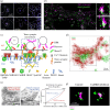
Synaptic nano-organization. (a) dSTORM images of endogenous glutamatergic receptors. From the left, AMPARs, NMDARs and mGluR5. Adapted from [92]. (b) Dual-colour dSTORM images of trans-synaptic nanocolumns. Images of GluA2-containing AMPAR labelled with Alexa 532 nm and regulating synaptic membrane exocytosis protein (RIM) labelled with Alexa 647 nm. Adapted from [93]. (c) Schematic of trans-synaptic nanocolumn molecular organization. Adapted from [94]. (d) Dual-colour dSTORM images of endogenous NR1-containing and GluA2-containing NMDARs and AMPARs in cultured hippocampal neurons. Adapted from [92]. (e) Immunogold labelling of AMPAR andl NMDAR with respect to glutamate release sites. Adapted from [95]. (f) Inhibition of CaMKII decreases the segregation of AMPAR and NMDAR sub-synaptic domains. Adapted from [96].
AMPAR surface mobility and overall AMPAR trafficking in and out synapses.
Figure 2.
AMPAR surface mobility and overall AMPAR trafficking in and out synapses. (a) Representative trajectories of AMPARs labelled with latex beads in the proximity of pre-synaptic sites labelled with FM1-43 (green) in cultured hippocampal neurons. Diffusive movements in blue and confined movements in red. Adapted from [48]. (b) Images of high-resolution uPAINT of AMPAR surface mobility with individual AMPAR trajectories of immobile receptors (left) mostly present in nanoclusters (yellow circles) and mobile receptors (right), enriched outside nanoclusters. Adapted from [87]. (c) Representative multi-photon image in vivo of a bleached spine and fluorescent recovery in a layer 2/3 visual cortex dendrite. The cell is expressing cytosolic DsRed (magenta) and SEP-GluA1 (green). Adapted from [136]. (d) Distribution of AMPAR diffusion coefficients in spines. Approximately 30–50% of receptors are mobile with a D > 0.01 μm2 s–1. Adapted from [135]. (e) Model of AMPAR trafficking in and out of the PSD [1]. Newly synthesized receptors are transported inside the cell in vesicles [2]. Vesicles are exocytosed within the extrasynaptic compartment [3]. On the surface, AMPARs move randomly by Brownian diffusion and can be [4] reversibly stabilized through diffusion trapping in the PSD [5]. Diffusing receptors are internalized in the extrasynaptic compartment through clathrin-dependent endocytosis [6]. Endocytosed receptors can be recycled back through exocytosis. Adapted from [135].
Model of the molecular mechanism of activity-dependent AMPAR diffusion trapping and role in early phases of LTP.
Figure 3.
Model of the molecular mechanism of activity-dependent AMPAR diffusion trapping and role in early phases of LTP. (a) Left inset, TARP γ2 and γ8 AMPAR auxiliary subunits allow stabilization of AMPAR complexes to PSD95 intracellular scaffold thanks to the interaction of their intracellular c-terminus PDZ ligand with the PDZ domains of PSD95. Right inset, theC-terminal domain of TARP γ2 and γ8 contains an upstream Ser- and Arg-rich domain (‘Sensor’) and a terminal PDZ domain ligand. The Ser can be phosphorylated by CaMKII and PKC. Bottom, upon activity-triggered calcium influx through NMDAR, CaMKII translocates to the spine and phosphorylates the TARP Ser-rich domain. This allows the repulsion of the TARP c-terminus from the negatively charged plasma membrane, its extension into the cytoplasm and binding to PDZ domains of PSD95. This results in AMPAR immobilization (b). Upon pre-synaptic tetanic stimulation, an immediate (seconds to minutes) dual potentiation of synaptic strength occurs, owing to both pre-synaptic post-tetanic potentiation (PTP) and post-synaptic diffusion trapping of AMPARs (STP). Both phenomena occur on similar time-frames and cannot be distinguished based solely on kinetics, resulting in potentation of Excitatory Post Synaptic Currents (EPSC) early LTP (eLTP). In a secondary timeframe, intracellular AMPARs are exocytosed and incorporated in the plasma membrane to allow re-equilibration of the fraction of mobile AMPARs in synapses, leading to exocytosis-dependent sustained LTP. The sum of all these components forms LTP.

Similar articles

Cited by

References

    1. Bliss TV, Lomo T. 1973. Long-lasting potentiation of synaptic transmission in the dentate area of the anaesthetized rabbit following stimulation of the perforant path. J. Physiol. 232 , 331–356. (10.1113/jphysiol.1973.sp010273) - DOI - PMC - PubMed
    1. Bear MF, Malenka RC. 1994. Synaptic plasticity: LTP and LTD. Curr. Opin. Neurobiol. 4 , 389–399. (10.1016/0959-4388(94)90101-5) - DOI - PubMed
    1. Enoki R, Hu YL, Hamilton D, Fine A. 2009. Expression of long-term plasticity at individual synapses in hippocampus is graded, bidirectional, and mainly presynaptic: optical quantal analysis. Neuron 62 , 242–253. (10.1016/j.neuron.2009.02.026) - DOI - PubMed
    1. Lisman J. 2003. Long-term potentiation: outstanding questions and attempted synthesis. Phil. Trans. R. Soc. Lond. B 358 , 829–842. (10.1098/rstb.2002.1242) - DOI - PMC - PubMed
    1. Bliss TV, Collingridge GL. 1993. A synaptic model of memory: long-term potentiation in the hippocampus. Nature 361 , 31–39. (10.1038/361031a0) - DOI - PubMed

Substances

Grants and funding

LinkOut - more resources