. 2024 Jul 29;379(1906):20230221.
doi: 10.1098/rstb.2023.0221. Epub 2024 Jun 10.

Adiponectin rescues synaptic plasticity in the dentate gyrus of a mouse model of Fragile X Syndrome

Affiliations

Adiponectin rescues synaptic plasticity in the dentate gyrus of a mouse model of Fragile X Syndrome

Jonathan S Thacker et al. Philos Trans R Soc Lond B Biol Sci. .

Abstract

Fragile X syndrome (FXS) is the most common inherited cause of intellectual disability and is the leading known single-gene cause of autism spectrum disorder. Patients with FXS display varied behavioural deficits that include mild to severe cognitive impairments in addition to mood disorders. Currently, there is no cure for this condition; however, there is an emerging focus on therapies that inhibit mechanistic target of rapamycin (mTOR)-dependent protein synthesis owing to the clinical effectiveness of metformin for alleviating some behavioural symptoms in FXS. Adiponectin (APN) is a neurohormone that is released by adipocytes and provides an alternative means to inhibit mTOR activation in the brain. In these studies, we show that Fmr1 knockout mice, like patients with FXS, show reduced levels of circulating APN and that both long-term potentiation (LTP) and long-term depression (LTD) in the dentate gyrus (DG) are impaired. Brief (20 min) incubation of hippocampal slices in APN (50 nM) was able to rescue both LTP and LTD in the DG and increased both the surface expression and phosphorylation of GluA1 receptors. These results provide evidence for reduced APN levels in FXS playing a role in decreasing bidirectional synaptic plasticity and show that therapies which enhance APN levels may have therapeutic potential for this and related conditions.This article is part of a discussion meeting issue 'Long-term potentiation: 50 years on'.

Keywords: adiponectin; dentate gyrus; fragile X syndrome; hippocampus; long-term potentiation; mTOR.


Conflict of interest statement

We declare we have no competing interests.

Figures
Fmr1 KO mice experience bidirectional synaptic plasticity deficits in the hippocampal DG.
Figure 1.
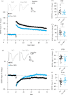
Fmr1 KO mice experience bidirectional synaptic plasticity deficits in the hippocampal DG. (a) EPSP recordings for LTP measured in WT (n = 19 slices/eight animals, black) and Fmr1 KO (n = 16 slices per eight animals, blue) mice 55–60 min after the application of a conditioning stimulus. Student’s t‐test on (b) PTP assessed in the first minute after conditioning stimulus and (c) LTP (average from 55–60 min after induction) revealed significant deficits (PTP: t 34 = 2.20, p = 0.04; LTP: t 33 = 2.31, p = 0.03). (d) EPSP recordings for LTD measure in WT (n = 25 slices per eight animals) and Fmr1 KO (n = 19 slices per six animals) mice 55–60 min after application of a conditioning stimulus. Student’s t‐test on (e) STD assessed in the first minute after conditioning stimulus remained unchanged (t 42 = 0.89, p = 0.38), whereas (f) LTD (averaged from 55–60 min after induction) was significantly affected (t 42 = 2.49, *p < 0.01).
APN rescues deficits in bidirectional synaptic plasticity in the DG of Fmr1 KO mice.
Figure 2.
APN rescues deficits in bidirectional synaptic plasticity in the DG of Fmr1 KO mice. Acute hippocampal slices prepared from Fmr1 KO mice exposed to APN for 20 min (APN20) rescued both (a) LTP (11 slices/five animals, mean difference = 36 ± 12%, p = 0.008) and (b) LTD (13 slices per four animals, mean difference = 12 ± 5%, p = 0.03) in the DG. (c) Paired-pulse ratio was reduced by approximately 20% in APN20 slices compared with untreated Fmr1 KO mice (mean difference = 0.21 ± 0.07, p = 0.02). (d) I/O curves were enhanced at higher stimulation intensities in APN20-treated slices (0.18: t 156 = 4.03, p = 0.0005; 0.24: t 156 = 5.75, p < 0.0001; 0.30: t 156 = 6.75, p < 0.0001). (e) Representative traces for I/O curves for WT and APN-treated slices. (f) Western blot experiments generated from slices under identical conditions revealed significant changes in (g) total surface (t 8 = 2.0, p = 0.04) and (h) phosphorylation at S845 (t 8 = 1.94, p = 0.04) of the AMPA-receptor subunit GluA1. *p < 0.05.
Prolonged APN exposure (>90 min) impairs rescue by APN but can be restored by blocking AMPK.
Figure 3.
Prolonged APN exposure (>90 min) impairs rescue by APN but can be restored by blocking AMPK. (a) Acute hippocampal slices prepared from Fmr1 KO mice exposed to APN for >90 min (APN90; 9 slices/four animals) do not significantly improve LTP in the DG (mean difference = 17 ± 13%, p = 0.36, blue open circles). However, simultaneous blocking of AMPK via CC significantly improves LTP outcomes in the DG (mean difference = 45 ± 13%, p = 0.003, 11 slices per four animals, orange open circles). (b) Paired-pulse ratio was significantly reduced (−9%) when APN90-treated slices were co-treated with CC (t 23 = 1.8, p = 0.04). (c) I/O curves were unremarkable in APN90-CC co-treated slices (F 1,23 = 1.3, p = 0.27). *p < 0.05; **p < 0.01.
Signalling cascades of synthetic and endogenous adipokines.
Figure 4.
Signalling cascades of synthetic and endogenous adipokines. Schematic representation showing how either endogenous APN can activate AdipoR1 and AdipoR2, engaging an intracellular cascade that can facilitate synaptic plasticity in the hippocampus. Note that the model predicts that APN receptor activation would also promote energy homeostasis and increase autophagy while reducing spine production. Abbreviations: AKT, protein kinase B; APPL1, adaptor protein, phosphotyrosine interacting with PH domain and leucine zipper 1; CaMKII, calcium-calmodulin-dependent protein kinase II; CREB, cAMP-response element binding protein;, ERK , extracellular signal-regulated kinase; GS3Kβ, glycogen synthase kinase-3 beta; mTORC1, mammalian target of rapamycin complex 1; PI3K, phosphoinositide 3-kinase; PPARα, peroxisome proliferator-activated receptor alpha; Rheb, Ras homologue enriched in brain.

References

    1. Contractor A, Klyachko VAA, Portera-Cailliau C. 2015. Altered neuronal and circuit excitability in fragile X syndrome. Neuron 87 , 699–715. ( 10.1016/j.neuron.2015.06.017) - DOI - PMC - PubMed
    1. Kelleher RJ, Bear MF. 2008. The autistic neuron: troubled translation? Cell 135 , 401–406. ( 10.1016/j.cell.2008.10.017) - DOI - PubMed
    1. Hernandez RN, Feinberg RL, Vaurio R, Passanante NM, Thompson RE, Kaufmann WE. 2009. Autism spectrum disorder in fragile X syndrome: a longitudinal evaluation. Am. J. Med. Genet. A 149 , 1125–1137. ( 10.1002/ajmg.a.32848) - DOI - PMC - PubMed
    1. Verkerk AJMH, et al. 1991. Identification of a gene (FMR-1) containing a CGG repeat coincident with a breakpoint cluster region exhibiting length variation in fragile X syndrome. Cell 65 , 905–914. ( 10.1016/0092-8674(91)90397-H) - DOI - PubMed
    1. Pieretti M, Zhang FP, Fu YH, Warren ST, Oostra BA, Caskey CT, Nelson DL. 1991. Absence of expression of the FMR-1 gene in fragile X syndrome. Cell 66 , 817–822. ( 10.1016/0092-8674(91)90125-i) - DOI - PubMed

MeSH terms

LinkOut - more resources