[Preprint]. 2024 Jun 1:2024.05.29.596333.
doi: 10.1101/2024.05.29.596333.

Neuronal segmentation in cephalopod arms

Affiliations

Neuronal segmentation in cephalopod arms

Cassady S Olson et al. bioRxiv. .

Update in

Abstract

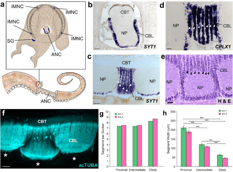
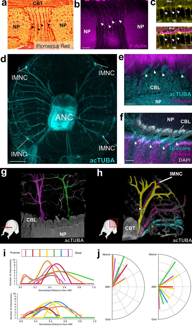
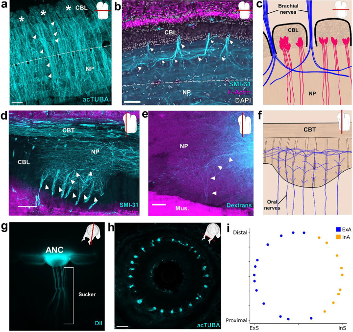
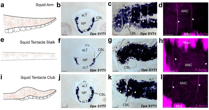
The prehensile arms of the cephalopod are among these animals most remarkable features, but the neural circuitry governing arm and sucker movements remains largely unknown. We studied the neuronal organization of the adult axial nerve cord (ANC) of Octopus bimaculoides with molecular and cellular methods. The ANCs, which lie in the center of every arm, are the largest neuronal structures in the octopus, containing four times as many neurons as found in the central brain. In transverse cross section, the cell body layer (CBL) of the ANC wraps around its neuropil (NP) with little apparent segregation of sensory and motor neurons or nerve exits. Strikingly, when studied in longitudinal sections, the ANC is segmented. ANC neuronal cell bodies form columns separated by septa, with 15 segments overlying each pair of suckers. The segments underlie a modular organization to the ANC neuropil: neuronal cell bodies within each segment send the bulk of their processes directly into the adjoining neuropil, with some reaching the contralateral side. In addition, some nerve processes branch upon entering the NP, forming short-range projections to neighboring segments and mid-range projections to the ANC segments of adjoining suckers. The septa between the segments are employed as ANC nerve exits and as channels for ANC vasculature. Cellular analysis establishes that adjoining septa issue nerves with distinct fiber trajectories, which across two segments (or three septa) fully innervate the arm musculature. Sucker nerves also use the septa, setting up a nerve fiber "suckerotopy" in the sucker-side of the ANC. Comparative anatomy suggests a strong link between segmentation and flexible sucker-laden arms. In the squid Doryteuthis pealeii, the arms and the sucker-rich club of the tentacles have segments, but the sucker-poor stalk of the tentacles does not. The neural modules described here provide a new template for understanding the motor control of octopus soft tissues. In addition, this finding represents the first demonstration of nervous system segmentation in a mollusc.


Figures

Figure 1:
Figure 1:. The axial nerve cord (ANC) is segmented.
a, Transverse diagram of O. bimaculoides arm with the location of neuronal cell bodies outlined in blue. b, In situ hybridization (ISH) for SYT1 in a transverse section of the ANC. Scale bar: 100 μm. c-e: Longitudinal sections through the ANC demonstrating segmentation in the cell body layer with (c) ISH for SYT1. Scale bar: 100 μm, (d) ISH for CPLX1 and (e) hematoxylin and eosin (H&E). Scale bars: 50 μm. f, Whole mount immunostaining with acetylated alpha tubulin (acTUBA) of a dissected ANC. Segmentation pattern is continuous as the ANC oscillates from sucker to sucker. * denotes individual suckers. Scale bar: 100 μm. g-h. Quantification of segmentation down the proximal-distal axis. Arm 1 (green) was stained with acTUBA and arm 2 (pink), with H&E. (g) The number of segments per sucker is maintained along the proximal-distal axis (average Arm 1 = 7.64; average Arm 2 = 7.88). (h) Segment width decreases down the proximal distal axis. n = 48 per condition, error bars +/− sem, ***p < 0.001. CBL, cell body layer; CBT, cerebrobrachial tract; IMNC, intramuscular nerve cord; NP, neuropil; SG, sucker ganglion.
Figure 2:
Figure 2:. Septa between segments are enriched for collagen, blood vessels and nerve fibers.
a, Picrosirius Red stain of an ANC longitudinal section. Red denotes the presence of collagen. b, F-actin (magenta) labeled by phalloidin is localized to the septa. Scale bar: 100 μm. c, Top- Trans-vascular dextran labeling demonstrates vasculature (yellow) in the septa. Bottom- Dextran labeling colocalizes with F-actin staining (magenta). Scale bar: 20 μm. d, Maximum projection of acetylated alpha-tubulin (acTUBA) whole mount immunostaining of a transverse slice. Nerve fibers exit the ANC at discrete locations. Scale bar: 500 μm. e, f, Longitudinal ANC sections of the brachial nerve exits in the septa. (e) acTUBA nerve labeling (cyan), (f) experimental dextran tracing of oral nerves. Injection site in central NP. Scale bars: 100 μm. g-j Brachial nerves exiting from neighboring septa have different targets. (g) Maximum projection of a horizontal whole mount stained with acTUBA. Two nerves (false colored in magenta and green) innervating similar territories of muscle are separated by multiple segments. (h) Maximum projection of a transverse whole mount stained with acTUBA. Three nerves (false colored with yellow, magenta, and cyan) exiting from neighboring septa target different brachial territories. (i) Branching profiles of nerves across multiple septa was characterized by Sholl analysis. Profiles differ from septum to septum. Top- Aboral nerves. Bottom- Central nerves. (j) Nerve fiber average trajectories vary across septa. Left- Aboral nerves. Right- Central nerves. Mus., brachial musculature.
Figure 3:
Figure 3:. Segmental organization of the ANC neuropil.
a-c, Aboral, or brachial, NP. (a) Maximum projection of a horizontal acTUBA wholemount (cyan) showing that the bulk of each cell body segment extends its processes into the NP of the segment. Some processes cross the midline (dashed line). * denotes individual segments. (b) Horizontal section immunostained with SMI-31 (cyan), phalloidin (magenta) and DAPI (gray). SMI-31-rich subpopulation of nerve fibers (cyan) branch proximally and distally upon entering the ANC. (c) Diagram of a horizontal section through the brachial NP. Nerve fibers (blue) pool across neurons (magenta) arranged in segments. d-f, Oral, or sucker, NP. (d) Longitudinal ANC section stained with SMI-31 (cyan) and phalloidin (magenta). NP fibers (cyan) collect into internal fascicles before exiting as oral nerves. (e) Longitudinal section of NP dextran tracer-deposit (cyan) demonstrating NP fibers forming a fascicle. (f) Diagram of NP fibers in the longitudinal plane. From oral to aboral, oral nerves enter the ANC, first showing local branching, then more extensive branching. g-i, Oral ANC nerves distributed around a sucker. (g) Longitudinal whole mount. DiI crystal (cyan) placed in a single ANC sucker enlargement targets a single sucker. (h) Horizontal acTUBA-stained section through a sucker demonstrates radially symmetric decoration. (i) Distribution of oral nerve fiber tips around a sucker. Nerves from the external ANC side (ExA) are tagged in blue; nerves from the internal side (InA), in orange. Data from a transversely imaged whole mount stained with acTUBA. The ExA covers 68% of the sucker; the InA, 32%. Scale bars: 100 μm.
Figure 4:
Figure 4:. Segmentation is a shared feature of flexible, sucker-laden cephalopod appendages.
a-d The ANC of the D. pealeii arm is segmented. (a) Cartoon depiction of D. pealeii arm, which has suckers. (b) ISH for Dpe SYT1 of an ANC transverse section demonstrating the CBL. (c) ISH for Dpe SYT1 of an arm longitudinal section demonstrates CBL segmentation. (d) F-actin (magenta) marks vasculature in the arm ANC septa. e-h The ANC of the tentacle stalk lacks CBL segmentation. (e) Cartoon depiction of D. pealeii tentacle stalk which is devoid of suckers. (f) Transverse ISH section for Dpe SYT1 demonstrates a clear, although reduced, tentacle CBL. (g) Dpe SYT1 ISH of a longitudinal ANC section does not show CBL segmentation in the tentacle stalk. (h) F-actin (magenta) demonstrates a regularly arranged vasculature in the tentacle stalk, but these blood vessels do not lie within the CBL. i-l, The ANC in the tentacle club has segments. (i) Cartoon depiction of D. pealeii tentacle club, which is located at the end of the stalk and has suckers. (j) Transverse ISH section for Dpe SYT1 in the tentacle club ANC demonstrates the CBL. (k) Longitudinal Dpe SYT1 section of the tentacle club CBL shows segmentation. (l) F-actin (magenta) identifies regularly in the tentacle club septa. White arrowheads in (c, k) point to CBL segments. Those in (d, h, l) indicate phalloidin-labeled blood vessels. aLT, aboral longitudinal tract; oLT, oral longitudinal tract. Scale bars: 100 μm.

Similar articles

References

    1. Hochner B., Zullo L., Shomrat T., Levy G. & Nesher N. Embodied mechanisms of motor control in the octopus. Curr. Biol. 33, R1119–R1125 (2023). - PubMed
    1. Olson C. S. & Ragsdale C. W. Toward an Understanding of Octopus Arm Motor Control. Integr. Comp. Biol. icad069 (2023) doi:10.1093/icb/icad069. - DOI - PMC - PubMed
    1. Kier W. M. & Stella M. P. The arrangement and function of octopus arm musculature and connective tissue. J. Morphol. 268, 831–843 (2007). - PubMed
    1. Kennedy E. B. L., Buresch K. C., Boinapally P. & Hanlon R. T. Octopus arms exhibit exceptional flexibility. Sci. Rep. 10, 20872 (2020). - PMC - PubMed
    1. Grasso F. W. Octopus sucker-arm coordination in grasping and manipulation*. Am. Malacol. Bull. 24, 13–23 (2008).

Publication types