[Preprint]. 2024 May 28:2024.05.28.596170.
doi: 10.1101/2024.05.28.596170.

Role of the CTCF Binding Site in Human T-Cell Leukemia Virus-1 Pathogenesis

Affiliations

Role of the CTCF Binding Site in Human T-Cell Leukemia Virus-1 Pathogenesis

Ancy Joseph et al. bioRxiv. .

Update in

Abstract

During HTLV-1 infection, the virus integrates into the host cell genome as a provirus with a single CCCTC binding protein (CTCF) binding site (vCTCF-BS), which acts as an insulator between transcriptionally active and inactive regions. Previous studies have shown that the vCTCF-BS is important for maintenance of chromatin structure, regulation of viral expression, and DNA and histone methylation. Here, we show that the vCTCF-BS also regulates viral infection and pathogenesis in vivo in a humanized (Hu) mouse model of adult T-cell leukemia/lymphoma. Three cell lines were used to initiate infection of the Hu-mice, i) HTLV-1-WT which carries an intact HTLV-1 provirus genome, ii) HTLV-1-CTCF, which contains a provirus with a mutated vCTCF-BS which abolishes CTCF binding, and a stop codon immediate upstream of the mutated vCTCF-BS which deletes the last 23 amino acids of p12, and iii) HTLV-1-p12stop that contains the intact vCTCF-BS, but retains the same stop codon in p12 as in the HTLV-1-CTCF cell line. Hu-mice were infected with mitomycin treated or irradiated HTLV-1 producing cell lines. There was a delay in pathogenicity when Hu-mice were infected with the CTCF virus compared to mice infected with either p12 stop or WT virus. Proviral load (PVL), spleen weights, and CD4 T cell counts were significantly lower in HTLV-1-CTCF infected mice compared to HTLV-1-p12stop infected mice. Furthermore, we found a direct correlation between the PVL in peripheral blood and death of HTLV-1-CTCF infected mice. In cell lines, we found that the vCTCF-BS regulates Tax expression in a time-dependent manner. The scRNAseq analysis of splenocytes from infected mice suggests that the vCTCF-BS plays an important role in activation and expansion of T lymphocytes in vivo. Overall, these findings indicate that the vCTCF-BS regulates Tax expression, proviral load, and HTLV pathogenicity in vivo.

Keywords: ATL; CTCF; DNA Methylation; Epigenome; HTLV.


Figures
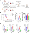
Figure 1.
Figure 1.. Infection of CD 34+ humanized mice resulted in decrease in pathogenicity in CTCF Infected mice.
A. Schematic representation of CD34+ and CD133+ hematopoietic stem cell humanization and experimental flow. B. Survival curve of HTLV-1-WT, HTLV-1-p12stop, and HTLV-1-CTCF infected CD34+ Hu-mice. C. Spleen weights at time of necropsy/death were significantly lower in HTLV-1-CTCF compared to HTLV-1-p12stop infected mice. D. Absolute lymphocyte counts at time of necropsy in peripheral blood. E. Percentage of CD4+ T cells among total CD45+ cells in blood, spleen, and bone marrow cells (* indicates p value lower than 0.05; ** lower than 0.01: *** lower than 0.001).
Figure 2.
Figure 2.. Repressed proviral loads in Hu-mice infected with HTLV-1-CTCF.
A. Individual Hu-mice infected with HTLV-1-WT or HTLV-1-p12stop had a high PVL at 2.5 wpi, whereas most mice infected with HTLV-1-CTCF had low PVL at all time points up to 10 wpi. B. Comparison of average PVL in HTLV-1-WT, HTLV-1-p12stop and HTLV-1-CTCF infected mice at 2.5 wpi. C. PVLs in blood, spleen, liver, and bone marrow cells of infected mice at time of necropsy. (* indicates p value lower than 0.05; ** lower than 0.01; *** lower than 0.001).
Figure 3.
Figure 3.. Histopathological changes in spleen and liver of infected Hu-mice.
A. Hematoxylin and Eosin staining (original magnification 10 X) of spleen showing infiltrating lymphocytes in spleen (top panel), and liver showing lymphoid infiltration into the periportal, midzonal and centrilobular region in the liver (lower panel) Infiltration of lymphoid cells are marked with arrow. B. IHC stain for human CD4 inpatient samples as well as humanized mice infected with HTLV-1(original magnification 20 X). Controls for IHC include human tonsil, and samples from two patients who had HTLV-1-associated lymphoma, including a paratracheal lymph node (HTLV-patient 1) and spheoid mass (HTLV-patient 2). The lower panel shows CD4 staining in HTLV-1-WT, HTLV-1-p12stop, and HTLV-1-CTCF infected Hu-mice.
Figure 4.
Figure 4.. Comparison of pathogenicity in HTLV-1-CTCF Hu-mice based on absolute lymphocyte count.
A. HTLV-1-CTCF infected Hu-mice were separated into two groups based on absolute lymphocyte count at time of necropsy (Group 1 > 400 cells/μl; Group 2 < 400 cells/ul). B. The mice in Group 2 survived until the end point of the experiment. C. Spleen weights were significantly lower in HTLV-1-CTCF-2 than HTLV-1-CTCF-1 infected mice. D. Proviral load in blood was significantly lower in HTLV-1-CTCF-2 than HTLV-1-CTCF-1 infected mice at 2.5 and 5 wpi. E. Total CD4+ T cell counts in blood and spleen were significantly lower in HTLV-1-CTCF-2 than HTLV-1-CTCF-1 infected Hu-mice. F. Correlation of peripheral blood PVL and survival in HTLV-1-CTCF infected Hu-mice. (* indicates p value lower than 0.05; ** lower than 0.01; *** lower than 0.001).
Figure 5.
Figure 5.. vCTCF-BS determines the effect of HTLV-1 on survival and expansion of lymphocytes in vivo.
A. Single cell RNAseq was performed on splenocytes harvested from Hu-mice infected with HTLV-1-CTCF or HTLV-1-p12stop. B. TSNE plots of a representative data set confirm that TCR+ clusters overlap with T cell markers and viral gene expression, and that human CD4+ and CD8+ T cells clusters can be readily identified. C. String network (string-db.org) of genes significantly upregulated in CD4+ T cells confirm that expanded lymphocyte populations in both HTLV-1-CTCF and HTLV-1-p12stop infected Hu-mice express genes associated with ATLL, including IL2RA, FOXP3, BATF3, CD28, and CTLA4. Symbols within circles represent schematic protein structures. D. Heatmap comparing the relative abundance of CD4+ and CD8+ T cells in each sample, normalized against total TCR+ cells in each sample. E. Expression of ALOX5AP shown as the ratio of ALOX5AP+/ALOX5AP− in CD8 vs. CD4 in each sample. F. Number of cells with GLAG peptide in TRB CDR3 of T cell clones.
Figure 6.
Figure 6.. Temporal expression of Tax in JET cells infected with HTLV-1-CTCF and HTLV-1-p12stop.
A. Schematic flow of the experiment, shows transfection of 293T cells with pHTLV-1(WT), pHTLV-1(p12stop), or pHTLV-1(CTCF) plasmid. Transfected cells were co-cultured with JET cells, carrying a Tax-dependent td tomato (RFP) indicator. After 72 hrs of infection the total number of td tomato+ cells in HTLV-1-CTCF infected cells was lower when compared to HTLV-1-p12stop infected cells. B. In order to examine the time course of infection, co-cultured cells were placed in the IncuCyte and were observed for 5 days. The total red object count is shown for duplicate samples of cells infected with HTLV-1-p12stop or HTLV-1-CTCF. C. The total red intensity in infected cells is shown. (** indicates p value lower than 0.01).
Figure 7.
Figure 7.. Working Hypothesis.
A. Schematic depiction of the HTLV-1 genome indicating the location of the vCTCF-BS and major transcripts. B. Working model of the role of the vCTCF-BS in viral gene expression and pathogenesis. Early infection, before methylation of the integrated provirus and suppression of (+) strand transcription from the 5’LTR promoter, the vCTCF-BS acts as an enhancer, Tax expression is elevated, and the effects of TAX protein (virus production, lymphocyte expansion, Tax antigen presentation, cytotoxicity) are elevated. Cytotoxicity resulting from virus production and Tax expression applies selective pressure for (+) sense-strand suppression (DNA hypermethylation) that converts the CTCF binding site from an enhancer to a barrier element and suppresses Tax expression and activity. This rhythm maximizes virus production early followed by entrance into latency to preserve cellular viability, and leads to a burst of initial viremia in vivo followed by lymphocyte expansion and death in the infected Hu-mouse with no adaptive immunity (rapid onset acute disease) or a low-level / undetectable steady state plateau in an infected immunocompetent human (latency). The loss of the vCTCF-BS dysregulates this rhythm, and leads to low level viremia, smoldering infection, and asymptomatic or delayed pathogenesis in the humanized mouse.

Similar articles

References

    1. Bangham CRM, Matsuoka M. Human T-cell leuekemia virus types 1 and 2. In: Howley PM, Knipe DM, Whelan S, Freed EO, Cohen JL, editors. Fields Virology. RMA Vorises. Philadelphia, PA: Lippincott Williams & Wilkins; 2023. p. 527–57.
    1. Matsuoka M, Mesnard J-M. HTLV-1 bZIP factor; the key viral gene for pathogenesis. Retrovirology. 2020;17:2. - PMC - PubMed
    1. Mohanty S, Harhaj EW. Mechanisms of oncogeneis by HTLV-1 Tax. Pathogens. 2020;9:543. - PMC - PubMed
    1. Billman MR, Rueda D, Bangham CRM. Single-cell heterogeneity and cell-cycle-related viral gene bursts in the human leukaemia virus HTLV-1. Welcome Open Research. 2017;2:87. - PMC - PubMed
    1. Mahgoub M, Yasunaga JI, Iwami S, Nakaoka S, Koizumi Y, Shimura K, et al. Sporadic on/off switching of HTLV-1 Tax expression is crucial to maintain the whole population of virus-induced leukemic cells. Proceedings of the National Academy of Sciences, USA. 2018;115:E1269–E78. - PMC - PubMed

Publication types