[Preprint]. 2024 May 30:2024.05.30.596101.
doi: 10.1101/2024.05.30.596101.

Phosphorylation of VapB antitoxins affects intermolecular interactions to regulate VapC toxin activity in Mycobacterium tuberculosis

Affiliations

Phosphorylation of VapB antitoxins affects intermolecular interactions to regulate VapC toxin activity in Mycobacterium tuberculosis

Basanti Malakar et al. bioRxiv. .

Update in

Abstract

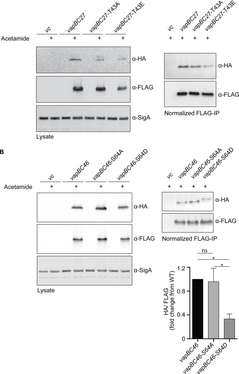
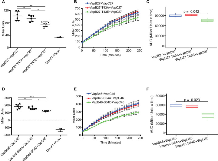
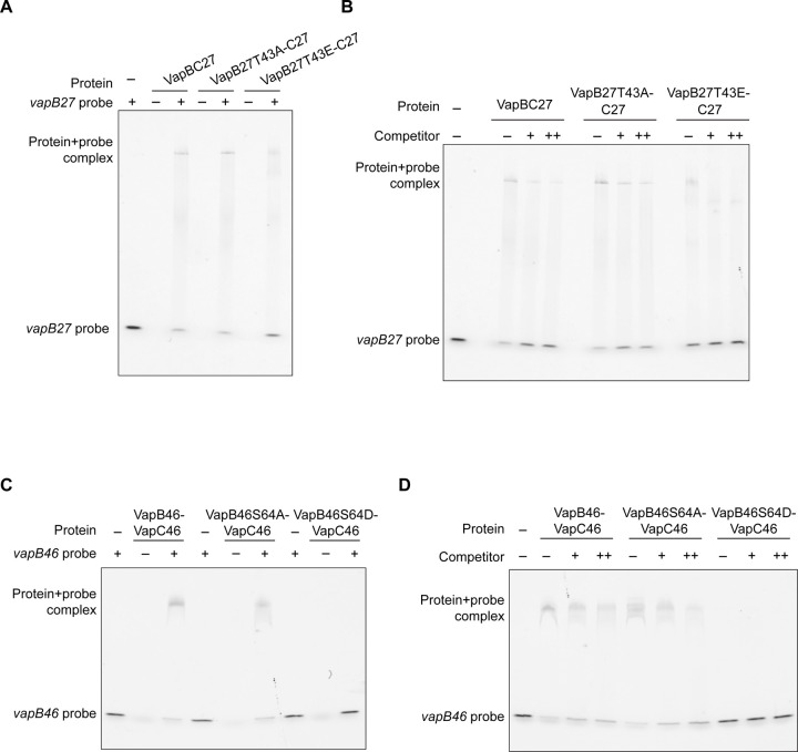
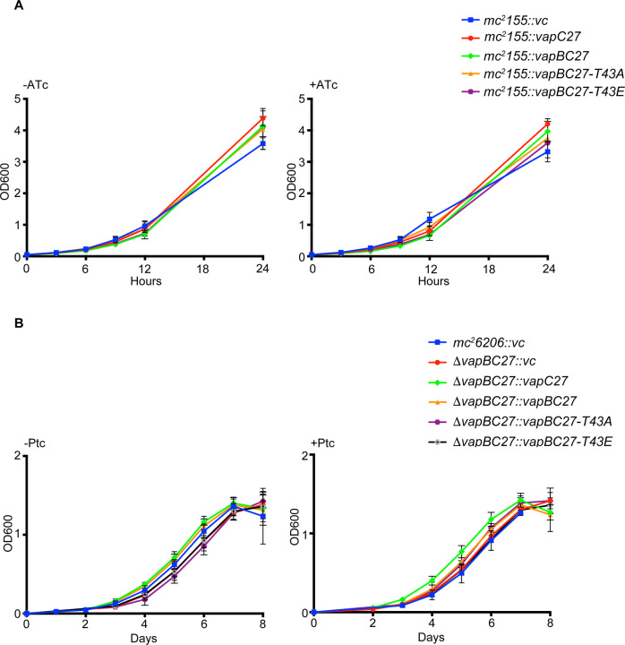
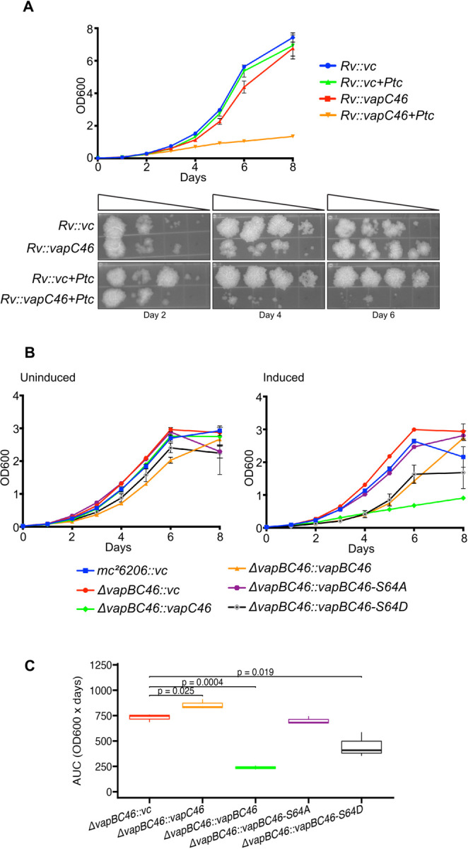
Toxin-antitoxin modules are present in many bacterial pathogens. The VapBC family is particularly abundant in members of the Mycobacterium tuberculosis complex, with 50 modules present in the M. tuberculosis genome. In type IIA modules the VapB antitoxin protein binds to and inhibits the activity of the co-expressed cognate VapC toxin protein. VapB proteins also bind to promoter region sequences and repress expression of the vapB-vapC operon. Though VapB-VapC interactions can control the amount of free VapC toxin in the bacterial cell, the mechanisms that affect this interaction are poorly understood. Based on our recent finding of Ser/Thr phosphorylation of VapB proteins in M. tuberculosis, we substituted phosphomimetic or phosphoablative amino acids at the phosphorylation sites of two VapB proteins. We found that phosphomimetic substitution of VapB27 and VapB46 resulted in decreased interaction with their respective cognate VapC proteins, whereas phosphoablative substitution did not alter binding. Similarly, we determined that phosphomimetic substitution interfered with VapB binding to promoter region DNA sequences. Both decreased VapB-VapC interaction and decreased VapB repression of vapB-vapC operon transcription would result in increased free VapC in the M. tuberculosis cell. M. tuberculosis strains expressing vapB46-vapC46 constructs containing a phosphoablative vapB mutation resulted in lower toxicity compared to a strain expressing native vapB46, whereas similar or greater toxicity was observed in the strain expressing the phosphomimetic vapB mutation. These results identify a novel mechanism by which VapC toxicity activity can be regulated by VapB phosphorylation, potentially in response to extracytoplasmic as well as intracellular signals.

Keywords: Mycobacterium tuberculosis; Toxin-antitoxin; VapB; VapC; bacterial stress response; latency; persistence; protein phosphorylation.


Figures

Figure 1.
Figure 1.. VapB-VapC interaction is negatively regulated by VapB containing phosphomimetic substitutions
A. Western blot analysis of M. smegmatis mc2-155 was performed using whole cell lysates from strains with or without acetamide-induced expression of HA-tagged M. tuberculosis VapB27 and FLAG-tagged M. tuberculosis VapC27 (left panel). Immunoprecipitation (IP) using an anti-FLAG antibody was performed, and FLAG-VapC-normalized immunoprecipitates were analyzed by western blotting using α-HA and α-FLAG antibodies (right panel). B. Western blot analysis of M. smegmatis whole cell lysates expressing HA-tagged VapB46 and FLAG-tagged VapC46 was performed as described above (left panel). FLAG-VapC-normalized immunoprecipitates were analyzed by western blotting using α-HA and α-FLAG antibodies (right panel, image). Band intensities were quantified using Li-Cor Image Studio (right panel, bar graph). Asterisk indicates a significant difference (P<0.05 by one way ANOVA) using data from 2 independent experiments. Error bars represent +/− 1 SD.
Figure 2.
Figure 2.. Bacterial two-hybrid assays indicate decreased interaction between M. tuberculosis VapC and phosphomimetic-substituted VapB versus native VapB
E. coli BTH101 cells were co-transformed with plasmids encoding VapB-T18 and T25-VapC fusion proteins, where T18 and T25 are fragments of adenylate cyclase that can form an active enzyme when they interact. The cells were grown on selective media, and the expression of the reporter gene was assayed by β-galactosidase activity. A-C. β-galactosidase activity of VapB27-VapC27 in endpoint and time course experiments. D-F. β-galactosidase reporter activity of VapB46-VapC46 interaction in endpoint and time course experiments. Data shown for endpoint analysis are representative of two experiments, each with 6 replicates. Statistical analysis of the endpoint assays (Panels A, D) was performed by ordinary one-way ANOVA using Graphpad Prism 9. ns, non-significant; *P<0.1; **P<0.01; ***P<0.001. Error bars represent +/ 1 SD. Data shown for the kinetic assays (Panels B, E) are representative of two experiments with triplicate cultures. Statistical analysis of kinetic data (Panels C, F) was performed using GrowthcurveR (56) and Student’s T-test as described in the Materials and Methods. The T18-CcmF1 and T25-PknA combination was used as the negative control.
Figure 3.
Figure 3.. A phosphomimetic substitution in VapB decreases binding of the VapB-VapC complex to promoter DNA
A. Electrophoretic Mobility Shift Assays (EMSA) were performed using 200 picomoles of purified VapB27-VapC27, VapB27(T43A)-VapC27, or VapB27(T43E)-VapC27 proteins and 5’-Cy3 labelled vapB27 promoter DNA as a fluorescent probe. B. The assays were performed in the presence of unlabeled specific competitor DNA at probe/competitor molar ratios of 1:25 and 1:50 (+ and ++, respectively). C. EMSA was performed using 100 picomoles of purified VapB46, VapB46(S64A), or VapB46(S64D) proteins in combination with 100 picomoles of VapC46, and a 5’-Cy3 labelled vapB46 promoter DNA as a fluorescent probe. D. The assays were performed in the presence of unlabeled specific competitor DNA as described above. Images are representative of 2 to 3 independent experiments.
Figure 4.
Figure 4.. VapC27 is not toxic when expressed in M. smegmatis or M. tuberculosis
A. M. smegmatis mc2-155 was electroporated with pMC1s-vapBC27 expressing wild type VapB or with VapB phosphorylation site mutant constructs. Strains were cultured in supplemented Middlebrook 7H9 medium with or without 100 ng/ml anhydrocycline (ATc). Growth was monitored by measuring the OD600 at regular intervals. The data shown are from one experiment with three cultures per strain. B. M. tuberculosis mc2-6206ΔvapBC27 was electroporated with pRH2046-vapBC27 wild type or phosphorylation site mutant constructs and grown in supplemented Middlebrook 7H9 medium with or without 0.5 μg /ml pristinamycin (ptc) inducer. OD600 was recorded daily. The data shown are from one experiment with with three cultures per strain.
Figure 5.
Figure 5.. VapC46 is toxic in M. tuberculosis in a manner that varies with co-expression of different vapB phosphorylation site alleles
A. M. tuberculosis H37Rv was electroporated with the Ptc-inducible vector pRH2046-vapC46 and grown in supplemented Middlebrook 7H9 medium in the presence or absence of 1 μg/ml Ptc inducer. OD600 was recorded daily. Serial dilutions (10−1-10−4) of all strains were spotted on 7H9 agar plates on days 2, 4, and 6. B. M. tuberculosis mc2-6206ΔvapBC46 was co-transformed with pRH2046-vapC46 and pMind-vapB46 wild type and phosphorylation site mutant constructs and grown in supplemented Middlebrook 7H9 medium with or without 0.25 μg /ml Ptc and 50 ng/ml tetracycline (tet) inducer. OD600 was recorded daily. The data shown are from one experiment with three cultures per strain. C. Box plot comparing AUCs of the growth curve of each strain to the the control mc2-6206ΔvapBC46::vc (vector control) strain. The central box represents the interquartile range (IQR), spanning from the 25th percentile (Q1) to the 75th percentile (Q3). The thick horizontal line inside the box represents the median AUC value. The whiskers extend from the box to the minimum and maximum non-outlier data points within 1.5 times the IQR. Decreased AUC indicates decreased growth and is a measuare of VapC toxicity.

Similar articles

References

    1. World Health Organization. 2023. Global tuberculosis report 2023., Geneva.
    1. Behr MA, Kaufmann E, Duffin J, Edelstein PH, Ramakrishnan L. 2021. Latent Tuberculosis: Two Centuries of Confusion. Am J Resp Crit Care 10.1164/rccm.202011-4239pp. - DOI - PMC - PubMed
    1. Keren I, Minami S, Rubin E, Lewis K. 2011. Characterization and Transcriptome Analysis of Mycobacterium tuberculosis Persisters. mBio 2:e00100–11–e00100–11. - PMC - PubMed
    1. . Tiwari P, Arora G, Singh M, Kidwai S, Narayan OP, Singh R. 2015. MazF ribonucleases promote Mycobacterium tuberculosis drug tolerance and virulence in guinea pigs. Nature Communications 6:6059. 6:6059. DOI: 10.1038/ncomms7059 - DOI - PubMed
    1. Keren I, Shah D, Spoering A, Kaldalu N, Lewis K. 2004. Specialized persister cells and the mechanism of multidrug tolerance in Escherichia coli. J Bacteriology 186:8172–8180. - PMC - PubMed

Publication types