[Preprint]. 2025 Mar 21:2024.05.29.596509.
doi: 10.1101/2024.05.29.596509.

Single neuron contributions to the auditory brainstem EEG

Affiliations

Single neuron contributions to the auditory brainstem EEG

Paula T Kuokkanen et al. bioRxiv. .

Update in

Abstract

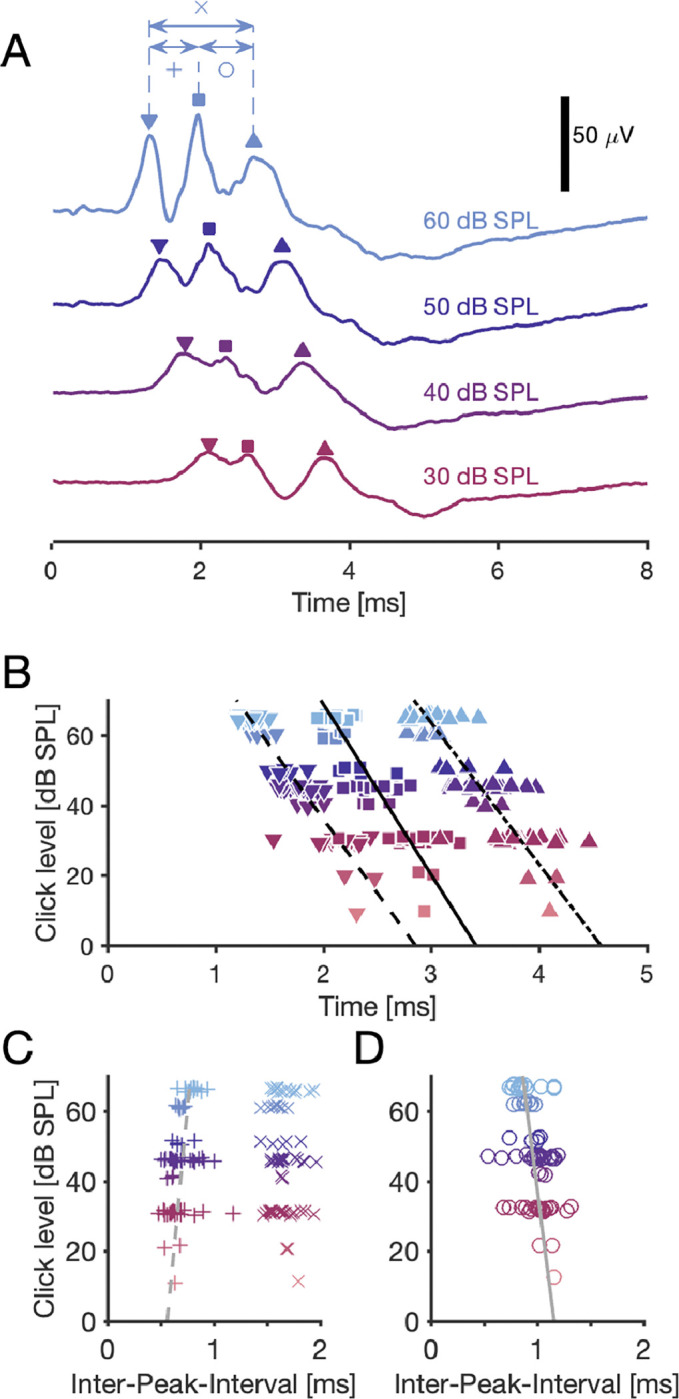
The auditory brainstem response (ABR) is an acoustically evoked EEG potential that is an important diagnostic tool for hearing loss, especially in newborns. The ABR originates from the response sequence of auditory nerve and brainstem nuclei, and a click-evoked ABR typically shows three positive peaks ('waves') within the first six milliseconds. However, an assignment of the waves of the ABR to specific sources is difficult, and a quantification of contributions to the ABR waves is not available. Here, we exploit the large size and physical separation of the barn owl first-order cochlear nucleus magnocellularis (NM) to estimate single-cell contributions to the ABR. We simultaneously recorded NM neurons' spikes and the EEG in owls of both sexes, and found that ≳ 5,000 spontaneous single-cell spikes are necessary to isolate a significant spike-triggered average response at the EEG electrode. An average single-neuron contribution to the ABR was predicted by convolving the spike-triggered average with the cell's peri-stimulus time histogram. Amplitudes of predicted contributions of single NM cells typically reached 32.9 ± 1.1 nV (mean ± SE, range: 2.5 - 162.7 nV), or 0.07 ± 0.02% (median ± SE; range from 0.01% to 1%) of the ABR amplitude. The time of the predicted peak coincided best with the peak of the ABR wave II, independent of the click sound level. Our results suggest that individual neurons' contributions to an EEG can vary widely, and that wave II of the ABR is shaped by NM units.

Significance statement: The auditory brainstem response (ABR) is a scalp potential used for the diagnosis of hearing loss, both clinically and in research. We investigated the contribution of single action potentials from auditory brainstem neurons to the ABR and provide direct evidence that action potentials recorded in a first order auditory nucleus, and their EEG contribution, coincide with wave II of the ABR. The study also shows that the contribution of single cells varies strongly across the population.


Conflict of interest statement

Conflict of interest: No conflicts of interest, financial or otherwise, are declared by the authors.

Figures

Figure 1:
Figure 1:. Recordings from NM cell body region.
A: Exemplar recording location (lesion, *) in a Nissl-stained coronal slice through the auditory brainstem. The nucleus laminaris (NL) is both ventral and lateral to NM. B, Top: Average waveform of 22641 spontaneous spikes (black line) ± SD (gray backgound); prepotential indicated by arrow. Bottom: Extracellular recordings from an NM neuron in response to tones at different frequencies (tone onsets indicated by vertical dashed line, detected spikes marked with *). C: Frequency-response tuning curve to pure tones at 50 dB SPL, with a maximum driven spike count rate of 376 spikes/s at 6750 Hz stimulus frequency. The best frequency (BF, marked with a blue triangle) of this unit was 7065 Hz. The dashed line indicates the spontaneous spike count rate 107 spikes/s. D: Spontaneous firing rates and BFs of all 53 units. Legend: NM: nucleus magnocellularis unit without a prepotential. MN/pp: nucleus magnocellularis unit with a prepotential. AN: auditory nerve fiber unit. pp: low-spontaneous rate unit with a prepotential. doublet: any unit with doublet-spiking. The NM/pp-unit shown in B and C is marked additionally with a blue triangle. Solid line: the decision boundary between NM and AN units (see Materials and Methods).
Figure 2:
Figure 2:. Click-response latency in NM is level- and BF-dependent.
A: Peri-stimulus time histograms (PSTHs) obtained at four different levels of click stimuli from an extracellularly recorded single NM unit. Each PSTH is a summary of responses to many clicks. At the top of each panel, we show two voltage traces from example trials with spike times marked by vertical bars, indicating the low number of spikes in each single trial and the high variability across trials, as expected (Köppl, 1997a; Fontaine et al., 2015). Bin width: 50 μs. The arrow-heads mark the click-response latency at each level. B: Click-response latency decreased with increasing stimulus level and with increasing BF. The examples in A are marked with filled diamonds. Dashed line: −19 ± 3 μs/dB · level + 3.3 ± 0.2 ms (the GLM for the mean BF = 5.58 kHz). 32 NM units, with 1–4 stimulus levels each, resulting in N = 91 click-response latencies.
Figure 3:
Figure 3:. Delay of ABR waves depends on sound level.
A: Examples of an ABR, recorded in response to four different levels of a click with onset at 0 ms. Each curve shows three main peaks (marked with symbols ‘∇’ for wave I, ‘□’ for wave II, and ‘△’ for wave III).e inter-peak-intervals are marked with symbols ‘x’, ‘+’, and ‘o’. B: ABR waves’ peak timing depended significantly on the stimulus level. Linear least-square fits (lines): Wave I peak: −24 μs/dB · level + 2.853 ms. Wave II peak: −21 μs/dB · level + 3.414 ms. Wave III peak: −25 μs/dB · level + 4.573 ms. All groups: Pearson correlation coefficients < −0.84 with p-values < 10−20, N = 75 for each wave. The markers are jittered within 1 dB to reduce overlap. C: The inter-peak-interval between peaks 1 and 2 depended on the stimulus level as 3.1 μs/dB · level + 0.561 ms (linear least-square fit), with Pearson correlation coefficient of 0.35 (p = 0.0022). The average inter-peak-interval (± SE) between peaks 1 and 3 it was 1.67±0.02 ms with no significant correlation with level (Pearson CC: −0.11; p = 0.34). D: The inter-peak-interval between peaks 2 and 3 depended on the stimulus level as −4 μs/dB · level + 1.159 ms (linear least-square fit), with Pearson correlation coefficient of −0.41 (p = 0.00034, N = 75). B–D: 24 ABR recordings, with 1–4 stimulus levels each, resulting in N = 75 delays and inter-peak-intervals per group.
Figure 4:
Figure 4:. Magnocellular single cell spikes make a detectable contribution at the scalp electrode.
Ai: Average spike waveform of 84 248 spontaneous spikes of an NM cell (green), recorded extracellularly, and a random selection of 100 spike waveforms thereof (gray). Aii: Average waveform at the EEG electrode (STA EEG, black) and SE (shaded), with EEG waveforms aligned to the peaks of the spikes of the NM cell in Ai (thin vertical black line). The parts of the STA EEG marked in orange have a significance level p < 0.01, and black portions are non-significant. B: Average spike waveform and STA EEG from a different NM unit. C: 24 STA EEGs, sorted by the significance of their peaks (vertical black bars) within ±1.0 ms with respect to the spikes of the respective NM units. Significant curves (SNRLB ≥ 0 dB) are highlighted by black numbers of the corresponding values of the SNRLB (N = 16); non-significant curves do not have values (N = 8). Asterisks indicate the maximum bootstrapped significance of the SDs of curves (*: p < 0.05, **: p < 0.01, ***: p < 0.001, see Materials and Methods), and significant parts of the waveforms are colored according to the colorbar at the top. Not significant parts are black. The grand average STA EEG (± SD in gray) of the significant curves is shown at the bottom, with the peak amplitude 40 ± 60 nV at −90 μs. D: Peak delays (STA EEG wrt. the spike waveform) and maximum STA EEG amplitudes (peak voltages) were not correlated (Pearson CC: 0.20, p = 0.35, N = 24). Significant data points (SNRLB ≥ 0 dB) are black (N = 16), and the non-significant ones are gray (N = 8). There was no difference between the two groups neither in the number of spikes, in the peak voltages, in the peak delays nor in the SNR of the STA EEG (see Table 2). Histogram on the top: distribution of the STA EEG peak voltages. Histogram on the right-hand side: distribution of the STA EEG peak delays. Population statistics: see Table 2.
Figure 5:
Figure 5:. Predicted NM single-cell contribution aligns best with peak of ABR wave II.
A, Top: PSTH (gray bars) in response to a click stimulus at 0 ms. Inset: STA EEG of the spontaneous spikes (N = 84 248; see Fig. 4Aii). Middle: Predicted single-unit contribution to the ABR (green), calculated as the convolution of the STA EEG with the PSTH (both shown above); peak amplitude of prediction: 162 nV (wrt. average level at click onset ±1 ms). Delay of peak indicated by ‘P’. Bottom: ABR (yellow) in response to the click stimulus; peak-to-peak amplitude of ABR wave II: 47 μV (wrt. lowest neighboring minimum). Delay of peak indicated by ‘P2’. All parts of this panel share the same time scale, and the click onset is marked with a vertical line at 0 ms. B–D: Population data from 38 EEG recordings (at variable click levels) and from 16 NM cells. Plots share the same color schema with respect to stimulus levels (see legend in C). B: Boxplots and data points of the relative delays wrt. each ABR peak and for each level group. The relative delay is the difference between the delay P of the predicted single-cell ABR contribution peak and one of the delays (P1, P2, or P3) of a peak of ABR waves I through III; we also show the relative delay of the predicted peak and the closest ABR wave’s peak (PcP; *: p = 0.011, ***: p < 0.0001, 2-population t-tests). The vertical red lines mark the medians of each relative delay across levels. C: Amplitude of predicted contribution peak vs. amplitude of ABR wave II. Short lines connect data points obtained from the same NM cell but at different click levels. Long diagonal lines indicate fixed relative amplitude, i.e. ratio of predicted and observed amplitudes of peaks. D: Histogram of relative amplitudes.

Similar articles

References

    1. Abdi H, Bonferroni and šidák corrections for multiple comparisons In: Encyclopedia of measurement and statistics, edited by Salkin N. Sage, Thousand Oaks, CA, US, 2007. p. 103 – 107.
    1. Achor LJ, Starr A. Auditory brain stem responses in the cat. I. intracranial and extracranial recordings. Electroencephalogr. Clin. Neurophysiol. 48: 154–173, 1980. - PubMed
    1. Ahlfors SP, Han J, Belliveau JW, Hämäläinen MS. Sensitivity of MEG and EEG to source orientation. Brain Topogr 23: 227–232, 2010a. - PMC - PubMed
    1. Ahlfors SP, Han J, Lin FH, Witzel T, Belliveau JW, Hämäläinen MS, Halgren E. Cancellation of EEG and MEG signals generated by extended and distributed sources. Hum Brain Mapp 31: 140–149, 2010b. - PMC - PubMed
    1. Ahn J, MacLeod KM. Target-specific regulation of presynaptic release properties at auditory nerve terminals in the avian cochlear nucleus. J Neurophysiol 115: 1679–1690, 2016. - PMC - PubMed

Publication types