[Preprint]. 2024 May 28:2024.05.24.595708.
doi: 10.1101/2024.05.24.595708.

Regeneration in the absence of canonical neoblasts in an early branching flatworm

Affiliations

Regeneration in the absence of canonical neoblasts in an early branching flatworm

Ludwik Gąsiorowski et al. bioRxiv. .

Update in

Abstract

The remarkable regenerative abilities of flatworms are closely linked to neoblasts - adult pluripotent stem cells that are the only division-competent cell type outside of the reproductive system. Although the presence of neoblast-like cells and whole-body regeneration in other animals has led to the idea that these features may represent the ancestral metazoan state, the evolutionary origin of both remains unclear. Here we show that the catenulid Stenostomum brevipharyngium, a member of the earliest-branching flatworm lineage, lacks conventional neoblasts despite being capable of whole-body regeneration and asexual reproduction. Using a combination of single-nuclei transcriptomics, in situ gene expression analysis, and functional experiments, we find that cell divisions are not restricted to a single cell type and are associated with multiple fully differentiated somatic tissues. Furthermore, the cohort of germline multipotency genes, which are considered canonical neoblast markers, are not expressed in dividing cells, but in the germline instead, and we experimentally show that they are neither necessary for proliferation nor regeneration. Overall, our results challenge the notion that canonical neoblasts are necessary for flatworm regeneration and open up the possibility that neoblast-like cells may have evolved convergently in different animals, independent of their regenerative capacity.


Figures

Fig. 1.
Fig. 1.. Regeneration and asexual reproduction in Stenostomum brevipharyngium.
a, distribution of the regenerative abilities, asexual reproduction, and adult pluripotent stem cells on the flatworm phylogeny; Catenulida, which includes S. brevipharyngium, are marked in red. b, asexual reproduction by paratomy; br, brain; in, intestine; lnc, longitudinal nerve cord; ph, pharynx; pn, protonephridium; rm, rostral muscles; sp, sensory pits; white bars indicate two zooids. c, anterior regeneration; red dotted lines indicate amputation plane. d–f, effects of irradiation on paratomy and survival (d), tissue homeostasis (e), and regeneration (f). Morphological structures at panels c, e, and f are labeled with arrowheads as follows: dark blue, rostral nerves; green, brain neuropile; red, sensory pits; cyan, rostral muscles; magenta, pharynx; yellow, protonephridium. Asterisks on panel d indicate significant differences from controls (at p-value <0.05) as inferred with the Mann-Whitney U-test. Red bars in panel e indicate the disorganization of posterior tissues. Scale bars on all panels represent 20 μm.
Fig. 2.
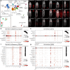
Fig. 2.. Effects of irradiation on gene expression and cell division in Stenostomum brevipharyngium.
a, Volcano plots showing statistical significance and magnitude of change for differentially expressed genes (DEG) in response to different irradiation doses. b, a heat map of the DEG that shows statistically significant changes (adjusted p-value <0.05) at all investigated conditions. c, a heat map and line plots showing changes in the expression levels of the components of the germline multipotency program (GMP) and cell-division genes in response to irradiation, gray boxes indicate insignificant changes (adjusted p-value>0.05). d, distribution of EdU+ cells. e, effects of irradiation on mitotic activity as inferred from EdU and H3P stainings. f, prominent nucleolus in division-competent cells as visualized with antibody staining against the nucleolar marker, fibrillarin. g–i, distribution of mitotically active cells (red arrowheads) in the head, epidermis, and protonephridium. MIP stands for maximum intensity projection. Bars with asterisks indicate significant differences (at p-value <0.05) as inferred with the Mann-Whitney U-test.
Fig. 3.
Fig. 3.. Single-cell atlas of Stenostomum brevipharyngium.
a, a two-dimensional uniform manifold approximation and projection (UMAP) showing cell clusters of S. brevipharyngium. b, expression patterns of the cluster-specific molecular markers (red) with an overlayed signal from nuclear staining with Hoechst (white), scale bars represent 20 μm. c, irradiation sensitivity of particular cell clusters. d–g, dotplots showing cell type-specific expression of the components of the germline multipotency program (d, f) and cell division genes (e, g) in Schmidtea mediterranea (d, e) and S. brevipharyngium (f, g).
Fig. 4.
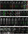
Fig. 4.. Gene expression and division competence in stem cells, somatic tissues, and germline of Stenostomum brevipharyngium.
a–d, expression of the stem cell markers derived from single-cell experiments combined with 2h EdU incorporation. e–g, co-expression of the epidermal and stem cell markers in the dividing cells of the epidermis. h–i, EdU+ cells (arrowheads) in protonephridium express protonephridial marker (h) but not stem cell marker (i). j, mutually exclusive expression of gut and stem cell markers. k–m, expression of the germline multipotency program (GMP) components and stem cell markers, the presumptive gonadal anlagen on the dorsal side of the animal express GMP components, piwiA and vasa (arrowheads, k), but not stem cell marker (l). n, cells expressing stem cell markers (arrowheads) but not GMP components are present in the blastema of head-regenerating worms. o, elevated expression of the GMP components and stem cell marker in gonadal anlagen of lethally irradiated worms. Cell nuclei are counterstained with Hoechst (white). Scale bars on all panels represent 20 μm, if not indicated otherwise.
Fig. 5.
Fig. 5.. Functional testing for the involvement of germline multipotency program and stem cell genes in asexual reproduction, and regeneration of Stenostomum brevipharyngium.
a–b, validation of the efficiency of the dsRNA-mediated knockdowns of piwiA (a) and piwiB (b), cell nuclei are counterstained with Hoechst (white), scale bars represent 20 μm. c–e, effects of gene knockdowns on mitotic activity (c), anterior regeneration (d), and survival and paratomy (e). Bars with asterisks indicate significant differences (at p-value <0.05) as inferred with the Mann-Whitney U-test. Asterisks indicate a significant difference from the eGFP RNAi control group (at p-value <0.05) as inferred with the Fisher’s exact test (d) and Mann-Whitney U-test (e).
Fig. 6.
Fig. 6.. Divergent stem cell systems of flatworms in the context of the metazoan stem cells and regeneration.
a–b, cross-section through the trunks of Stenostomum brevipharyngium (a) and Schmidtea mediterranea (b) allows comparison of their respective stem cell systems, the arrows indicate the dorso-ventral axis of the animals. S. brevipharyngium relies on a complex stem cell system with multiple division-competent cell types, while in S. mediterranea all somatic cell types are derived from the neoblast (green), which represents the only dividing cell type and expresses components of the germline multipotency program. c, distribution of the germline multipotency program positive stem cells and regenerative abilities across metazoan phylogeny, the flatworm clade is indicated in green.

Similar articles

References

    1. Egger B, Gschwentner R, Rieger R: Free-living flatworms under the knife: past and present. Development genes and evolution 2007, 217:89–104. - PMC - PubMed
    1. Baguñà J: The planarian neoblast: the rambling history of its origin and some current black boxes. International Journal of Developmental Biology 2012, 56(1–2-3):19–37. - PubMed
    1. Peter R, Gschwentner R, Schürmann W, Rieger RM, Ladurner P: The significance of stem cells in free-living flatworms: one common source for all cells in the adult. Journal of Applied Biomedicine 2004, 2(1):21–35.
    1. Reddien PW, Alvarado AS: Fundamentals of planarian regeneration. Annu Rev Cell Dev Biol 2004, 20:725–757. - PubMed
    1. Wagner DE, Wang IE, Reddien PW: Clonogenic neoblasts are pluripotent adult stem cells that underlie planarian regeneration. Science 2011, 332(6031):811–816. - PMC - PubMed

Publication types