[Preprint]. 2024 May 30:2024.05.30.596597.
doi: 10.1101/2024.05.30.596597.

2'-Fucosyllactose Inhibits Human Norovirus Replication in Human Intestinal Enteroids

Affiliations

2'-Fucosyllactose Inhibits Human Norovirus Replication in Human Intestinal Enteroids

Ketki Patil et al. bioRxiv. .

Update in

Abstract

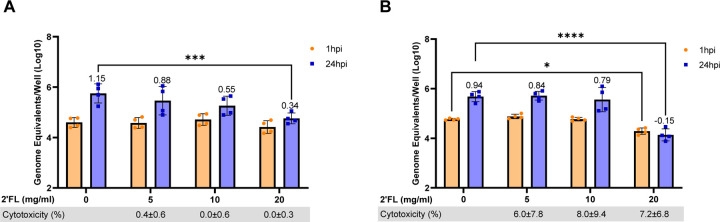
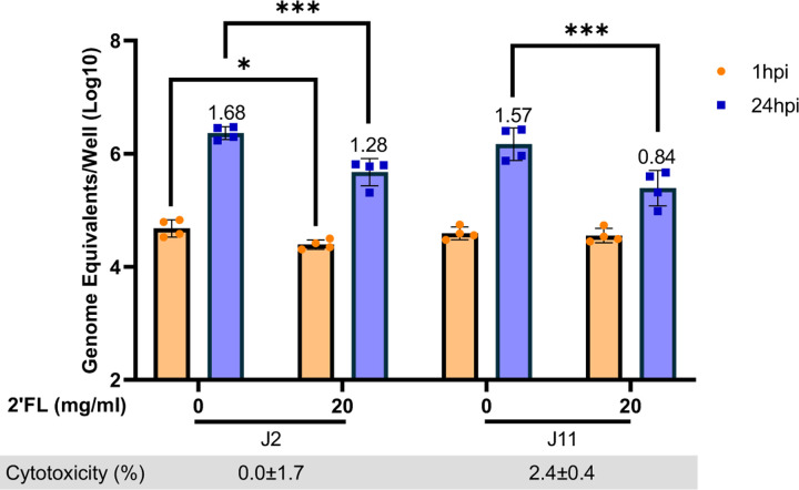
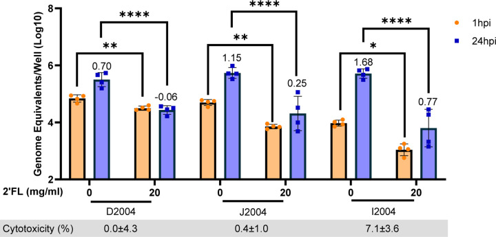
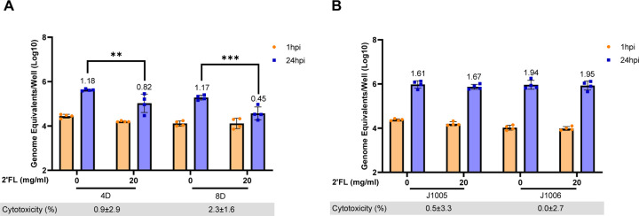
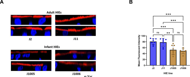
Human noroviruses (HuNoVs) are the leading cause of acute gastroenteritis worldwide. Currently, there are no targeted antivirals for the treatment of HuNoV infection. Histo-blood group antigens (HBGAs) on the intestinal epithelium are cellular attachment factors for HuNoVs; molecules that block the binding of HuNoVs to HBGAs thus have the potential to be developed as antivirals. Human milk oligosaccharides (HMOs) are glycans in human milk with structures analogous to HBGAs. HMOs have been shown to act as decoy receptors to prevent the attachment of multiple enteric pathogens to host cells. Previous X-ray crystallography studies have demonstrated the binding of HMO 2'-fucosyllactose (2'FL) in the same pocket as HBGAs for some HuNoV strains. We evaluated the effect of 2'FL on the replication of a globally dominant GII.4 Sydney [P16] HuNoV strain using human intestinal enteroids (HIEs) from adults and children. A significant reduction in GII.4 Sydney [P16] replication was seen in duodenal and jejunal HIEs from multiple adult donors, all segments of the small intestine from an adult organ donor and in two pediatric duodenal HIEs. However, 2'FL did not inhibit HuNoV replication in two infant jejunal HIEs that had significantly lower expression of α1-2-fucosylated glycans. 2'FL can be synthesized in large scale, and safety and tolerance have been assessed previously. Our data suggest that 2'FL has the potential to be developed as a therapeutic for HuNoV gastroenteritis.

Keywords: 2’-Fucosyllactose; Antiviral; Enteroids; Human Milk Oligosaccharide; Norovirus; Therapeutic.


Figures

Figure 1:
Figure 1:. 20 mg/ml 2’FL significantly reduces GII.4 Sydney 2012 VLP binding to PGM.
Dose-response studies testing 1.25 mg/ml, 2.5 mg/ml, 5 mg/ml, 10 mg/ml and 20 mg/ml of 2’FL with 2.5 ug/ml VLPs. All comparisons were made to the condition where 2’FL was not present (0 mg/ml). Data represented are from n=3 independent experiments with averages from 3 technical replicates per experiment. The P-values were calculated using Student’s t-test. *P ≤ 0.05.
Figure 2:
Figure 2:. 20 mg/ml 2’FL significantly reduces GII.4 Sydney [P16] HuNoV replication in adult duodenal HIE lines.
Dose response assays were carried out in adult duodenal HIE lines (A) D109 and (B) D2004 using 5 mg/ml, 10 mg/ml and 20 mg/ml of 2’FL. GE/well were determined by RT-qPCR at 1 hour post infection (hpi) and 24 hpi. Numbers above the bars indicate log10 fold change comparing GE/well at 24 hpi to 1 hpi. Cytotoxicity (measured by lactase dehydrogenase assay) is represented in percentage below each graph. Data represented are means ± standard deviation (SD) from n=2 independent experiments with 2 technical replicates per experiment. The P-values were calculated using ANOVA, Sidak’s Multiple Comparisons Test. *P ≤ 0.05, ***P ≤ 0.001, ****P ≤ 0.0001.
Figure 3:
Figure 3:. 2’FL significantly reduces GII.4 Sydney [P16] HuNoV replication in adult jejunal HIE lines.
20 mg/ml of 2’FL was tested in two adult jejunal HIEs J2 and J11. GE/well were determined by RT-qPCR at 1 hpi and 24 hpi. Numbers above the bars indicate log10 fold change comparing GE/well at 24 hpi to 1 hpi. Cytotoxicity is represented in percentage below each graph. Data represented are means ± standard deviation (SD) from n=2 independent experiments with 2 technical replicates per experiment. The P-values were calculated using ANOVA, Sidak’s Multiple Comparisons Test. *P ≤ 0.05, ***P ≤ 0.001.
Figure 4:
Figure 4:. 2’FL significantly reduces GII.4 Sydney [P16] HuNoV binding and replication in all segments from the same adult donor.
Studies testing 20 mg/ml of 2’FL in duodenal (D2004), jejunal (J2004) and ileal (I2004) from an adult donor line. GE/well were determined by RT-qPCR at 1 hpi and 24 hpi. Numbers above the bars indicate log10 fold change comparing GE/well at 24 hpi to 1 hpi. Cytotoxicity is represented in percentage below each graph. Data represented are means ± standard deviation (SD) from n=2 independent experiments with 2 technical replicates per experiment. The P-values were calculated using ANOVA, Sidak’s Multiple Comparisons Test. *P ≤ 0.05, **P ≤ 0.01, ****P ≤ 0.0001.
Figure 5:
Figure 5:. 2’FL significantly reduces GII.4 Sydney [P16] HuNoV replication in pediatric duodenal but not infant jejunal HIE lines.
20 mg/ml of 2’FL was tested in (A) two pediatric duodenal HIEs (4D and 8D) and (B) two infant jejunal HIEs (J1005 and J1006). GEs per well were determined by RT-qPCR at 1 hpi and 24 hpi. Numbers above the bars indicate log10 fold change comparing GEs at 24 hpi to 1 hpi. Cytotoxicity is represented in percentage below each graph. Data represented are means ± standard deviation (SD) from n=2 independent experiments with 2 technical replicates per experiment. The P-values were calculated using ANOVA, Sidak’s Multiple Comparisons Test. **P ≤ 0.01, ***P ≤ 0.001.
Figure 6:
Figure 6:. Level of HBGA expression is lower in infant jejunal HIE lines as compared to adult jejunal HIE lines.
(A) Infant jejunal lines (J1005 and J1006) and adult jejunal lines (J2 and J11) stained with Ulex europaeus Agglutinin-1 (UEA-1) were imaged using confocal microscopy. Two representative images are shown per HIE line. (B) Fluorescence intensity was measured for each line using FIJI/Image J. Two-four fields per well were analyzed. Mean fluorescence data from 5 identical regions of interest (ROIs) per 2–4 fields were averaged. The P-values were calculated using ANOVA, Holm-Sidak’s Multiple Comparisons Test. **P ≤ 0.01, ***P ≤ 0.001. N=2 independent experiments.

Similar articles

References

    1. Ahmed SM, Hall AJ, Robinson AE, Verhoef L, Premkumar P, Parashar UD, Koopmans M, Lopman BA. 2014. Global prevalence of norovirus in cases of gastroenteritis: a systematic review and meta-analysis. Lancet Infect Dis 14:725–730. - PMC - PubMed
    1. Pires SM, Fischer-Walker CL, Lanata CF, Devleesschauwer B, Hall AJ, Kirk MD, Duarte AS, Black RE, Angulo FJ. 2015. Aetiology-Specific Estimates of the Global and Regional Incidence and Mortality of Diarrhoeal Diseases Commonly Transmitted through Food. PLoS One 10:e0142927. - PMC - PubMed
    1. Lopman BA, Steele D, Kirkwood CD, Parashar UD. 2016. The Vast and Varied Global Burden of Norovirus: Prospects for Prevention and Control. PLoS Med 13:e1001999. - PMC - PubMed
    1. Gaythorpe KAM, Trotter CL, Lopman B, Steele M, Conlan AJK. 2018. Norovirus transmission dynamics: a modelling review. Epidemiol Infect 146:147–158. - PMC - PubMed
    1. Bartsch SM, Lopman BA, Ozawa S, Hall AJ, Lee BY. 2016. Global Economic Burden of Norovirus Gastroenteritis. PLoS One 11:e0151219. - PMC - PubMed

Publication types