[Preprint]. 2024 May 30:2024.05.30.596714.
doi: 10.1101/2024.05.30.596714.

The Iroquois (Iro/Irx) homeobox genes are conserved Hox targets involved in motor neuron development

Affiliations

The Iroquois (Iro/Irx) homeobox genes are conserved Hox targets involved in motor neuron development

Catarina Catela et al. bioRxiv. .

Update in

Abstract

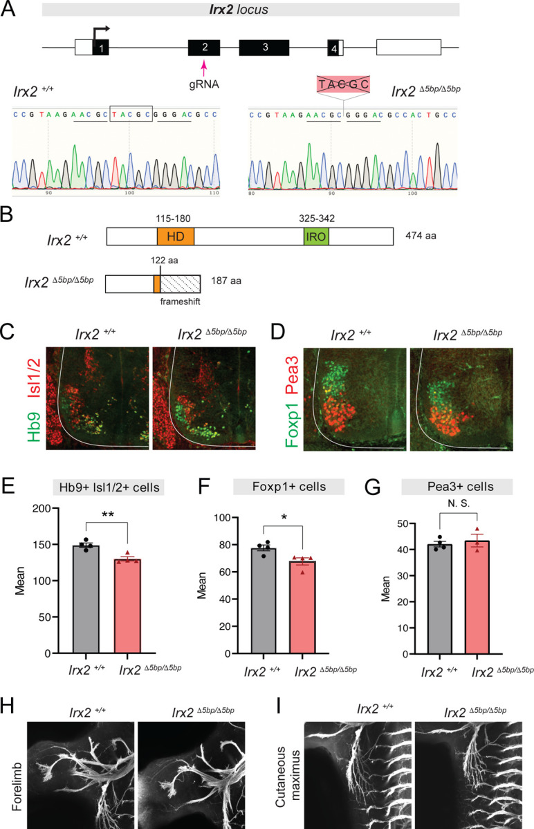
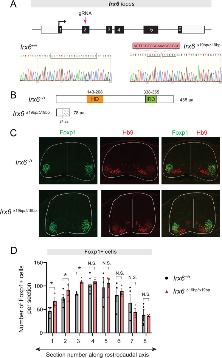
The Iroquois (Iro/Irx) homeobox genes encode transcription factors with fundamental roles in animal development. Despite their link to various congenital conditions in humans, our understanding of Iro/Irx gene expression, function, and regulation remains incomplete. Here, we conducted a systematic expression analysis of all six mouse Irx genes in the embryonic spinal cord. We found five Irx genes (Irx1, Irx2, Irx3, Irx5, and Irx6) to be confined mostly to ventral spinal domains, offering new molecular markers for specific groups of post-mitotic motor neurons (MNs). Further, we engineered Irx2, Irx5, and Irx6 mouse mutants and uncovered essential but distinct roles for Irx2 and Irx6 in MN development. Last, we found that the highly conserved regulators of MN development across species, the HOX proteins, directly control Irx gene expression both in mouse and C. elegans MNs, critically expanding the repertoire of HOX target genes in the developing nervous system. Altogether, our study provides important insights into Iro/Irx expression and function in the developing spinal cord, and uncovers an ancient gene regulatory relationship between HOX and Iro/Irx genes.


Conflict of interest statement

DECLARATION OF INTERESTS The authors declare no competing interests.

Figures

Figure 1.
Figure 1.. Expression pattern of all six Irx genes in mouse spinal motor neurons.
(A) Phylogenetic tree of the Iro/Irx gene family. Adapted from . Scale bar: percentage of sequence divergence. Numbers above branches: percentage of times the branch was found in 200 bootstrap replicates. Brackets highlight the clustering of vertebrate genes into six orthologous groups. (B) Schematic of the two Irx chromosomal clusters in mice. (C) Schematic of the mouse Irx protein domains. Each protein has a highly conserved 63 amino acid-long homeodomain and a distinct 16 amino acid-long Iro box domain. (D) RNA ISH analysis of Irx1, Irx2, Irx3, Irx4, Irx5, and Irx6 on cross-sections of e12.5 wild-type mouse spinal cords. Brachial (C4-T1) domain. N = 4.
Figure 2.
Figure 2.. Expression of Irx1, Irx2, Irx3, Irx5, and Irx6 in specific motor columns.
(A) Brachial spinal domain (C4-T1) in an e12.5 mouse. Cross-section: LMC and MMC neurons at the ventral region of the spinal cord. (B) Irx1 FISH coupled with Lhx3 antibody staining shows Irx1 expression (red) is detected in Lhx3-expressing MNs (green) of the MMC. (C) Schematic of Irx1 expression. (D-E) Irx2 FISH combined with immunostaining for Foxp1 (D) or Lhx3 (E) reveals Irx2 expression (red, also converted to white for better contrast) is detected in LMC (D) and MMC (E) neurons. (F) Schematic of Irx2 expression. (G) Double immunostaining for Irx3 and Lhx3 shows Irx3 (red) is expressed in a small population of Lhx3-positive MNs (green). (H) Irx3 antibody staining combined with Pea3 RNA FISH reveals almost no co-localization of Irx3 protein (red) to Pea3-expressing MNs (green) of the ventral LMC but expression in a small population of dorsal LMC MNs. (I) Schematic of Irx3 expression. (J) Double immunofluorescence reveals Irx5 expression (red) in Hb9-positive cells (green). (K) Two-color RNA FISH shows Irx5 expression (red) is detected in Pea3-expressing MNs of the ventral LMC. (L) Schematic of Irx5 expression. (M) Irx6 RNA (red) colocalizes with Foxp1 protein (green) in the ventral LMC. (N) Two-color RNA FISH reveals Irx6 expression (red) in Pea3-expressing MNs. (O) Schematic of Irx3 expression. (P) Representative images of HUES3 HB9::GFP-derived MNs upon immunostaining for HB9/MNX1 and IRX6 on day 21 of differentiation. Scale bar, 50 μm.
Figure 3.
Figure 3.. Generation and characterization of Irx2 mutant mice.
(A) A gRNA targets exon 2 of Irx2. Representative chromatograms of genotyped mice demonstrate the 5bp (TACGC) deletion in Irx2 Δ5bp/ Δ5bp mice. (B) Wild-type and truncated Irx2 proteins in the Irx2 Δ5bp mouse. See text for details. (C-D) Double immunostaining for Hb9 (green) and Isl1/2 (red) (panel C) or Foxp1 (green) and Pea3 (red) (panel D) in e12.5 wild-type and Irx2 Δ5bp/ Δ5bp spinal cords. (N=4). (E-G) Quantification of Hb9+/Isl1/2+, Foxp1+, or Pea3+ MNs in Irx2 Δ5bp/ Δ5bp mice at e12.5 (N=4). * p < 0.05, ** p < 0.01, *** p < 0.001, N. S.: Not significant. (H-I) MN axons labeled with Hb9::GFP in control and Irx2 Δ5bp/ Δ5bp mice at e12.5 (N=4). Projections to forelimb (H) and cutaneous maximus (I) muscle.
Figure 4.
Figure 4.. Generation and characterization of Irx5 mutant mice.
(A) A gRNA targets exon 1 of Irx5. Representative chromatograms of genotyped mice illustrate the 5 bp (CTCGC) deletion in Irx5 Δ5bp/ Δ5bp. (B) Wild-type and truncated Irx5 proteins produced in the Irx5 Δ5bp mouse. Both HD and Iro domains are removed in the Irx5 Δ5bp protein. (C) Immunohistochemistry shows strong reduction in Irx5 expression in Irx5 Δ5bp/ Δ5bp spinal cords at e12.5. (D) Double immunohistochemistry for Hb9 and Lhx3 in control and Irx5 Δ5bp/Δ5bp mice at e12.5. (E) Double immunohistochemistry for Foxp1 and Pea3 in control and Irx5 Δ5bp/Δ5bp mice at e12.5. (F-H) Quantification of Hb9+, Foxp1+, or Pea3+ MNs in Irx5 Δ5bp/ Δ5bp mice at e12.5 (N=4). * p < 0.05, ** p < 0.01, *** p < 0.001, N. S.: Not significant. (I-J) MN axons labeled with Hb9::GFP in control and Irx5 Δ5bp/ Δ5bp mice at e12.5 (N=4). Projections to forelimb (H) and cutaneous maximus (I) muscle.
Figure 5.
Figure 5.. Generation and characterization of Irx6 mutant mice.
(A) A gRNA targets exon 2 of Irx6 locus. Representative chromatograms of genotyped mice illustrate the 19 bp deletion in the Irx6 Δ19bp/ Δ19bp mouse. (B) Wildtype and truncated Irx6 proteins produced in the Irx6Δ19bp mouse. HD and Iro domains are removed in the Irx6 Δ19bp protein. (C) Antibody staining for Foxp1-expressing MNs (green) and Hb9-positive MNs (red) in control (Irx6 +/+) and Irx6 Δ19bp/Δ19bp spinal cords. (D) Quantifications of Foxp1+ MNs along the rostro-caudal axis reveal an increased number of LMC MNs in rostral sections of the Irx6 Δ19bp/Δ19bp spinal cord (N=4). * p < 0.05, ** p < 0.01, *** p < 0.001, N. S.: Not significant.
Figure 6.
Figure 6.. Hoxc8 controls Irx expression in spinal motor neurons.
(A) RNA ISH shows reduced Irx2, Irx5 and Irx6 expression in e12.5 Hoxc8 MNΔearly spinal cords. (B) At p8, Irx5 and Irx6 expression is reduced in Hoxc8 MNΔlate spinal cords, but Irx2 expression is unaffected (N=4). (C) Analysis of ChIP-Seq data from iHoxc8 MNs shows Hoxc8 directly binds to the cis-regulatory region of Irx genes. GEO accession numbers: Input (GSM4226461) and iHoxc8 replicates (GSM4226436, GSM4226437). Snapshots of each gene locus were generated with Integrative Genomics Viewer (IGV, Broad Institute).
Figure 7.
Figure 7.. LIN-39/Hox controls irx-1 expression in C. elegans motor neurons.
(A) MN cell body position along the C. elegans ventral nerve cord. Expression of an irx-1::eGFP reporter (wgIs536) is decreased in MNs of C. elegans animals carrying the lin-39 (n1760) LOF allele. A 300µm region of the ventral nerve cord (VNC) was analyzed at the fourth larval stage (L4). Anterior is left, dorsal is up. Arrowheads point to MN nuclei. Green fluorescence signal is shown in white for better contrast. (B) Quantification of total number of MNs expressing irx-1 in WT and lin-39 null mutants. Box and whiskers plot show the min, max and quartiles with single data points annotated. N > 23. (C) ChIP-Seq tracks are shown for LIN-39 on irx-1. ChIP-Seq data for LIN-39 come from the modENCODE project. (D) Schematic of known LIN-39 functions and target genes in C. elegans MNs. See Discussion for details. A complete list of terminal differentiation genes regulated by LIN-39 in C. elegans MNs can be found in ,. (E) Schematic of known Hoxc8 functions and target genes in brachial MNs. See Discussion for details. Target genes of Hoxc8 with roles in axon guidance and terminal differentiation have been previously described,.

Similar articles

References

    1. Cavodeassi F., Modolell J. & Gomez-Skarmeta J. L. The Iroquois family of genes: from body building to neural patterning. Development 128, 2847–2855 (2001). - PubMed
    1. Gomez-Skarmeta J. L. & Modolell J. Iroquois genes: genomic organization and function in vertebrate neural development. Curr Opin Genet Dev 12, 403–408, doi:10.1016/s0959-437x(02)00317-9 (2002). - DOI - PubMed
    1. Leyns L., Gomez-Skarmeta J. L. & Dambly-Chaudiere C. iroquois: a prepattern gene that controls the formation of bristles on the thorax of Drosophila. Mech Dev 59, 63–72, doi:10.1016/0925-4773(96)00577-1 (1996). - DOI - PubMed
    1. Burglin T. R. Analysis of TALE superclass homeobox genes (MEIS, PBC, KNOX, Iroquois, TGIF) reveals a novel domain conserved between plants and animals. Nucleic Acids Res 25, 4173–4180, doi:10.1093/nar/25.21.4173 (1997). - DOI - PMC - PubMed
    1. Gomez-Skarmeta J. L. & Modolell J. araucan and caupolican provide a link between compartment subdivisions and patterning of sensory organs and veins in the Drosophila wing. Genes Dev 10, 2935–2945, doi:10.1101/gad.10.22.2935 (1996). - DOI - PubMed

Publication types