[Preprint]. 2024 May 31:rs.3.rs-4438544.
doi: 10.21203/rs.3.rs-4438544/v1.

Targeted TGF-βR2 Knockdown in the Retrotrapezoid Nucleus Mitigates Respiratory Dysfunction and Cognitive Decline in a Mouse Model of Cerebral Amyloid Angiopathy with and without Stroke

Affiliations

Targeted TGF-βR2 Knockdown in the Retrotrapezoid Nucleus Mitigates Respiratory Dysfunction and Cognitive Decline in a Mouse Model of Cerebral Amyloid Angiopathy with and without Stroke

Ahmad El Hamamy et al. Res Sq. .

Update in

Abstract

Introduction: Cerebral amyloid angiopathy (CAA) is characterized by the deposition of amyloid-beta peptides within cerebral blood vessels, leading to neurovascular complications. Ischemic strokes result from acute disruptions in cerebral blood flow, triggering metabolic disturbances and neurodegeneration. Both conditions often co-occur and are associated with respiratory dysfunctions. The retrotrapezoid nucleus (RTN), which is crucial for CO2 sensing and breathing regulation in the brainstem, may play a key role in breathing disorders seen in these conditions. This study aims to investigate the role of Transforming Growth Factor Beta (TGF-β) signaling in the RTN on respiratory and cognitive functions in CAA, both with and without concurrent ischemic stroke.

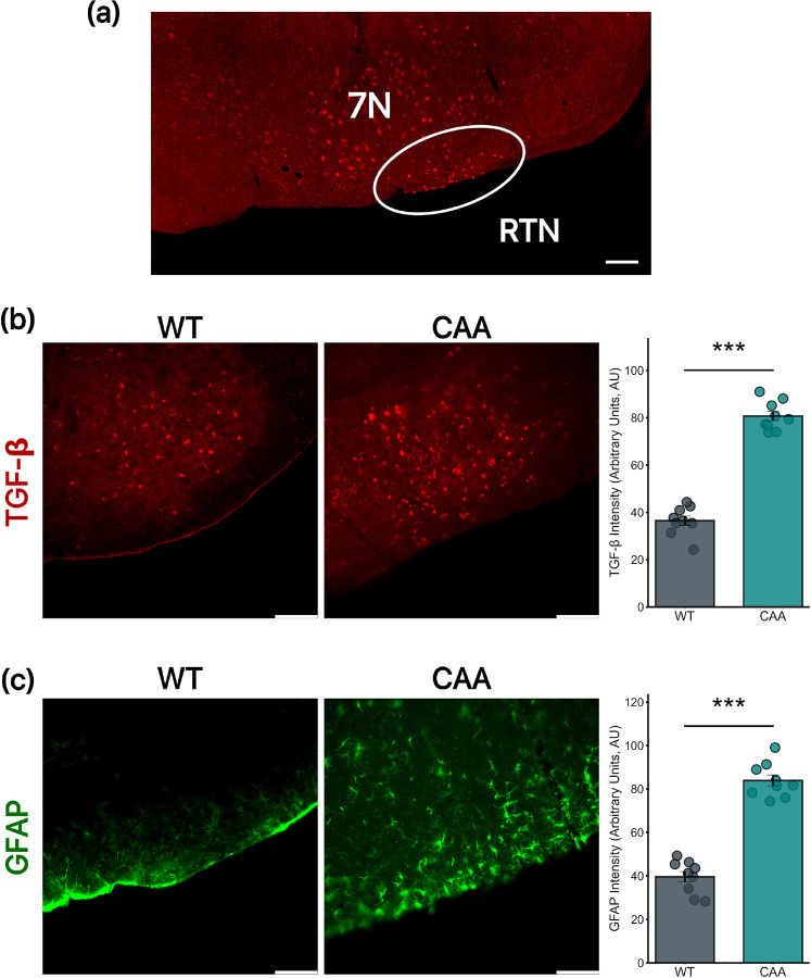
Methods: Adult male Tg-SwDI (CAA model) mice and C57BL/6 wild-type controls underwent stereotaxic injections of lentivirus targeting TGF-β2R2 in the RTN. Stroke was induced by middle cerebral artery occlusion using a monofilament. Respiratory functions were assessed using whole-body plethysmography, while cognitive functions were evaluated through the Barnes Maze and Novel Object Recognition Test (NORT). Immunohistochemical analysis was conducted to measure TGF-βR2 and GFAP expressions in the RTN.

Results: CAA mice exhibited significant respiratory dysfunctions, including reduced respiratory rates and increased apnea frequency, as well as impaired cognitive performance. TGF-βR2 knockdown in the RTN improved respiratory functions and cognitive outcomes in CAA mice. In CAA mice with concurrent stroke, TGF-βR2 knockdown similarly enhanced respiratory and cognitive functions. Immunohistochemistry confirmed reduced TGF-βR2 and GFAP expressions in the RTN following knockdown.

Conclusions: Our findings demonstrate that increased TGF-β signaling and gliosis in the RTN contribute to respiratory and cognitive dysfunctions in CAA and CAA with stroke. Targeting TGF-βR2 signaling in the RTN offers a promising therapeutic strategy to mitigate these impairments. This study is the first to report a causal link between brainstem gliosis and both respiratory and cognitive dysfunctions in CAA and stroke models.


Conflict of interest statement

Disclosures The authors declare no potential conflicts of interest with respect to research, authorship, and/or publication of this article.

Figures

Figure 1
Figure 1. Baseline differences in physiological and cognitive parameters between naive CAA and WT mice
(a) Respiratory frequency analysis shows a significant decrease in CAA mice compared to WT mice (Mean: 204±6.9 vs. 231±6.4; p=0.016). (b) Apnea rate comparison reveals a significant increase in CAA mice versus WT mice (Mean: 10±1 vs. 4.4±0.5; p=0.0002). (c) Barnes Maze test results demonstrate significantly poorer performance in CAA mice compared to WT mice (Mean: 65±4.56 vs. 31±5.95; p=0.0006). (d) Novel Object Recognition Test (NORT) outcomes indicate reduced cognitive engagement in CAA mice versus WT mice (Mean: 51±2 vs. 62.8±3; p=0.007). All groups consist of 9 mice each.
Figure 2
Figure 2. Localization and Comparative Analysis of RTN, TGF-βR2, and GFAP in the Brain Stem of WT and CAA Mice
(a) Histological representation of the RTN location within the brainstem, illustrating the specific area analyzed in both WT and CAA mice, in relation to the 7N. (b) TGF-βR2 expression is significantly increased in CAA mice compared to WT mice (Mean = 36.48±1.93 vs. 80.71±1.94, p = <0.0001). (c) GFAP expression is significantly elevated in CAA mice compared to WT mice (Mean = 39.64±2.38 vs. 80.71±1.94, p = <0.0001). All groups consist of 9 mice each. Scale bar = 100 μm.
Figure 3
Figure 3. Impact of TGF-βR2 KD on CAA Mice’s Respiratory function and cognition
(a) Respiratory rate comparison showing a significant increase in the KD group versus the vehicle group (Mean: 216±4 vs. 200±5; p=0.04). (b) Apnea rate analysis reveals a substantial decrease in the KD group versus the vehicle group (Mean: 3.4±0.3 vs. 8±0.6; p<0.0001). (c) Barnes Maze test results demonstrate significant cognitive improvements in KD-treated mice compared to vehicle-treated mice (Mean: 29.5±3.7 vs. 58.4±3.9; p=0.0001). (d) Novel Object Recognition Test (NORT) outcomes highlight enhanced engagement with novel objects in KD-treated mice versus vehicle-treated mice (Mean: 61±2.98 vs. 45±3.64; p=0.006). All groups consist of 9 mice each.
Figure 4
Figure 4. TGF-βR2 KD reduced GFAP in CAA mice
(a) TGF-βR2 abundance is reduced in CAA TGF-βR2 KD mice (Right) compared to CAA Vehicle-treated mice (Left) (Mean: 81.9±2.56 vs. m71.180±1.85; p=0.006). (b) GFAP signaling, indicating reactive astrocytic activity, is decreased in CAA TGF-βR2 KD mice (Right) compared to CAA Vehicle-treated mice (Left) (Mean: 81.9±2.56 vs 71.180±1.85, p<0.0001). All groups consist of 9 mice each. Scale bar = 100 μm.
Figure 5
Figure 5. Impact of TGF-βR2 KD on Respiratory and Cognitive Parameters in CAA Mice with concurrent MCAO
(a) Respiratory frequency analysis demonstrates a significant increase in MCAO mice treated with TGF-βR2 KD compared to those treated with vehicle (Mean: 212±8 vs. 165±16; p=0.02). This highlights the improved respiratory function in the KD-treated group. (b) Apnea rate comparison shows a significant reduction in MCAO mice treated with TGF-βR2 KD versus those treated with vehicle (Mean: 9.8±0.6 vs. 15.7±1; p=0.0002). This suggests a beneficial effect of TGF-βRII KD on reducing apnea episodes. (c) Barnes Maze test results indicate a significant improvement in cognitive performance in MCAO mice treated with TGF-βR2 KD compared to vehicle-treated mice (Mean: 50±4.9 vs. 71±6; p=0.016, Mann-Whitney U test). (d) Novel Object Recognition Test (NORT) outcomes reveal enhanced cognitive engagement in MCAO mice treated with TGF-βR2 KD compared to those treated with vehicle (Mean: 56±2 vs. 48±1.8; p=0.01). (N=9 for MCAO + Veh and 10 for MCAO+KD)
Figure 6
Figure 6. TGF-βR2 KD reduced GFAP in CAA mice with stroke
Immunohistochemistry analysis was performed to evaluate the effects of MCAO CAA TGF-βR2 treatment on KD mice. (a) TGF-βR2 abundance is significantly reduced in MCAO CAA TGF-βR2 KD mice (Right) compared to MCAO CAA Vehicle-treated mice (Left) (115±2.95 vs 97.18±3.931, p=0.005). (b) GFAP signaling, indicative of reactive astrocytic activity, is significantly decreased in MCAO CAA TGF-βR2 KD mice (Right) compared to MCAO CAA Vehicle-treated mice (Left) (Mean: 108.3±2.744 vs 85.5±1.768, p<0.0001). (N=8 for MCAO + Veh and 7 for MCAO+KD). Scale bar = 100 μm.

References

    1. Smith EE. Cerebral amyloid angiopathy as a cause of neurodegeneration. J Neurochem. 2018;144:651–8. - PubMed
    1. Deng W, Guo S, van Veluw SJ, Yu Z, Chan SJ, Takase H, et al. Effects of cerebral amyloid angiopathy on the brain vasculome. Aging Cell. 2022;21:e13503. - PMC - PubMed
    1. Fotiadis P, van Rooden S, van der Grond J, Martinez-Ramirez SA, Auriel S. Cortical atrophy in patients with cerebral amyloid angiopathy: A case-control study. Lancet Neurol. 2016;15:811–9. - PMC - PubMed
    1. Yamada M. Cerebral amyloid angiopathy: Emerging concepts. J Stroke Cerebrovasc Dis. 2015;17:17–30. - PMC - PubMed
    1. Dirnagl U, Iadecola C, Moskowitz MA. Pathobiology of ischaemic stroke: An integrated view. Trends Neurosci. 1999;22:391–7. - PubMed

Publication types

LinkOut - more resources