[Preprint]. 2024 May 28:2024.05.28.596234.
doi: 10.1101/2024.05.28.596234.

Active E. coli heteromeric acetyl-CoA carboxylase forms polymorphic helical tubular filaments

Affiliations

Active E. coli heteromeric acetyl-CoA carboxylase forms polymorphic helical tubular filaments

Xueyong Xu et al. bioRxiv. .

Abstract

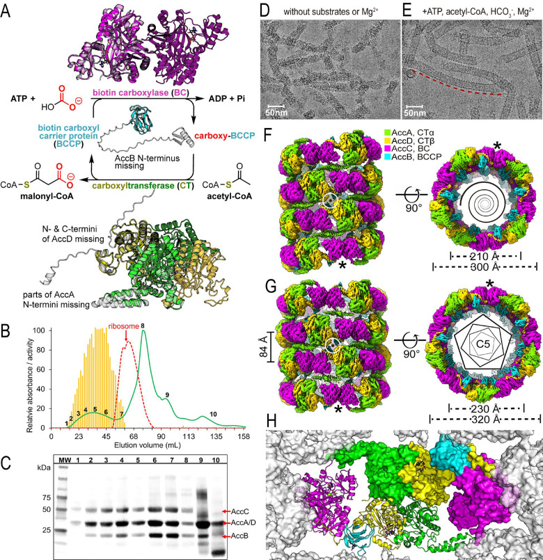
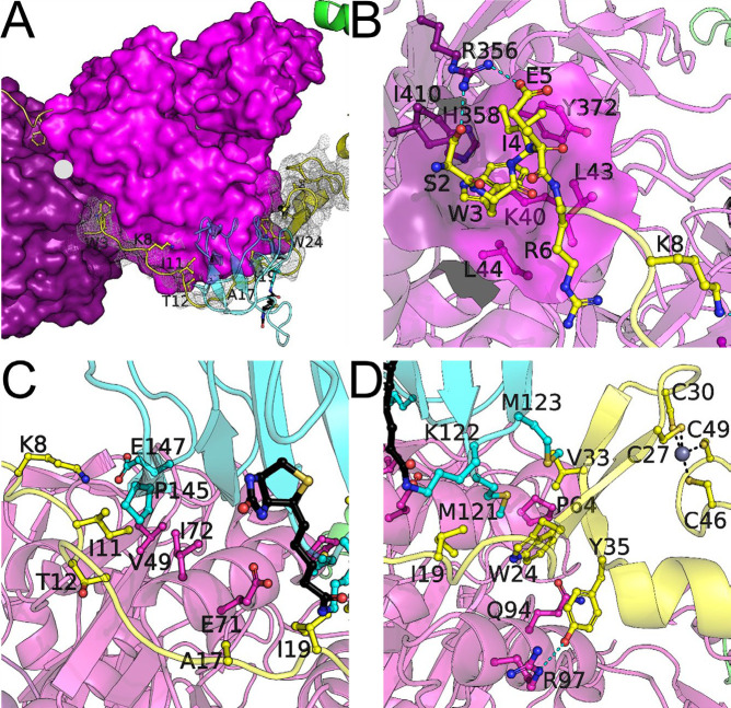
The Escherichia coli heteromeric acetyl-CoA carboxylase (ACC) has four subunits assumed to form an elusive catalytic complex and are involved in allosteric and transcriptional regulation. The E. coli ACC represents almost all ACCs from pathogenic bacteria making it a key antibiotic development target to fight growing antibiotic resistance. Furthermore, it is a model for cyanobacterial and plant plastid ACCs as biofuel engineering targets. Here we report the catalytic E. coli ACC complex surprisingly forms tubes rather than dispersed particles. The cryo-EM structure reveals key protein-protein interactions underpinning efficient catalysis and how transcriptional regulatory roles are masked during catalysis. Discovering the protein-protein interaction interfaces that facilitate catalysis, allosteric and transcriptional regulation provides new routes to engineering catalytic activity and new targets for drug discovery.


Conflict of interest statement

Competing interests: Authors declare that they have no competing interests.

Figures

Fig. 1.
Fig. 1.. Catalytic activity, purification and cryo-EM structures of E. coli ACC.
(A) Catalytic activity and structures of isolated E. coli ACC subunits. Two distinct catalytic activities of the ACC system are shown in the center. Previous crystal structures of AccC (PDB 1DV2) are colored magenta, AccB (PDB 1BDO) colored cyan and AccA:AccD system (PDB 2F9Y) colored green/yellow, respectively and their respective AlphaFold models (shown in light gray). Colors are carried through subsequent figures. The N-termini of AccA/B/D and C-termini of AccD are predicted in the AlphaFold models but missing in published crystal structures. The dimer complements of AccC and AccA:AccD are shown in a darker shade. (B) Size exclusion chromatography revealing the most active form of ACC is a complex larger than the E. coli ribosome. Absorbance at 280 nm is shown in green for E. coli ACC purification over a Sephacryl S-500 HR column (20 MDa mass cutoff), and relative activity is shown in bar format. The red dashed line is for the E. coli ribosome (2500 kDa). (C) SDS-PAGE of fractions from SEC purification showing the presence of the ACC proteins. The numbers for the lanes correspond to numbered fractions in the chromatogram above. The amount of material in each lane is normalized to constant absorbance at 280 nm. (D and E) Representative micrographs of helical assemblies formed in the absence (D) and in the presence (E) of substrates and MgCl2, respectively. The tube highlighted by a red dashed line is longer than 0.3 μm. (F and G), Cryo-EM density maps of the narrow and the wide tubes, respectively. C5 point group symmetry of the wide tube is shown as regular pentagons and the axes of dihedral symmetries of both tubes are perpendicular to the helical axes and go through the centers of the white circles. Equivalent positions are indicated by *. (H) Inside the tube looking out at one of the five sets of protomers making up a ring of (G).
Figure 2.
Figure 2.. Detailed AccD interactions with BC and BCCP.
(A) Overview of the AccD interactions with BC and BCCP, cryo-EM density is shown as a gray mesh and the attached biotin is shown in black. Notice the two-fold symmetry at the BC interface with the two N-termini of AccD, indicated by a ●. (B) The AccD Trp-3 binding pocket on the BC interface. (C) Hydrophobic cluster near the middle of the extended AccD N-terminus. (D) Hydrophobic cluster between the AccD zinc-finger domain and BC/BCCP.
Figure 3.
Figure 3.. AccA N-terminus domain swapped model and BC interactions.
One of the possible sets of asymmetric interactions at the interface between stacks of rings. This figure illustrates how the tubes can be stabilized by novel interactions likely due to the amphipathic nature of the AccA N-terminal helices. AccA interactions with the BC domain C-terminus likely alter catalysis, since the BC C-terminal helix would need to locally unfold into an extended conformation.
Figure 4.
Figure 4.. BCCP structure and interactions.
(A) The interface between the BC and BCCP is shown for our cryo-EM structure, BCCP is in cyan with the attached biotin in black and BC in magenta, and the previous crystal structure in light gray. (B) The BCCP is near a central mass of density in our maps that can be explained by the BCCP N-terminus, which is modeled in the end on view. Linkers between the BCCP N-terminus and biotin-attachment domain are invisible in the cryo-EM maps, but shown here for scale. (C) An AlphaFold multimer model for 10 copies of the BCCP N-terminal 32 residues, which are predicted to fold into a β-barrel.

Similar articles

References

    1. Tong L. Structure and function of biotin-dependent carboxylases. Cell Mol Life Sci 70, 863–891, doi:10.1007/s00018-012-1096-0 (2013). - DOI - PMC - PubMed
    1. Heath R. J. & Rock C. O. Regulation of Fatty Acid Elongation and Initiation by Acyl-Acyl Carrier Protein inEscherichia coli. Journal of Biological Chemistry 271, 1833–1836, doi:10.1074/jbc.271.4.1833 (1996). - DOI - PubMed
    1. Evans A., Ribble W., Schexnaydre E. & Waldrop G. L. Acetyl-CoA carboxylase from Escherichia coli exhibits a pronounced hysteresis when inhibited by palmitoyl-acyl carrier protein. Archives of biochemistry and biophysics 636, 100–109, doi:10.1016/j.abb.2017.10.016 (2017). - DOI - PubMed
    1. Davis M. S. & Cronan J. E. Jr. Inhibition of Escherichia coli acetyl coenzyme A carboxylase by acyl-acyl carrier protein. Journal of bacteriology 183, 1499–1503, doi:10.1128/JB.183.4.1499-1503.2001 (2001). - DOI - PMC - PubMed
    1. James E. S. & Cronan J. E. Expression of two Escherichia coli acetyl-CoA carboxylase subunits is autoregulated. The Journal of biological chemistry 279, 2520–2527, doi:10.1074/jbc.M311584200 (2004). - DOI - PubMed

Publication types