Review
. 2023 Dec 22;4(1):20230092.
doi: 10.1002/EXP.20230092. eCollection 2024 Feb.

Emerging strategies for combating Fusobacterium nucleatum in colorectal cancer treatment: Systematic review, improvements and future challenges

Affiliations
Review

Emerging strategies for combating Fusobacterium nucleatum in colorectal cancer treatment: Systematic review, improvements and future challenges

Hongyu Liu et al. Exploration (Beijing). .

Abstract

Colorectal cancer (CRC) is generally characterized by a high prevalence of Fusobacterium nucleatum (F. nucleatum), a spindle-shaped, Gram-negative anaerobe pathogen derived from the oral cavity. This tumor-resident microorganism has been closely correlated with the occurrence, progression, chemoresistance and immunosuppressive microenvironment of CRC. Furthermore, F. nucleatum can specifically colonize CRC tissues through adhesion on its surface, forming biofilms that are highly resistant to commonly used antibiotics. Accordingly, it is crucial to develop efficacious non-antibiotic approaches to eradicate F. nucleatum and its biofilms for CRC treatment. In recent years, various antimicrobial strategies, such as natural extracts, inorganic chemicals, organic chemicals, polymers, inorganic-organic hybrid materials, bacteriophages, probiotics, and vaccines, have been proposed to combat F. nucleatum and F. nucleatum biofilms. This review summarizes the latest advancements in anti-F. nucleatum research, elucidates the antimicrobial mechanisms employed by these systems, and discusses the benefits and drawbacks of each antimicrobial technology. Additionally, this review also provides an outlook on the antimicrobial specificity, potential clinical implications, challenges, and future improvements of these antimicrobial strategies in the treatment of CRC.

Keywords: Fusobacterium nucleatum; antibacterial; anti‐biofilm; colorectal cancer; drug resistance.


Conflict of interest statement

The authors declare no conflict of interest.

Figures

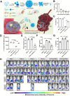
SCHEME 1
SCHEME 1
Cartoon depiction showing the proposed strategies based on natural extracts, inorganic chemicals, organic chemicals, polymers, inorganic–organic hybrid materials, bacteriophages, probiotics, and vaccines for combating F. nucleatum and F. nucleatum biofilms.
FIGURE 1
FIGURE 1
Design of CCHs: (A) Synthetic route of CCHs. Reproduced with permission.[ 85 ] Copyright 2021, Taylor & Francis. Structure of PTS and its anti‐F. nucleatum properties: B) Chemical structures of RES, PIC, OXY, and PTS. PTS triggered leakage of bacterial proteins and nucleic acids. The content of extracellular (C) proteins and (D) nucleic acid in F. nucleatum after treatment with PTS for 2, 4, and 6 h. (E) Cell viability of F. nucleatum after treatment with PTS. Reproduced with permission.[ 108 ] Copyright 2020, Springer Nature. Destructive effects of three EOs on F. nucleatum: (F) TEM images of F. nucleatum after different treatments for 1 h. Reproduced with permission.[ 111 ] Copyright 2020, MDPI.
FIGURE 2
FIGURE 2
LA‐loaded supramolecular nanosystem for addressing drug‐resistance CRC based on the inhibition of F. nucleatum: (A) Schematic diagram of supramolecular nanomedicine for enhanced systemic chemotherapy against drug‐resistant CRC. (B) The spread plate and quantification of intratumor F. nucleatum exposure to LA at various concentrations. (C) The quantification of intratumor F. nucleatum after various treatments. (D) Quantitative real‐time PCR detection of intratumor F. nucleatum 16S rRNA expression. (E–G) Expression of NF‐κB, TNF‐α, and IL‐6 in tumor tissues infected by F. nucleatum measured by enzyme‐linked immunosorbent assay (ELISA), where (+) and (−) indicates incubation microenvironments in the presence or absence of F. nucleatum, respectively. (H) In vivo bioluminescence images of mice following treatment with PBS (−), PBS (+), OxPt (−), OxPt (+), PG‐Pt‐LA/CB[7] (−), and PG‐Pt‐LA/CB[7] (+) (where (+) and (−) referred to incubation in the presence or absence of F. nucleatum, respectively). Reproduced with permission.[ 135 ] Copyright 2022, Elsevier.
FIGURE 3
FIGURE 3
Structure, antimicrobial mechanism, and anti‐F.nucleatum property of pAgNCs. (A) Structure diagram of antibacterial pAgNCs. (B) Schematic illustration showing the multifaceted antibacterial mechanism of pAgNCs by penetrating and rupturing bacterial membranes. (C) Live/dead staining of F. nucleatum observed by CLSM. Scale bar: 10 µm. (D) Changes of MIC against F. nucleatum over number of cycles after treatment with pAgNCs, AgNPs and kanamycin. (E) Detailed MIC changes between pAgNCs and AgNPs. Reproduced with permission.[ 152 ] Copyright 2021, American Chemical Society. The effects of ZIF‐8 or ZIF‐8:Ce NPs on inhibition of F. nucleatum biofilms: F) Representative CLSM‐3D live/dead photographs of F. nucleatum biofilms after treatment with ZIF‐8 or ZIF‐8:Ce NPs. Reproduced with permission.[ 153 ] Copyright 2019, Royal Society of Chemistry.
FIGURE 4
FIGURE 4
Physical properties and inhibition effect against F. nucleatum biofilms of UCNPs@TiO2: (A) TEM image of UCNPs@TiO2. (B) High‐resolution TEM image of UCNPs@TiO2. (C) Hydrodynamic diameters of UCNPs and UCNPs@TiO2. (D) Zeta potential of UCNPs@TiO2. (E) F. nucleatum biofilm CFU counts after four days. (F) Metabolism activity of four‐day F. nucleatum biofilms. Reproduced with permission.[ 190 ] Copyright 2019, Elsevier. Inhibition of F. nucleatum biofilms by l‐lysine: (G) Effects of l‐lysine with different concentrations on the formation of F. nucleatum biofilms by CV staining. (J) CLSM‐3D live/dead fluorescence imaging of F. nucleatum biofilms. Reproduced with permission.[ 201 ] Copyright 2022, Elsevier. Peptide effects on the development of a single‐species F. nucleatum biofilms: (H) AmyI‐1‐18 and (I) G12R. The remaining quantity of biofilm was measured using CV staining after incubation with each peptide. Reproduced with permission.[ 205 ] Copyright 2020, Elsevier.
FIGURE 5
FIGURE 5
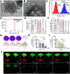
Design and therapeutic outcome of Q‐P‐A@CP[5]A for F. nucleatum‐induced drug‐resistant CRC: (A) Schematic illustration showing the construction of Q‐P‐A@CP[5]A and the treatment process for drug‐resistant CRC. (B) CLSM‐3D imaging of F. nucleatum biofilms following treatment with PBS, Q‐P‐A, and Q‐P‐A@CP[5]A (C) Flow cytometry analysis of F. nucleatum‐co‐cultured HT29 cells after being treated with PBS (i), Q‐P‐A (ii), and Q‐P‐A@CP[5]A (iii). (D) CLSM images of F. nucleatum‐co‐cultured HT29 cells after being treated with Q‐P‐A and Q‐P‐A@CP[5]A TUNEL‐positive cells exhibited green fluorescence, and DAPI‐stained nuclei showed blue fluorescence. Scale bar: 20 µm. (E) Representative photographs of HT29 tumor‐bearing nude mice treated by PBS (control), oxaliplatin, F. nucleatum, oxaliplatin, and F. nucleatum, Q‐P‐A@CP[5]A, and Q‐P‐A@CP[5]A and F. nucleatum. (F) Number of intratumor bacteria after various treatments. Inset: corresponding colony counts in tumor tissues. Reproduced with permission.[ 53 ] Copyright 2021, Royal Society of Chemistry.
FIGURE 6
FIGURE 6
Effect of different proportions of Mn‐doped NaYF4@Ce6@silane on F. nucleatum biofilms under NIR irradiation: (A) CLSM‐3D images of live/dead cells of 4‐day biofilms of F. nucleatum NIR control, NIR + NaYF4@Ce6@silane, NIR + NaYF4‐Mn10%@Ce6@silane, NIR + NaYF4‐Mn20%@Ce6@silane, NIR + NaYF4‐Mn30%@Ce6@silane. Reproduced with permission.[ 220 ] Copyright 2019, MDPI. Antimicrobial mechanism and efficacy of CuTCPP‐Fe2O3 against F. nucleatum. (B) Schematic diagram showing the antibacterial process of photodynamic ion therapy, which relies on the interaction of released ions and ROS. (C) CFU counts of F. nucleatum treated with CuTCPP and CuTCPP‐Fe2O3 for 20 min followed by 2‐h irradiation by 660 nm laser in the dark. (D) Scanning electron microscope images of F. nucleatum treated with CuTCPP‐Fe2O3 and 660 nm laser irradiation (scale bar: 500 nm). (E) TEM images and accompanying EDS curves of F. nucleatum treated with CuTCPP‐Fe2O3 and 660 laser irradiation. Scale bar: 200 nm. Reproduced with permission.[ 57 ] Copyright 2021, American Chemical Society. The combined effect of CeCyan and Cu5.4O system on the PDT against F. nucleatum. (F) Representative photographs of F. nucleatum colonies following treatment in various ways (+ L indicates the process of exposure to a 660 nm laser at 200 mW cm−2). Reproduced with permission.[ 58 ] Copyright 2022, Elsevier.
FIGURE 7
FIGURE 7
P2 phage inhibiting F. nucleatum and suppressing F. nucleatum‐induced drug‐resistant CRC in coordination with IDNP. (A) Schematic illustration showing the detailed process of the phage‐guided biotic‐abiotic hybrid nanosystem for tumor suppression. (B) In vitro lysis of various bacterial species by P2 phages. (C) CLSM images of various species of bacteria after being bound with P2 phages. Bacteria with green fluorescence were labeled with FITC, whereas phages with red fluorescence were stained with RhB. (D) In vitro anticancer impact of phage and chemotherapy (IDNP) against F. nucleatum‐co‐cultured CRC cells. Results for the mice bearing orthotopic CT26luc tumors. Reproduced with permission.[ 227 ] Copyright 2019, Springer Nature. Morphology and anti‐biofilm capability of FNU1. (E) TEM image of FNU1 showing the morphology and size of Siphoviridae bacteriophage. (F) CLSM images of SYBR gold and PI staining following FNU1 bacteriophage treated (left) and untreated (right) F. nucleatum biofilms. Reproduced with permission.[ 60 ] Copyright 2019, Springer Nature.

Similar articles

Cited by

References

    1. Biller L. H., Schrag D., JAMA 2021, 325, 669. - PubMed
    1. Siegel R. L., Miller K. D., Fuchs H. E., Jemal A., CA Cancer J. Clin. 2021, 71, 7. - PubMed
    1. Xi Y., Xu P., Transl. Oncol. 2021, 14, 101174. - PMC - PubMed
    1. Li Y.‐Y., Ge Q.‐X., Cao J., Zhou Y.‐J., Du Y.‐L., Shen B., Wan Y.‐J. Y., Nie Y.‐Q., World J. Gastroenterol. 2016, 22, 3227. - PMC - PubMed
    1. Yang Y., Weng W., Peng J., Hong L., Yang L., Toiyama Y., Gao R., Liu M., Yin M., Pan C., Li H., Guo B., Zhu Q., Wei Q., Moyer M. P., Wang P., Cai S., Goel A., Qin H., Ma Y., Gastroenterology 2017, 152, 851. - PMC - PubMed

LinkOut - more resources