. 2024 May 22;9(22):23468-23475.
doi: 10.1021/acsomega.4c00517. eCollection 2024 Jun 4.

General Characterization of Properties of Ordered and Disordered Proteins by Wide-Line 1H NMR

Affiliations

General Characterization of Properties of Ordered and Disordered Proteins by Wide-Line 1H NMR

Mónika Bokor et al. ACS Omega. .

Abstract

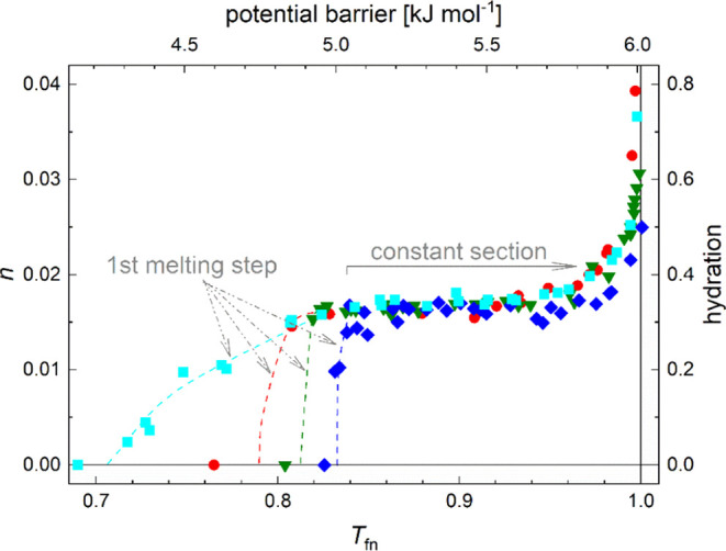
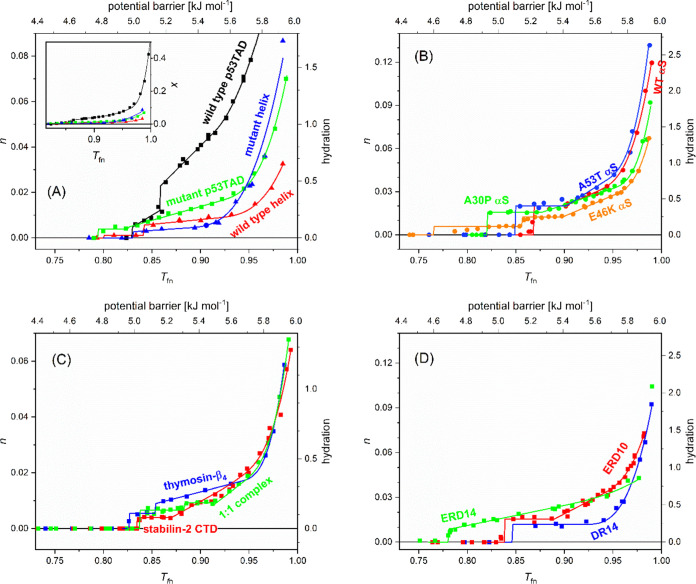
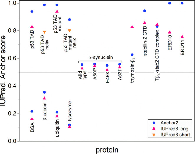
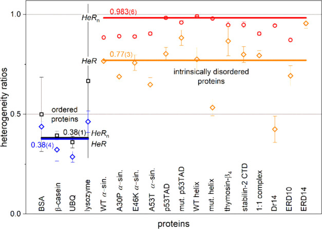
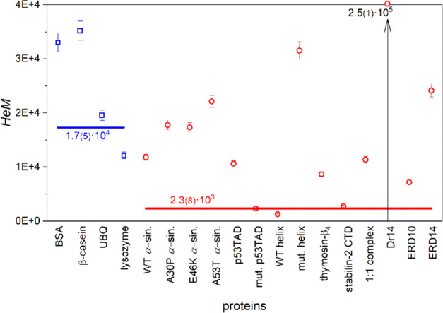
Wide-line 1H NMR is an efficient spectroscopic method to determine the disorder tendency of a protein. It directly measures the properties of the hydration shell of proteins, delivering exact and measurable values of their disorder/order content. A comparison is performed between several globular and disordered proteins. The common properties of the subzero mobile hydration water of these two groups were investigated. The amount of the mobile hydration water and the shape of the melting diagram at subzero temperatures together provide a possibility to distinguish globular proteins from disordered proteins. The shape of the melting diagram also gives information about the presence of secondary structural elements. The disordered and globular protein regions' fundamentally different structures are reflected in their melting diagrams, allowing one to directly determine the level of disorder in a specific protein structure. Intrinsically disordered proteins bind water more strongly than globular proteins, which is shown by the somewhat higher temperature values where mobile hydration water first appears but with a significantly higher heterogeneity in the energy distributions of protein-water interactions.


Conflict of interest statement

The authors declare no competing financial interest.

Figures

Figure 1
Figure 1
Melting diagrams of globular/ordered proteins: BSA (red circles), β-casein (green triangles), ubiquitin (dark blue diamonds), and lysozyme (light blue squares).
Figure 2
Figure 2
Melting diagrams of intrinsically disordered proteins. (A) wild-type p53 TAD (black), mutant p53 TAD (green), helical part of p53 TAD: wild-type (red) and mutant (blue); (B) α-synucleins: wild-type (red), A30P (green), E46K (orange), A53T (blue); (C) thymosin-β4 (blue), stabilin-2 CTD residues 2501–2551 (red), and their 1:1 complex (green); (D) Dr14 (blue), ERD10 (red), and ERD14 (green) proteins. The line is the analytical description (eq 1) applied.
Figure 3
Figure 3
IUPred3 identifies Intrinsically Disordered Protein Regions (IDPRs) and returns a score between 0 and 1, corresponding to the probability of a disordered region. IDPRs often harbor binding regions identified by the ANCHOR2 prediction algorithm, which assigns a score representing the probability of a disordered binding region.
Figure 4
Figure 4
Trend changing points of the melting diagrams (MDs) according to the type of protein order. Tfn1 values denote the MD point where linear elevation starts after a constant section. Similarly, the cubic MD section starts at Tfn3. The lines represent error-weighted averages with values marked at the lines.
Figure 5
Figure 5
Dynamic parameters HeR (ratio of homogeneity) and HeRn (ratio of the amount of heterogeneously bound water to the total number of bound water) for ordered and disordered proteins. The lines represent error-weighted averages with values marked at the lines.
Figure 6
Figure 6
Dynamic parameter HeM (measure of heterogeneity) for ordered (blue squares) and disordered (red circles) proteins. The lines represent error-weighted averages with values marked at the lines.

Similar articles

Cited by

References

    1. Habchi J.; Tompa P.; Longhi S.; Uversky V. N. Introducing protein intrinsic disorder. Chem. Rev. 2014, 114, 6561–6588. 10.1021/cr400514h. - DOI - PubMed
    1. Berman H. M.; Westbrook J.; Feng Z.; Gilliland G.; Bhat T. N.; Weissig H.; Shindyalov I. N.; Bourne P. E. The Protein Data Bank. Nucleic Acids Res. 2000, 28, 235–242. 10.1093/nar/28.1.235. - DOI - PMC - PubMed
    1. Tompa K.; Bokor M.; Verebélyi T.; Tompa P. Water rotation barriers on protein molecular surfaces. Chem. Phys. 2015, 448, 15–25. 10.1016/j.chemphys.2014.12.008. - DOI
    1. CRC Handbook of Chemistry and Physics, 103rd ed.; Rumble J., Ed.; CRC Press, 2022–2023. 9781032121710.
    1. Bokor M.; Tantos Á.; Tompa P.; Han K.-H.; Tompa K. WT and A53T α-synuclein systems: melting diagram and its new interpretation. Int. J. Mol. Sci. 2020, 21, 399710.3390/ijms21113997. - DOI - PMC - PubMed

LinkOut - more resources