Nomogram based on dual-energy CT-derived extracellular volume fraction for the prediction of microsatellite instability status in gastric cancer
- PMID: 38854729
- PMCID: PMC11156999
- DOI: 10.3389/fonc.2024.1370031
Nomogram based on dual-energy CT-derived extracellular volume fraction for the prediction of microsatellite instability status in gastric cancer
Abstract
Purpose: To develop and validate a nomogram based on extracellular volume (ECV) fraction derived from dual-energy CT (DECT) for preoperatively predicting microsatellite instability (MSI) status in gastric cancer (GC).
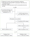
Materials and methods: A total of 123 patients with GCs who underwent contrast-enhanced abdominal DECT scans were retrospectively enrolled. Patients were divided into MSI (n=41) and microsatellite stability (MSS, n=82) groups according to postoperative immunohistochemistry staining, then randomly assigned to the training (n=86) and validation cohorts (n=37). We extracted clinicopathological characteristics, CT imaging features, iodine concentrations (ICs), and normalized IC values against the aorta (nICs) in three enhanced phases. The ECV fraction derived from the iodine density map at the equilibrium phase was calculated. Univariate and multivariable logistic regression analyses were used to identify independent risk predictors for MSI status. Then, a nomogram was established, and its performance was evaluated by ROC analysis and Delong test. Its calibration performance and clinical utility were assessed by calibration curve and decision curve analysis, respectively.
Results: The ECV fraction, tumor location, and Borrmann type were independent predictors of MSI status (all P < 0.05) and were used to establish the nomogram. The nomogram yielded higher AUCs of 0.826 (0.729-0.899) and 0.833 (0.675-0.935) in training and validation cohorts than single variables (P<0.05), with good calibration and clinical utility.
Conclusions: The nomogram based on DECT-derived ECV fraction has the potential as a noninvasive biomarker to predict MSI status in GC patients.
Keywords: dual-energy CT; extracellular volume fraction; gastric cancer; microsatellite instability; nomogram.
Copyright © 2024 Hu, Zhao, Ji, Chen, Xu, Liu, Zhang and Liu.
Conflict of interest statement
The authors declare that the research was conducted in the absence of any commercial or financial relationships that could be construed as a potential conflict of interest.
Figures


Similar articles
-
Radiomics Analysis of Iodine-Based Material Decomposition Images With Dual-Energy Computed Tomography Imaging for Preoperatively Predicting Microsatellite Instability Status in Colorectal Cancer.Front Oncol. 2019 Nov 22;9:1250. doi: 10.3389/fonc.2019.01250. eCollection 2019. Front Oncol. 2019. PMID: 31824843 Free PMC article.
-
Development and validation of a radiomics-based nomogram for the preoperative prediction of microsatellite instability in colorectal cancer.BMC Cancer. 2022 May 9;22(1):524. doi: 10.1186/s12885-022-09584-3. BMC Cancer. 2022. PMID: 35534797 Free PMC article.
-
Dual-layer spectral-detector CT for predicting microsatellite instability status and prognosis in locally advanced gastric cancer.Insights Imaging. 2023 Sep 19;14(1):151. doi: 10.1186/s13244-023-01490-x. Insights Imaging. 2023. PMID: 37726599 Free PMC article.
-
Spectral CT-based nomogram for preoperative prediction of perineural invasion in locally advanced gastric cancer: a prospective study.Eur Radiol. 2023 Jul;33(7):5172-5183. doi: 10.1007/s00330-023-09464-9. Epub 2023 Feb 24. Eur Radiol. 2023. PMID: 36826503
-
Nomogram based on CT-derived extracellular volume for the prediction of post-hepatectomy liver failure in patients with resectable hepatocellular carcinoma.Eur Radiol. 2022 Dec;32(12):8529-8539. doi: 10.1007/s00330-022-08917-x. Epub 2022 Jun 9. Eur Radiol. 2022. PMID: 35678856
Cited by
-
Application and progress of nomograms in gastric cancer.Front Med (Lausanne). 2025 Jan 29;12:1510742. doi: 10.3389/fmed.2025.1510742. eCollection 2025. Front Med (Lausanne). 2025. PMID: 39944483 Free PMC article. Review.
-
Predictive value of extracellular volume fraction determined using enhanced computed tomography for pathological grading of clear cell renal cell carcinoma: a preliminary study.Cancer Imaging. 2025 Apr 4;25(1):49. doi: 10.1186/s40644-025-00866-0. Cancer Imaging. 2025. PMID: 40186299 Free PMC article.
-
Incremental value of extracellular volume fraction based on CT for microsatellite status in colorectal cancer.Jpn J Radiol. 2025 Jul 4. doi: 10.1007/s11604-025-01825-2. Online ahead of print. Jpn J Radiol. 2025. PMID: 40613851
References
-
- Arora S, Balasubramaniam S, Zhang W, Zhang L, Sridhara R, Spillman D, et al. . Fda approval summary: pembrolizumab plus lenvatinib for endometrial carcinoma, a collaborative international review under project orbis. Clin Cancer Res. (2020) 26:5062–7. doi: 10.1158/1078-0432.Ccr-19-3979 - DOI - PubMed
LinkOut - more resources
Full Text Sources
Miscellaneous

