The diverse liver viromes of Australian geckos and skinks are dominated by hepaciviruses and picornaviruses and reflect host taxonomy and habitat
- PMID: 38854849
- PMCID: PMC11160328
- DOI: 10.1093/ve/veae044
The diverse liver viromes of Australian geckos and skinks are dominated by hepaciviruses and picornaviruses and reflect host taxonomy and habitat
Abstract
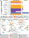
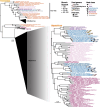
Lizards have diverse ecologies and evolutionary histories, and represent a promising group to explore how hosts shape virome structure and virus evolution. Yet, little is known about the viromes of these animals. In Australia, squamates (lizards and snakes) comprise the most diverse order of vertebrates, and Australia hosts the highest diversity of lizards globally, with the greatest breadth of habitat use. We used meta-transcriptomic sequencing to determine the virome of nine co-distributed, tropical lizard species from three taxonomic families in Australia and analyzed these data to identify host traits associated with viral abundance and diversity. We show that lizards carry a large diversity of viruses, identifying more than thirty novel, highly divergent vertebrate-associated viruses. These viruses were from nine viral families, including several that contain well known pathogens, such as the Flaviviridae, Picornaviridae, Bornaviridae, Iridoviridae, and Rhabdoviridae. Members of the Flaviviridae were particularly abundant across species sampled here, largely belonging to the genus Hepacivirus: fourteen novel hepaciviruses were identified, broadening the known diversity of this group and better defining its evolution by uncovering new reptilian clades. The evolutionary histories of the viruses studied here frequently aligned with the biogeographic and phylogenetic histories of the hosts, indicating that exogenous viruses may help infer host evolutionary history if sampling is strategic and sampling density high enough. Notably, analysis of alpha and beta diversity revealed that virome composition and richness in the animals sampled here was shaped by host taxonomy and habitat. In sum, we identified a diverse range of reptile viruses that broadly contributes to our understanding of virus-host ecology and evolution.
Keywords: Hepacivirus; evolution; meta-transcriptomics; metagenomics; one health; viral ecology.
© The Author(s) 2024. Published by Oxford University Press.
Conflict of interest statement
None declared.
Figures


Similar articles
-
The radiation of New Zealand's skinks and geckos is associated with distinct viromes.BMC Ecol Evol. 2024 Jun 13;24(1):81. doi: 10.1186/s12862-024-02269-4. BMC Ecol Evol. 2024. PMID: 38872095 Free PMC article.
-
Virome composition in marine fish revealed by meta-transcriptomics.Virus Evol. 2021 Feb 4;7(1):veab005. doi: 10.1093/ve/veab005. eCollection 2021 Jan. Virus Evol. 2021. PMID: 33623709 Free PMC article.
-
Extensive Diversity of RNA Viruses in Australian Ticks.J Virol. 2019 Jan 17;93(3):e01358-18. doi: 10.1128/JVI.01358-18. Print 2019 Feb 1. J Virol. 2019. PMID: 30404810 Free PMC article.
-
Making sense of the virome in light of evolution and ecology.Proc Biol Sci. 2025 Apr;292(2044):20250389. doi: 10.1098/rspb.2025.0389. Epub 2025 Apr 2. Proc Biol Sci. 2025. PMID: 40169018 Free PMC article. Review.
-
Avian picornaviruses: molecular evolution, genome diversity and unusual genome features of a rapidly expanding group of viruses in birds.Infect Genet Evol. 2014 Dec;28:151-66. doi: 10.1016/j.meegid.2014.09.027. Epub 2014 Sep 30. Infect Genet Evol. 2014. PMID: 25278047 Review.
Cited by
-
A trove of antiviral TRIM family E3 ligases in reptiles.bioRxiv [Preprint]. 2025 Jun 25:2025.06.23.661133. doi: 10.1101/2025.06.23.661133. bioRxiv. 2025. PMID: 40667304 Free PMC article. Preprint.
-
Host ecology and phylogeny shape the temporal dynamics of social bee viromes.Nat Commun. 2025 Mar 5;16(1):2207. doi: 10.1038/s41467-025-57314-7. Nat Commun. 2025. PMID: 40044660 Free PMC article.
-
The radiation of New Zealand's skinks and geckos is associated with distinct viromes.BMC Ecol Evol. 2024 Jun 13;24(1):81. doi: 10.1186/s12862-024-02269-4. BMC Ecol Evol. 2024. PMID: 38872095 Free PMC article.
-
First Report on Detection and Molecular Characterization of Astroviruses in Mongooses.Viruses. 2024 Aug 8;16(8):1269. doi: 10.3390/v16081269. Viruses. 2024. PMID: 39205243 Free PMC article.
-
Two Avastrovirus Species Discovered in Psittaciformes Expand the Host Range of the Family Astroviridae.Viruses. 2025 Mar 20;17(3):450. doi: 10.3390/v17030450. Viruses. 2025. PMID: 40143376 Free PMC article.
References
-
- Altschul S. F. et al. (1990) ‘Basic Local Alignment Search Tool’, Journal of Molecular Biology, 215: 403–10. - PubMed
LinkOut - more resources
Full Text Sources

