. 2024 Jun 4:17:1309-1319.
doi: 10.2147/CCID.S449987. eCollection 2024.

Ergothioneine Protects Against UV-Induced Oxidative Stress Through the PI3K/AKT/Nrf2 Signaling Pathway

Affiliations

Ergothioneine Protects Against UV-Induced Oxidative Stress Through the PI3K/AKT/Nrf2 Signaling Pathway

Yongchao Li et al. Clin Cosmet Investig Dermatol. .

Abstract

Background: Ergothioneine (EGT) is an antioxidant, which could be detected in human tissues, and human skin cells could utilize EGT and play an anti-oxidative role in keratinocytes. And in this study we are going to elucidate whether EGT could protect the skin from photoaging by Ultraviolet (UV) exposure in mice and its molecule pathway.

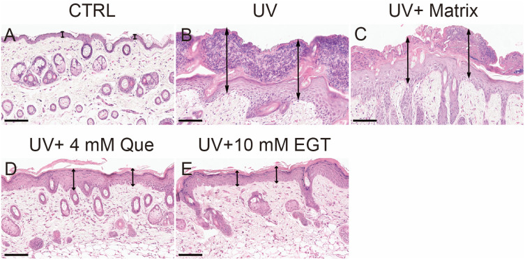
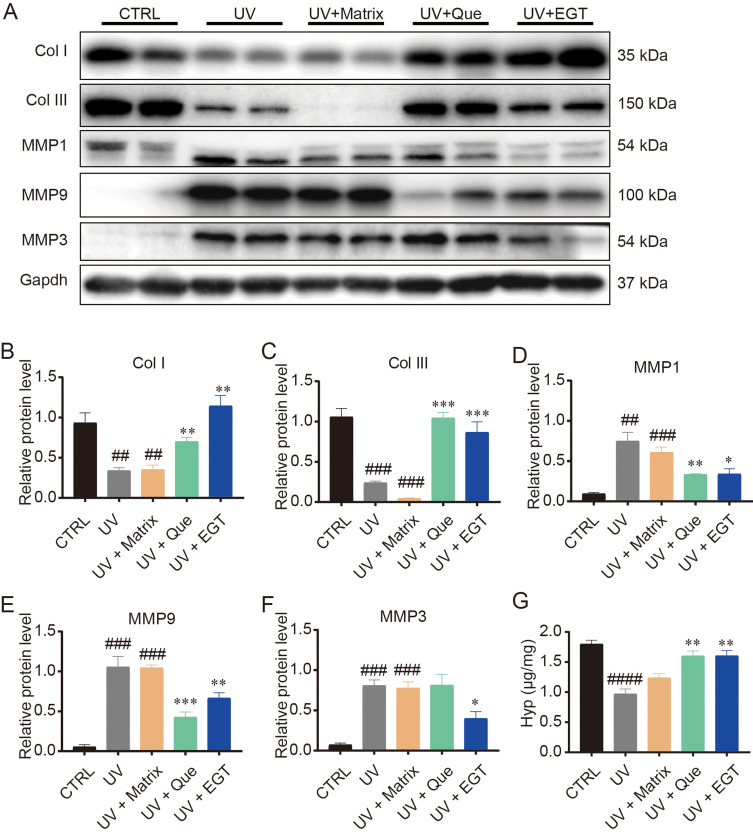
Methods: Histological analysis was performed for evaluating the skin structure change. Malondialdehyde (MDA) and superoxide dismutase (SOD) levels were measured with biological assay for evaluating oxidative and antioxidative ability of skin exposed to UV light. And the level of marker molecules in mouse skin were detected by hydroxyproline (Hyp) assay, immunohistochemical analysis, Western blot, and quantitative real-time PCR (qRT-PCR). The markers of skin aging and cell death were tested by cell culture and treatment, Western blot and qRT-PCR.

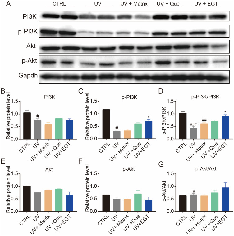
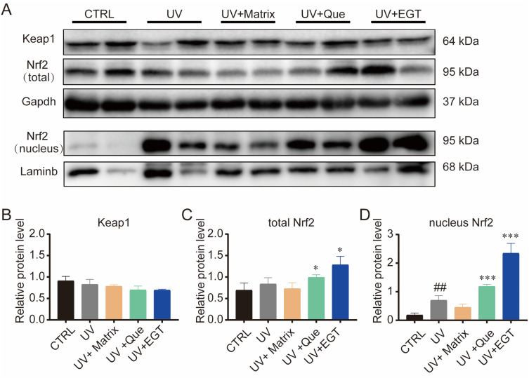
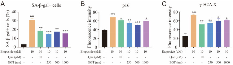
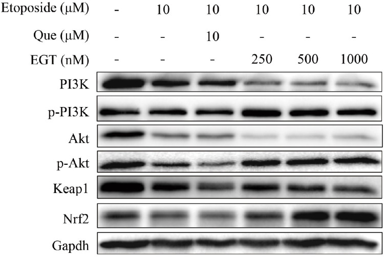
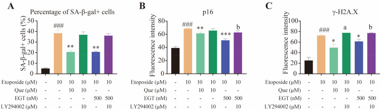
Results: EGT decreased the levels of inflammatory factors induced by UV exposure in mouse skin. MDA and SOD activity detection showed that EGT decreased MDA levels, increased SOD activity, and upregulated PI3K/Akt/Nrf2 signals in mouse skin exposed to UV, which further activated Nrf2 in the nucleus and enhanced the expression of Nrf2 target genes. In the cell model, we revealed that EGT could inhibit the increase in senescence-associated β-galactosidase-positive cells and p16 and γ-H2A.X positive cells induced by etoposide and activate PI3K/Akt/Nrf2 signaling. Moreover, a PI3K inhibitor blocked EGT protection against etoposide-induced cell death.

Conclusion: The study showed EGT may play an important protective role against cell damage or death through the PI3K/Akt/Nrf2 signaling pathway in skin.

Keywords: Nrf2; PI3K/Akt; ergothioneine; skin aging.


Conflict of interest statement

Ergothioneine production patent (WO-2014100752-A1) has been assigned from Boston University to Ergo-health LLC and Pinghua Liu is one of the co-founders of Ergo-health. The authors declare that they have no other competing interests in this work.

Figures

Figure 1
Figure 1
EGT reduce the increase of epidermis thickness under UV irradiation. H&E staining of back skin of control (CTRL) group (A), UV group (B), UV+Matrix group (C), UV+Que group (D), UV+EGT group (E). The scale bar was 100 μm, and the black double arrows indicate the thickness of epidermis.
Figure 2
Figure 2
EGT significantly could reduce MMPs level and maintain collagen protein in mouse skin exposed to UV. (A) The protein levels of collagens and MMPs, the protein levels relative to Gapdh of collagen I (B), collagen III (C), MMP1 (D), MMP9 (E) and MMP3 (F). (G) Hyp level was detected using Hyp kit. Control group vs UV group or UV + Matrix group: ##P < 0.01, ###P < 0.001, ####P < 0.0001. Que group or EGT group vs UV + Matrix group: *P < 0.05, **P < 0.01, ***P < 0.001.
Figure 3
Figure 3
EGT could upregulate the levels of p-PI3K and p-Akt, and activate PI3K/Akt signals. (A) Western blot bands of PI3K, p-PI3K, Akt and p-Akt. (B, C, E and F) The histograms of PI3K, p-PI3K, Akt and p-Akt relative to Gapdh, and (D and G) the ratio value of p-PI3K/PI3K and p-Akt/Akt. Control group vs UV group or UV + Matrix group: #P < 0.05, ##P < 0.01, ###P < 0.001. Que group or EGT group vs UV + Matrix group: *P < 0.05.
Figure 4
Figure 4
Ergothioneine promoted Nrf2 expression into the nucleus. (A) The levels of protein Keap1, total Nrf2, nucleus Nrf2. (A) The bands of Keap1, Nrf2 (total), Nrf2 (nucleus) and Lamin b. The gray values of Keap1 (B), total Nrf2 (C) and nucleus Nrf2 (D) relative to Gapdh or Lamin b. Control group vs UV group or UV + Matrix group: ##P < 0.01. Que group or EGT group vs UV + Matrix group: *P < 0.05, ***P < 0.001.
Figure 5
Figure 5
EGT could reduce the number of SA-β-Gal positive cells, and downregulate p16 and γ-H2A.X in HaCaT cells treated with etoposide. (A) the number of SA-β-gal positive cells, (B) the fluorescence intensity of p16 in HaCaT cells, (C) the fluorescence intensity of γ-H2A.X in HaCaT cells. Control group vs etoposide group, ###P < 0.001. Etoposide group vs Que or EGT groups, *P < 0.05, **P < 0.01, ***P < 0.001.
Figure 6
Figure 6
EGT could increase p-PI3K and p-Akt, Nrf2 levels in HaCaT cells treated by etoposide. The HaCaT cells were treated by etoposide for 48 h, then adding Que and EGT for 48 h. Finally, the cells were harvested for Western blot. DMSO was used as a control treatment.
Figure 7
Figure 7
PI3K inhibitor (LY294002) could repress EGT protection in etoposide-induced cell death. The percentage of SA-β-gal cells (A) were based on SA-β-gal staining, and the fluorescence intensity of p16 (B) and γ-H2A.X (C) were from the immunofluorescence of p16 and γ-H2A.X. Control group vs etoposide group: ###P < 0.001. Etoposide group vs Que or EGT groups: *P < 0.05, **P < 0.01, ***P < 0.001. Que group vs Que+LY294002 group, aP < 0.05. EGT group vs EGT+LY294002 group, bP < 0.05.

Similar articles

Cited by

References

    1. Hseu YC, Lo HW, Korivi M, Tsai YC, Tang MJ, Yang HL. Dermato-protective properties of ergothioneine through induction of Nrf2/ARE-mediated antioxidant genes in UVA-irradiated Human keratinocytes. Free Radic Biol Med. 2015;86:102–117. doi:10.1016/j.freeradbiomed.2015.05.026 - DOI - PubMed
    1. Cheah IK, Halliwell B. Ergothioneine; antioxidant potential, physiological function and role in disease. BBA. 2012;1822(5):784–793. doi:10.1016/j.bbadis.2011.09.017 - DOI - PubMed
    1. Hartman PE. Ergothioneine as antioxidant. Methods Enzymol. 1990;186:310. - PubMed
    1. Markova NG, Karaman-Jurukovska N, Dong KK, Damaghi N, Smiles KA, Yarosh DB. Skin cells and tissue are capable of using L-ergothioneine as an integral component of their antioxidant defense system. Free Radic Biol Med. 2009;46(8):1168–1176. doi:10.1016/j.freeradbiomed.2009.01.021 - DOI - PubMed
    1. Hseu YC, Vudhya Gowrisankar Y, Chen XZ, Yang YC, Yang HL. The antiaging activity of ergothioneine in UVA-irradiated human dermal fibroblasts via the inhibition of the AP-1 pathway and the activation of Nrf2-mediated antioxidant genes. Oxid Med Cell Longev. 2020;2020(10):2576823. doi:10.1155/2020/2576823 - DOI - PMC - PubMed