

Implications of lung shunt fraction calculation discrepancy in Yttrium-90 radioembolization treatment from 2D planar vs 3D single photon emission CT imaging
- PMID: 38854889
- PMCID: PMC11162746
- DOI: 10.1093/bjrcr/uaae016
Implications of lung shunt fraction calculation discrepancy in Yttrium-90 radioembolization treatment from 2D planar vs 3D single photon emission CT imaging
Abstract
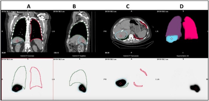
The safety and efficacy of Yttrium-90 (Y-90) radio-embolization therapy is partly dependent on the lung shunt fraction (LSF). There may be a notable disparity between LSF when calculated using 2D planar imaging vs 3D single photon emission CT (SPECT); this can affect the total allowable Y-90 dose delivered and therefore change the effectiveness of the procedure. The case presented demonstrates an 81% decrease in LSF when calculated by SPECT as compared to 2D planar imaging. This case highlights the importance of considering the imaging technique and the potential discrepancies that can arise between planar and SPECT imaging in LSF assessment.
Keywords: Y-90; hepatocellular carcinoma; lung shunt fraction; planar imaging; single photon emission CT (SPECT); trans-arterial radioembolization.
Published by Oxford University Press on behalf of the British Institute of Radiology 2024.
Conflict of interest statement
None declared.
Figures


Similar articles
-
Clinical and Dosimetric Implications of Calculating Lung Shunt Fraction for Hepatic 90Y Radioembolization Using SPECT/CT Versus Planar Scintigraphy.AJR Am J Roentgenol. 2022 Apr;218(4):728-737. doi: 10.2214/AJR.21.26663. Epub 2021 Oct 27. AJR Am J Roentgenol. 2022. PMID: 34704460
-
Radioembolization lung shunt estimation based on a 90 Y pretreatment procedure: A phantom study.Med Phys. 2018 Oct;45(10):4744-4753. doi: 10.1002/mp.13168. Epub 2018 Sep 21. Med Phys. 2018. PMID: 30179259
-
The value of 99mTc-MAA SPECT/CT for lung shunt estimation in 90Y radioembolization: a phantom and patient study.EJNMMI Res. 2018 Jun 15;8(1):50. doi: 10.1186/s13550-018-0402-8. EJNMMI Res. 2018. PMID: 29904808 Free PMC article.
-
Lung shunt and lung dose calculation methods for radioembolization treatment planning.Q J Nucl Med Mol Imaging. 2021 Mar;65(1):32-42. doi: 10.23736/S1824-4785.20.03287-2. Epub 2021 Jan 4. Q J Nucl Med Mol Imaging. 2021. PMID: 33393753 Review.
-
Lung shunt fraction quantification methods in radioembolization: What you need to know.Br J Radiol. 2022 Oct 1;95(1139):20220470. doi: 10.1259/bjr.20220470. Epub 2022 Aug 10. Br J Radiol. 2022. PMID: 35848755 Free PMC article. Review.
References
-
- Salem R, Padia SA, Lam M, et al.Clinical, dosimetric, and reporting considerations for Y-90 glass microspheres in hepatocellular carcinoma: updated 2022 recommendations from an international multidisciplinary working group. Eur J Nucl Med Mol Imaging. 2023;50(2):328-343. 10.1007/s00259-022-05956-w - DOI - PMC - PubMed
Publication types
LinkOut - more resources
Full Text Sources
