

Characteristics of optic disc hemorrhage and optic nerve changes following acute primary angle closure
- PMID: 38854957
- PMCID: PMC11157090
- DOI: 10.3389/fneur.2024.1333091
Characteristics of optic disc hemorrhage and optic nerve changes following acute primary angle closure
Abstract
Introduction: Acute primary angle closure (APAC) is an emergency ophthalmic presentation and a major cause of irreversible blindness in China. However, only a few studies have focused on the characteristics of optic disc hemorrhage (ODH) during an APAC attack, including its shape, depth, location, scope, and duration after intraocular pressure (IOP) control, along with changes in the optic nerve. This study aimed to analyze the characteristics of ODH and optic nerve changes in patients during their first APAC episode.
Methods: This retrospective study involved 32 eyes from 32 patients with APAC who received sequential treatment and analyzed the following parameters: the highest IOP and its duration, ODH, retinal nerve fiber layer thickness (RNFLT), and mean deviation (MD). We compared parameters obtained from the affected eye (ODH group) and contralateral unaffected eye (control group), as well as intragroup comparisons.
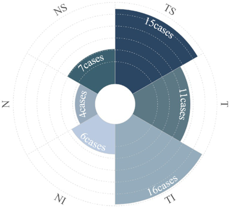
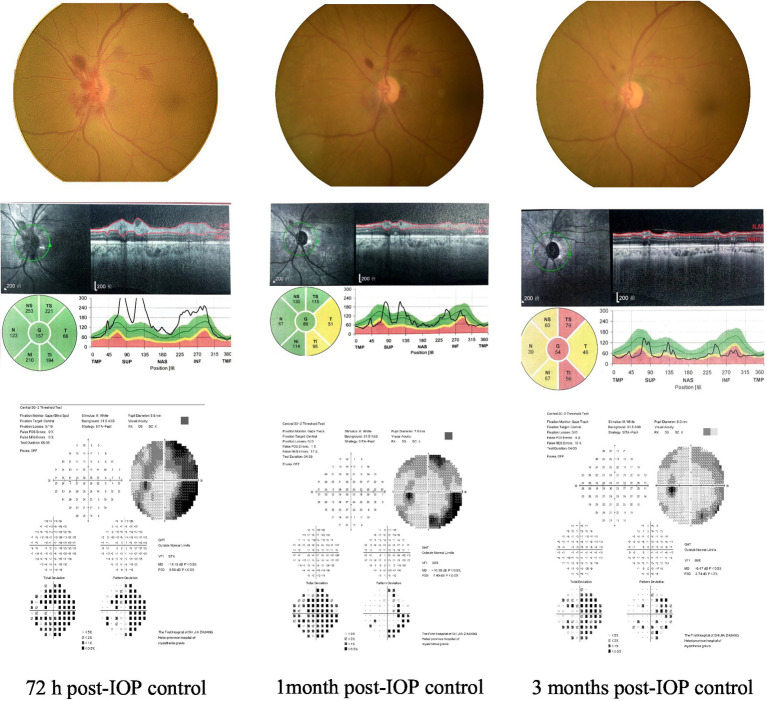
Results: The mean IOP in the ODH group was 64.28 ± 10.36 mmHg, with a duration of 4.44 ± 2.35 days. Flame and splinter shapes accounted for 84.38% of the ODH. The mean ODH duration was 4.81 ± 3.25 weeks. ODH during APAC was isolated to one sector in 59.38% of cases, mostly occurring in the temporal superior and temporal inferior (each accounting for 21.88% of the cases). There was a positive correlation between the extent of hemorrhage and the highest IOP duration (p < 0.001). RNFLT was significantly thickened within 72 h post-IOP control but was thinned by 2 weeks. By 6 months, the thinning stabilized, and there was no difference noted between the ODH and control groups at 12 months. MD partly improved at 6 months post-IOP control, and ODH scope significantly affected the MD (p < 0.001). The duration of high IOP was positively correlated to the ODH scope and MD damage.
Discussion: Timely and effective IOP management is essential for recovering visual function following an APAC attack.
Keywords: acute primary angle closure; intraocular pressure; optic disc hemorrhage; retinal nerve fiber layer; visual field.
Copyright © 2024 Zhang, Li, Tang, Yan, Geng, Li, Shi, Tang and Guo.
Conflict of interest statement
The authors declare that the research was conducted in the absence of any commercial or financial relationships that could be construed as a potential conflict of interest.
Figures


Similar articles
-
Quantification of retinal nerve fiber layer thickness after unilateral acute primary angle closure in Asian Indian eyes.J Glaucoma. 2013 Jan;22(1):26-30. doi: 10.1097/IJG.0b013e3182311d9f. J Glaucoma. 2013. PMID: 21946556
-
Bruch's membrane opening - minimum rim width measurement after acute primary angle-closure.Eur J Ophthalmol. 2022 Jul;32(4):2234-2240. doi: 10.1177/11206721211054967. Epub 2021 Nov 6. Eur J Ophthalmol. 2022. PMID: 34747240
-
Long-term outcomes in fellow eyes after acute primary angle closure in the contralateral eye.Ophthalmology. 2006 Jul;113(7):1087-91. doi: 10.1016/j.ophtha.2006.02.016. Ophthalmology. 2006. PMID: 16815398
-
Lens extraction versus laser peripheral iridotomy for acute primary angle closure.Cochrane Database Syst Rev. 2023 Mar 8;3(3):CD015116. doi: 10.1002/14651858.CD015116.pub2. Cochrane Database Syst Rev. 2023. PMID: 36884304 Free PMC article. Review.
-
[Aiming for zero blindness].Nippon Ganka Gakkai Zasshi. 2015 Mar;119(3):168-93; discussion 194. Nippon Ganka Gakkai Zasshi. 2015. PMID: 25854109 Review. Japanese.
References
LinkOut - more resources
Full Text Sources