

Super resolution using sparse sampling at portable ultra-low field MR
- PMID: 38854960
- PMCID: PMC11157107
- DOI: 10.3389/fneur.2024.1330203
Super resolution using sparse sampling at portable ultra-low field MR
Abstract
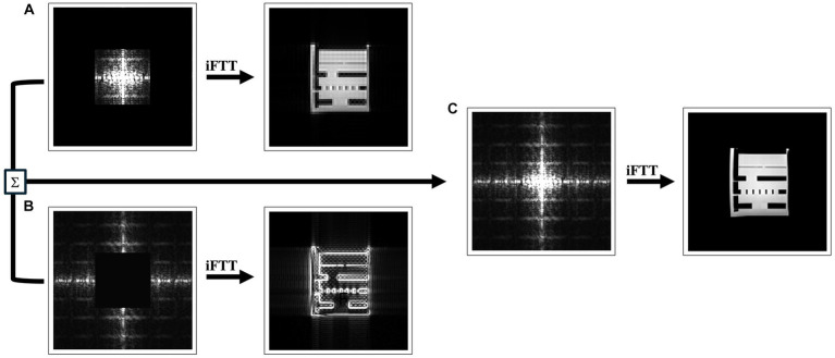
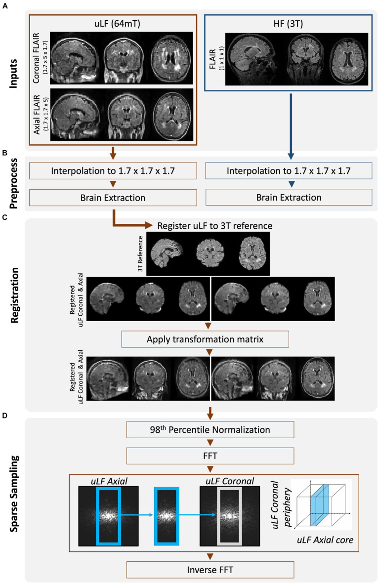
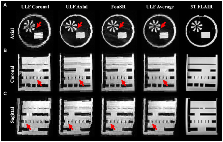
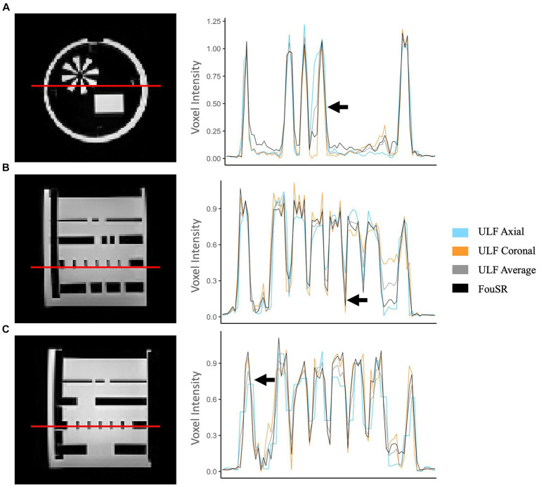
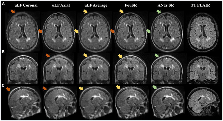
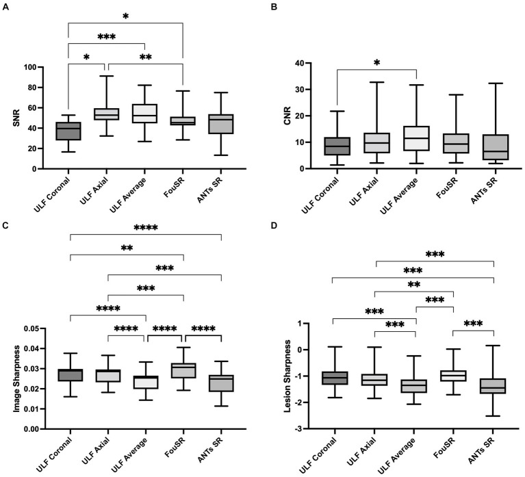
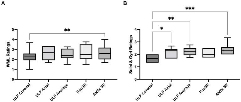
Ultra-low field (ULF) magnetic resonance imaging (MRI) holds the potential to make MRI more accessible, given its cost-effectiveness, reduced power requirements, and portability. However, signal-to-noise ratio (SNR) drops with field strength, necessitating imaging with lower resolution and longer scan times. This study introduces a novel Fourier-based Super Resolution (FouSR) approach, designed to enhance the resolution of ULF MRI images with minimal increase in total scan time. FouSR combines spatial frequencies from two orthogonal ULF images of anisotropic resolution to create an isotropic T2-weighted fluid-attenuated inversion recovery (FLAIR) image. We hypothesized that FouSR could effectively recover information from under-sampled slice directions, thereby improving the delineation of multiple sclerosis (MS) lesions and other significant anatomical features. Importantly, the FouSR algorithm can be implemented on the scanner with changes to the k-space trajectory. Paired ULF (Hyperfine SWOOP, 0.064 tesla) and high field (Siemens, Skyra, 3 Tesla) FLAIR scans were collected on the same day from a phantom and a cohort of 10 participants with MS or suspected MS (6 female; mean ± SD age: 44.1 ± 4.1). ULF scans were acquired along both coronal and axial planes, featuring an in-plane resolution of 1.7 mm × 1.7 mm with a slice thickness of 5 mm. FouSR was evaluated against registered ULF coronal and axial scans, their average (ULF average) and a gold standard SR (ANTs SR). FouSR exhibited higher SNR (47.96 12.6) compared to ULF coronal (36.7 12.2) and higher lesion conspicuity (0.12 0.06) compared to ULF axial (0.13 0.07) but did not exhibit any significant differences contrast-to-noise-ratio (CNR) compared to other methods in patient scans. However, FouSR demonstrated superior image sharpness (0.025 0.0040) compared to all other techniques (ULF coronal 0.021 0.0037, q = 5.9, p-adj. = 0.011; ULF axial 0.018 0.0026, q = 11.1, p-adj. = 0.0001; ULF average 0.019 0.0034, q = 24.2, p-adj. < 0.0001) and higher lesion sharpness (-0.97 0.31) when compared to the ULF average (-1.02 0.37, t(543) = -10.174, p = <0.0001). Average blinded qualitative assessment by three experienced MS neurologists showed no significant difference in WML and sulci or gyri visualization between FouSR and other methods. FouSR can, in principle, be implemented on the scanner to produce clinically useful FLAIR images at higher resolution on the fly, providing a valuable tool for visualizing lesions and other anatomical structures in MS.
Keywords: Fourier-transform; multiple sclerosis; reconstruction; super-resolution; ultra-low field.
Copyright © 2024 Donnay, Okar, Tsagkas, Gaitán, Poorman, Reich and Nair.
Conflict of interest statement
MP was employed by Hyperfine, Inc. The remaining authors declare that the research was conducted in the absence of any commercial or financial relationships that could be construed as a potential conflict of interest.
Figures
Similar articles
-
Sensitivity of portable low-field magnetic resonance imaging for multiple sclerosis lesions.Neuroimage Clin. 2022;35:103101. doi: 10.1016/j.nicl.2022.103101. Epub 2022 Jun 27. Neuroimage Clin. 2022. PMID: 35792417 Free PMC article.
-
Combined Deep Learning-based Super-Resolution and Partial Fourier Reconstruction for Gradient Echo Sequences in Abdominal MRI at 3 Tesla: Shortening Breath-Hold Time and Improving Image Sharpness and Lesion Conspicuity.Acad Radiol. 2023 May;30(5):863-872. doi: 10.1016/j.acra.2022.06.003. Epub 2022 Jul 6. Acad Radiol. 2023. PMID: 35810067
-
FLAIR imaging for multiple sclerosis: a comparative MR study at 1.5 and 3.0 Tesla.Eur Radiol. 2006 Apr;16(4):915-21. doi: 10.1007/s00330-005-0070-8. Epub 2005 Dec 20. Eur Radiol. 2006. PMID: 16365731
-
Applications, limitations and advancements of ultra-low-field magnetic resonance imaging: A scoping review.Surg Neurol Int. 2024 Jun 28;15:218. doi: 10.25259/SNI_162_2024. eCollection 2024. Surg Neurol Int. 2024. PMID: 38974534 Free PMC article.
-
MRI at low field: A review of software solutions for improving SNR.NMR Biomed. 2025 Jan;38(1):e5268. doi: 10.1002/nbm.5268. Epub 2024 Oct 7. NMR Biomed. 2025. PMID: 39375036 Free PMC article. Review.
Cited by
-
Proof of concept: Portable ultra-low-field magnetic resonance imaging for the diagnosis of epileptogenic brain pathologies.Epilepsia. 2024 Dec;65(12):3607-3618. doi: 10.1111/epi.18171. Epub 2024 Oct 29. Epilepsia. 2024. PMID: 39470733 Free PMC article.
-
Opportunities for Artificial Intelligence in Operational Medicine: Lessons from the United States Military.Bioengineering (Basel). 2025 May 14;12(5):519. doi: 10.3390/bioengineering12050519. Bioengineering (Basel). 2025. PMID: 40428137 Free PMC article. Review.
-
High-Field-Blinded Assessment of Portable Ultra-Low-Field Brain MRI for Multiple Sclerosis.J Neuroimaging. 2025 Jan-Feb;35(1):e70005. doi: 10.1111/jon.70005. J Neuroimaging. 2025. PMID: 39815369 Free PMC article.
References
-
- Van Reeth E, Tham IWK, Tan CH, Poh CL. Super-resolution in magnetic resonance imaging: a review. Concept Magn Reson A. (2012) 40A:306–25. doi: 10.1002/cmr.a.21249 - DOI
-
- Deoni SCL, O’Muircheartaigh J, Ljungberg E, Huentelman M, Williams SCR. Simultaneous high-resolution T2-weighted imaging and quantitative T2 mapping at low magnetic field strengths using a multiple TE and multi-orientation acquisition approach. Magn Reson Med. (2022) 88:1273–81. doi: 10.1002/mrm.29273, PMID: - DOI - PMC - PubMed
LinkOut - more resources
Full Text Sources
Research Materials