. 2024 May 28:39:392-405.
doi: 10.1016/j.bioactmat.2024.05.038. eCollection 2024 Sep.

C176-loaded and phosphatidylserine-modified nanoparticles treat retinal neovascularization by promoting M2 macrophage polarization

Affiliations

C176-loaded and phosphatidylserine-modified nanoparticles treat retinal neovascularization by promoting M2 macrophage polarization

An Shao et al. Bioact Mater. .

Abstract

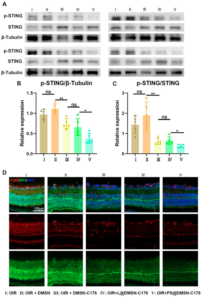
Retinal neovascularization (RNV), a typical pathological manifestation involved in most neovascular diseases, causes retinal detachment, vision loss, and ultimately irreversible blindness. Repeated intravitreal injections of anti-VEGF drugs were developed against RNV, with limitations of incomplete responses and adverse effects. Therefore, a new treatment with a better curative effect and more prolonged dosage is demanding. Here, we induced macrophage polarization to anti-inflammatory M2 phenotype by inhibiting cGAS-STING signaling with an antagonist C176, appreciating the role of cGAS-STING signaling in the retina in pro-inflammatory M1 polarization. C176-loaded and phosphatidylserine-modified dendritic mesoporous silica nanoparticles were constructed and examined by a single intravitreal injection. The biosafe nanoparticles were phagocytosed by retinal macrophages through a phosphatidylserine-mediated "eat me" signal, which persistently release C176 to suppress STING signaling and thereby promote macrophage M2 polarization specifically. A single dosage can effectively alleviate pathological angiogenesis phenotypes in murine oxygen-induced retinopathy models. In conclusion, these C176-loaded nanoparticles with enhanced cell uptake and long-lasting STING inhibition effects might serve as a promising way for treating RNV.

Keywords: Macrophage polarization; Nanocarrier; Retinal neovascularization; cGAS-STING pathway.


Conflict of interest statement

The authors declare that they have no known competing financial interests or personal relationships that could have appeared to influence the work reported in this paper.

Figures

Scheme 1
Scheme 1
The schematic illustration of the synthetic procedure and therapeutic mechanism of PS@DMSN-C176.
Fig. 1
Fig. 1
The characterization of PS@DMSN-C176. TEM images of (A) DMSN and (B) PS@DMSN-C176. (C) Hydrodynamic diameter and (D) Zeta potential of DMSN, L@DMSN-C176 and PS@DMSN-C176 measured by DLS (n = 3). (E) The entrapment efficiency and loading efficiency with different C176 feeding concentrations (n = 3). (F) Cumulative release of C-176 from PS@DMSN-C176 NPs (n = 3). Data were presented as mean ± SD.
Fig. 2
Fig. 2
Cell uptake and cytotoxicity of PS@DMSN-C176. (A) Phagocytic ratios of PS@DMSN-DID contain 0 %, 20 % and 40 % PS (n = 3). (B) Mean fluorescence intensity of cells after incubation with PS@DMSN-DID for 2 h, 4 h and 6 h (n = 3). (C) Cell viability of RAW264.7 cells incubated with different concentrations of PS@DMSN-C176 for 24 h (n = 3). (D) Representative images of cell uptake after 6 h incubation with PS@DMSN-DID. Data were presented as mean ± SD. ns means no significant difference. *p < 0.05, ****p < 0.0001.
Fig. 3
Fig. 3
PS@DMSN-C176 promotes the M2 polarization and inhibits M1 polarization in vitro. (A) Typical flow cytometry data and corresponding quantification of (B) iNos + CD206- (M1 marker) and (C) iNos-CD206+ (M2 marker) cell populations after LPS stimulation and indicated treatments (n = 3). The mRNA expression levels of anti-inflammatory cytokines (D) Arg-1, (E) IL-4, and pro-inflammatory cytokines (F) IL-1β, (G) IL-6. (H) Immunofluorescence of IL-10 and TNFα in RAW264.7 cells (n = 3). Data were presented as mean ± SD. ns means no significant difference. *p < 0.05, **p < 0.01, ***p < 0.001, ****p < 0.0001.
Fig. 4
Fig. 4
PS@DMSN-C176 inhibits cGAS-Sting pathway in vitro. (A) Western blot and protein levels of (B) p-STING, (C) p-TBK1, (D) TBK1, (E) p-IRF3, (F) IRF3 in RAW264.7 cells quantified by densitometry and normalized by β-tubulin levels (n = 3 to 5). (G) Immunofluorescence of IRF3 in Raw264.7 cells after different treatments. Data were presented as mean ± SD. ns means no significant difference. *p < 0.05, **p < 0.01, ***p < 0.001.
Fig. 5
Fig. 5
PS@DMSN-C176 attenuates neovascularization, vaso-obliteration and vascular leakage in the OIR model. (A) The schematic illustration of the OIR model. (B) Representative images of IB4-stained retinal flat mounts and the corresponding vaso-obliteration areas and neovascular areas (white areas within retinas). (C) H&E staining and histological analysis of infiltration of neovascular nuclei in the OIR retinas. Black arrows indicate neovascular nuclei on the vitreal side of the inner limiting membrane. (D) and (E) Vaso-obliteration and neovascular areas quantified at P17 (n = 6 to 7). (F) Quantitative analysis of the number of neovascular nuclei in the OIR retinas at P17 (n = 7 to 9). (G), (H) and (I) Western blot and protein levels of albumin and VEGF in mice retina at P17 quantified by densitometry and normalized by β-tubulin levels (n = 4). Data were presented as mean ± SD. ns means no significant difference. *p < 0.05, **p < 0.01, and ***p < 0.001.
Fig. 6
Fig. 6
PS@DMSN-C176 promotes M2 polarization and inhibits M1 polarization in vivo. (A) and (C) Representative images of retinal flat mounts stained with IB4, F4/80, TNF-α and IL-10. (B) and (D) Fluorescence intensity of F4/80, TNF-α, IL-10 staining was calculated by ImageJ software (n = 5 to 8). (E to H) qRT-PCR analysis of IL-1β, IL-6, iNos and CD206 mRNA levels in mice retina at P17 (n = 3). Data were presented as mean ± SD. ns means no significant difference. *p < 0.05, **p < 0.01, and ***p < 0.001.
Fig. 7
Fig. 7
PS@DMSN-C176 inhibits the cGAS-STING pathway in OIR mice retina. (A) Western blot and protein levels of (B) p-STING and (C) STING in mice retina at P17 quantified by densitometry and normalized by β-tubulin levels (n = 5). (D) Retinal sections of OIR mice stained with F4/80, IRF3 and DAPI. Data were presented as mean ± SD. ns means no significant difference. *p < 0.05, **p < 0.01.

References

    1. Hartnett M.E. Pathophysiology and mechanisms of severe retinopathy of prematurity. Ophthalmology. 2015 Jan;122(1):200–210. - PMC - PubMed
    1. Wong T.Y., Cheung C.M.G., Larsen M., Sharma S., Simó R. Diabetic retinopathy. Nat. Rev. Dis. Prim. 2016 Mar;2 - PubMed
    1. Cheung N., Mitchell P., Wong T.Y. Diabetic retinopathy. Lancet. 2010;376(9735):124–136. - PubMed
    1. Li X., Xu G., Wang Y., Xu X., Liu X., Tang S., et al. Safety and efficacy of conbercept in neovascular age-related macular degeneration: results from a 12-month randomized phase 2 Study: AURORA study. Ophthalmology. 2014;121(9):1740–1747. - PubMed
    1. Heier J.S., Brown D.M., Chong V., Korobelnik J.F., Kaiser P.K., Nguyen Q.D., et al. Intravitreal aflibercept (VEGF trap-eye) in wet age-related macular degeneration. Ophthalmology. 2012 Dec;119(12):2537–2548. - PubMed