. 2024 May 24:15:1393096.
doi: 10.3389/fimmu.2024.1393096. eCollection 2024.

Interleukin-1 regulates follicular T cells during the germinal center reaction

Affiliations

Interleukin-1 regulates follicular T cells during the germinal center reaction

Aude Belbezier et al. Front Immunol. .

Abstract

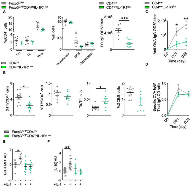
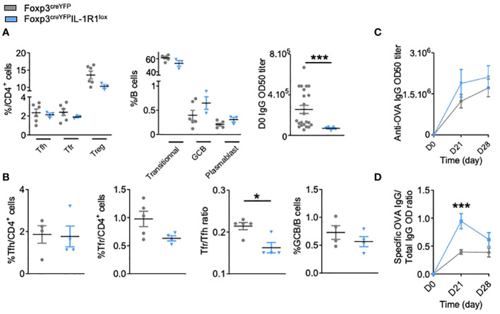
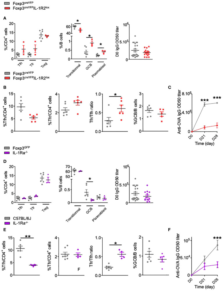
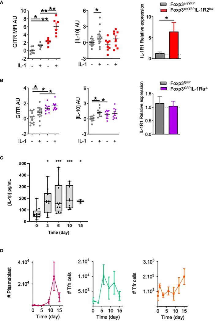
Introduction: Antibody production and the generation of memory B cells are regulated by T follicular helper (Tfh) and T follicular regulatory (Tfr) cells in germinal centers. However, the precise role of Tfr cells in controlling antibody production is still unclear. We have previously shown that both Tfh and Tfr cells express the IL-1R1 agonist receptor, whereas only Tfr cells express the IL-1R2 decoy and IL-1Ra antagonist receptors. We aimed to investigate the role of IL-1 receptors in the regulation of B cell responses by Tfh and Tfr.

Methods: We generated mice with IL-1 receptors inactivated in Tfh or Tfr and measured antibody production and cell activation after immunisation.

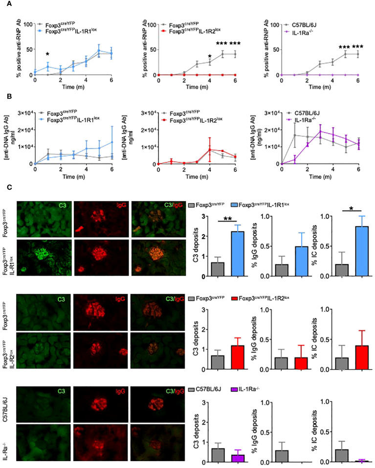
Results: While IL-1β levels are increased in the draining lymph node after immunisation, antigen-specific antibody levels and cell phenotypes indicated that IL-1β can activate both Tfh and Tfr cells through IL-1R1 stimulation. Surprisingly, expression of IL-1R2 and IL-1Ra on Tfr cells does not block IL-1 activation of Tfh cells, but rather prevents IL-1/IL-1R1-mediated early activation of Tfr cells. IL-1Rs also regulate the antibody response to autoantigens and its associated pathophysiology in an experimental lupus model.

Discussion: Collectively, our results show that IL-1 inhibitory receptors expressed by Tfr cells prevent their own activation and suppressive function, thus licensing IL-1-mediated activation of Tfh cells after immunisation. Further mechanistic studies should unravel these complex interactions between IL-1β and follicular helper and regulatory T cells and provide new avenues for therapeutic intervention.

Keywords: adaptive immunity; autoimmune diseases; humoral response; immunotherapy; vaccination.


Conflict of interest statement

The authors declare that the research was conducted in the absence of any commercial or financial relationships that could be construed as a potential conflict of interest.

Figures

Figure 1
Figure 1
IL-1 regulates Tfh cell but also Tfr cell activation. (A) Spectral flow cytometry quantification of the percentages of CD4+ T and B cell populations within SLOs and circulating IgG antibody levels before any immunization in CD4creIL-1R1lox (green triangle) and CD4cre (grey circle) mice. (B) Flow cytometry quantification of Tfh, Tfr, GCB cells and Tfr/Tfh ratio 28 days after intraperitoneal immunization with OVA-Alum in CD4creIL-1R1lox (green triangle) and CD4cre mice (grey circle). (C) Circulating anti-OVA IgG antibody level after intraperitoneal immunization with OVA-Alum in CD4creIL-1R1lox (green triangle) and CD4cre (grey circle) mice from D0 to D28. (D) Ratio of specific anti-OVA IgG Ab/total IgG Ab after OVA immunization in CD4creIL-1R1lox (green triangle) and CD4cre (grey circle) mice from D0 to D28. (E, F) Histograms comparing the MFI of GITR (E) and the level of IL-10 production (F) of Tfr cells from Foxp3GFPCD4creIL-1R1lox (green triangle) and Foxp3GFP (grey circle) mice cultured with or without IL-1. GITR MFI and IL-10 levels are expressed in arbitrary units that correspond to data centered and reduced relative to Tfr from Foxp3GFP (grey) mice grown without IL-1. *P < 0.05, **P < 0.01, ***P < 0.005, Mann-Whitney U test for unpaired data and Wilcoxon paired test for paired data. (B–F) Data are representative of three independent experiments.
Figure 2
Figure 2
IL-1R1 receptor expressed by Tfr cells is functional and critical for the regulation of the humoral immune response. (A) Spectral flow cytometry quantification of CD4+ T and B cell populations within SLOs and circulating IgG antibody levels before any immunization in Foxp3creYFPIL-1R1lox (blue triangle) and Foxp3creYFP mice (grey circle). (B) Flow cytometry quantification of Tfh, Tfr, GCB cells and Tfr/Tfh ratio 28 days after intraperitoneal immunization with OVA-Alum in Foxp3creYFPIL-1R1lox (blue triangle) and Foxp3creYFP mice (grey circle). (C) Circulating anti-OVA IgG antibody level after intraperitoneal immunization with OVA-Alum in Foxp3creYFPIL-1R1lox (blue triangle) and Foxp3creYFP mice (grey circle) from D0 to D28. (D) Ratio of specific anti-OVA IgG Ab/total IgG Ab after OVA immunization in Foxp3creYFPIL-1R1lox (blue triangle) and Foxp3creYFP mice (grey circle) at the times indicated. *P < 0.05, ***P < 0.005, Mann-Whitney U test. (B–D) Data are representative of three independent experiments.
Figure 3
Figure 3
IL-1R2 and IL-1Ra antagonist receptor are functional and critical for the regulation of the humoral immune response. (A) Spectral flow cytometry quantification of CD4+ T and B cell populations within SLOs and circulating IgG antibody levels before any immunization in Foxp3creYFPIL-1R2lox (red square) and Foxp3creYFP (grey circle) mice. (B) Flow cytometry quantification of Tfh, Tfr, GCB cells and Tfr/Tfh ratio 28 days after intraperitoneal immunization with OVA-Alum and (C) circulating anti-OVA IgG antibody level after intraperitoneal immunization with OVA-Alum in Foxp3creYFPIL-1R2lox (red square) and Foxp3creYFP (grey circle) mice from D0 to D28. (D) Quantification of CD4+ T, B cell subsets within SLOs and circulating IgG antibody levels before any immunization in Foxp3GFPIL-1Ra-/- (purple diamond) and Foxp3GFPIL-1raWT (grey circle) mice. (E) Proportion of Tfh, Tfr, GCB cells and Tfr/Tfh ratio 28 days after intraperitoneal immunization with OVA-Alum and (F) circulating anti-OVA IgG antibody level after intraperitoneal immunization with OVA-Alum in IL-1Ra-/- (purple diamond) and Foxp3GFP (grey circle) mice. *P < 0.05, **P < 0.01, ***P < 0.005, Mann-Whitney U test. (B, C, E, F) Data are representative of three independent experiments.
Figure 4
Figure 4
IL-1R2 and IL-1Ra inhibitory receptors prevent IL-1β-mediated activation of Tfr cells. Quantification of GITR expression, IL-10 production and IL-1R1 expression by Tfr cells from (A) Foxp3creYFPIL-1R2lox (red square) and Foxp3creYFP (B) Foxp3GFPIL-1Ra-/- mice (purple diamond) and Foxp3GFP mice (B) (grey circle), activated with or without IL-1. GITR MFI and IL-10 levels are expressed in arbitrary units that correspond to data centered and reduced relative to Tfr from respective control mice cultivated without IL-1. IL-1R1 expression is evaluated as the relative expression corresponding to the IL-1R1 expression level of the Tfr cell population compared to its relative level in control mice. (C, D) Kinetics of IL-1β production (grey) (C), plasmablasts (pink), Tfh cells (green) and Tfr cells (orange) (D) in draining lymph nodes from C57BL/6 mice at D0, D3, D6, D9, D12 and D15 after subcutaneous Alum-OVA immunization. Data represent the median of 3 mice ± SEM. *P < 0.05, **P < 0.01, ***P < 0.005, Mann-Whitney U test for unpaired data and Wilcoxon paired test for paired data. (A, B) Data are representative of three independent experiments. #: number of indicated cells.
Figure 5
Figure 5
Deletion of IL-1R1 on Tfr cells does not alter the risk of autoimmunity, but deletion of IL-1R2 and IL-1Ra on Tfr cells prevents autoimmune disease. (A) Proportion of mice with positive anti-RNP Ab after pristane immunization in Foxp3creYFPIL-1R1lox (blue, n= 6), Foxp3creYFPIL-1R2lox (red, n= 6), IL-1Ra-/- (purple, n= 6) and control (grey, Foxp3YFP, Foxp3YFP or and Foxp3GFP respectively, n= 6) mice. (B) Circulating anti-DNA IgG antibody level after pristane immunization in Foxp3creYFPIL-1R1lox (blue, n= 6), Foxp3creYFPIL-1R2lox (red, n= 6), IL-1Ra-/- (purple, n= 6) and control (grey, Foxp3YFP, Foxp3YFP or and Foxp3GFP respectively, n= 6) mice. (C) Deposition of C3 (green) and IgG (red) in glomeruli of kidney tissues from Foxp3creYFPIL-1R1lox (blue, n=6), Foxp3creYFPIL-1R2lox (red, n=5), IL-1Ra-/- (purple, n=5) and control (grey, Foxp3creYFPand Foxp3GFP, n=10) mice exposed to pristane. Intensity of C3 deposition is evaluated using a scale ranging from 0: absent, 1: weak, 2: moderate to 3: intense and IgG deposition is defined as an IgG immunofluorescence score greater than 2. The proportion of mice with immune complex deposition is defined as IgG and C3 immunofluorescence score greater than 2 in Foxp3creYFPIL-1R1lox (blue), Foxp3creYFPIL-1R2lox (red), IL-1Ra-/- (purple) and control (grey, Foxp3YFP and Foxp3GFP) mice after 6 months of exposure to pristane. *P < 0.05, **P < 0.01, ***P < 0.005, Mann-Whitney U test. (A, B) Data are representative of three independent experiments. .

Similar articles

Cited by

  • IL-1β signaling modulates T follicular helper and regulatory cells in human lymphoid tissues.
    Vaineau R, Jeger-Madiot R, Ali-Moussa S, Prudhomme L, Debarnot H, Coatnoan N, Dubois J, Binvignat M, Vantomme H, Gouritin B, Fourcade G, Engeroff P, Belbézier A, Luscan R, Denoyelle F, Lorenzon R, Ribet C, Rosenzwajg M, Bellier B, Klatzmann D, Tchitchek N, Graff-Dubois S. Vaineau R, et al. JCI Insight. 2025 May 20;10(12):e188724. doi: 10.1172/jci.insight.188724. eCollection 2025 Jun 23. JCI Insight. 2025. PMID: 40392614 Free PMC article.

References

    1. Chan AH, Schroder K. Inflammasome signaling and regulation of interleukin-1 family cytokines. J Exp Med. (2020) 217:e20190314. doi: 10.1084/jem.20190314 - DOI - PMC - PubMed
    1. Dinarello CA. A clinical perspective of IL-1β as the gatekeeper of inflammation. Eur J Immunol. (2011) 41:1203–17. doi: 10.1002/eji.201141550 - DOI - PubMed
    1. Garlanda C, Dinarello CA, Mantovani A. The interleukin-1 family: back to the future. Immunity. (2013) 39:1003–18. doi: 10.1016/j.immuni.2013.11.010 - DOI - PMC - PubMed
    1. Cohen I, Rider P, Carmi Y, Braiman A, Dotan S, White MR, et al. . Differential release of chromatin-bound IL-1α discriminates between necrotic and apoptotic cell death by the ability to induce sterile inflammation. Proc Natl Acad Sci. (2010) 107:2574–9. doi: 10.1073/pnas.0915018107 - DOI - PMC - PubMed
    1. Dower SK, Kronheim SR, March CJ, Conlon PJ, Hopp TP, Gillis S, et al. . Detection and characterization of high affinity plasma membrane receptors for human interleukin 1. J Exp Med. (1985) 162:501–15. doi: 10.1084/jem.162.2.501 - DOI - PMC - PubMed

MeSH terms

LinkOut - more resources