. 2024 May 24:15:1363457.
doi: 10.3389/fimmu.2024.1363457. eCollection 2024.

Oral curcumin ameliorates acute murine campylobacteriosis

Affiliations

Oral curcumin ameliorates acute murine campylobacteriosis

Markus M Heimesaat et al. Front Immunol. .

Abstract

Introduction: Human infections with the food-borne enteropathogen Campylobacter jejuni are responsible for increasing incidences of acute campylobacteriosis cases worldwide. Since antibiotic treatment is usually not indicated and the severity of the enteritis directly correlates with the risk of developing serious autoimmune disease later-on, novel antibiotics-independent intervention strategies with non-toxic compounds to ameliorate and even prevent campylobacteriosis are utmost wanted. Given its known pleiotropic health-promoting properties, curcumin constitutes such a promising candidate molecule. In our actual preclinical placebo-controlled intervention trial, we tested the anti-microbial and anti-inflammatory effects of oral curcumin pretreatment during acute experimental campylobacteriosis.

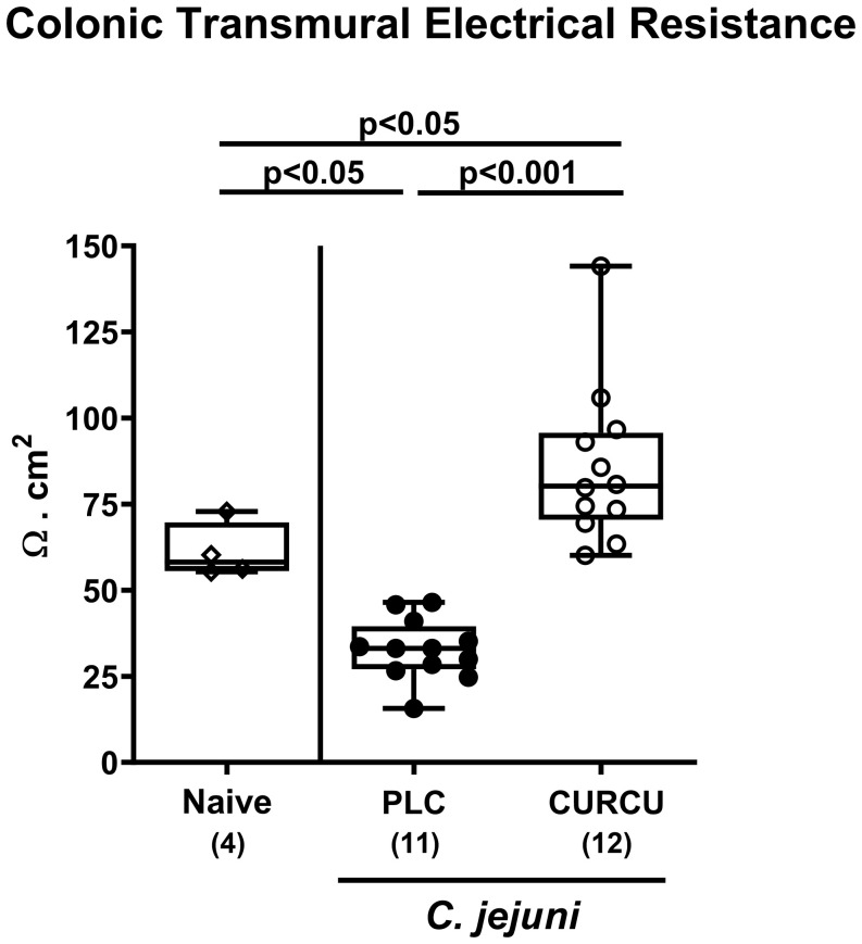
Methods: Therefore, secondary abiotic IL-10-/- mice were challenged with synthetic curcumin via the drinking water starting a week prior oral C. jejuni infection. To assess anti-pathogenic, clinical, immune-modulatory, and functional effects of curcumin prophylaxis, gastrointestinal C. jejuni bacteria were cultured, clinical signs and colonic histopathological changes quantitated, pro-inflammatory immune cell responses determined by in situ immunohistochemistry and intestinal, extra-intestinal and systemic pro-inflammatory mediator measurements, and finally, intestinal epithelial barrier function tested by electrophysiological resistance analysis of colonic ex vivo biopsies in the Ussing chamber.

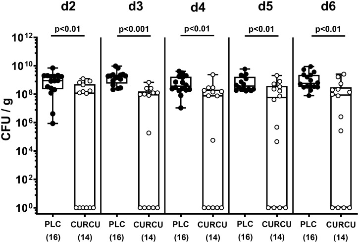
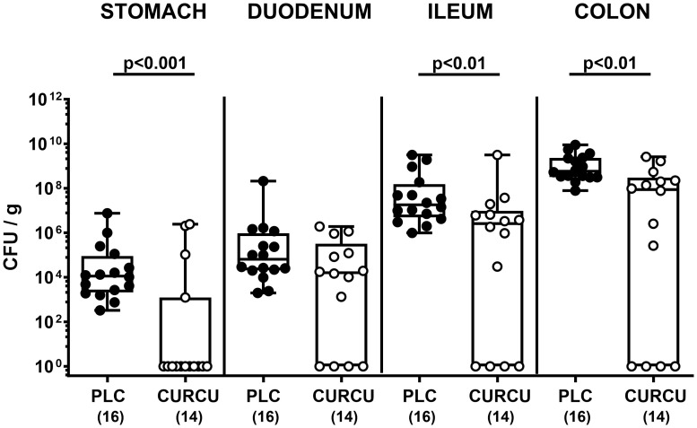
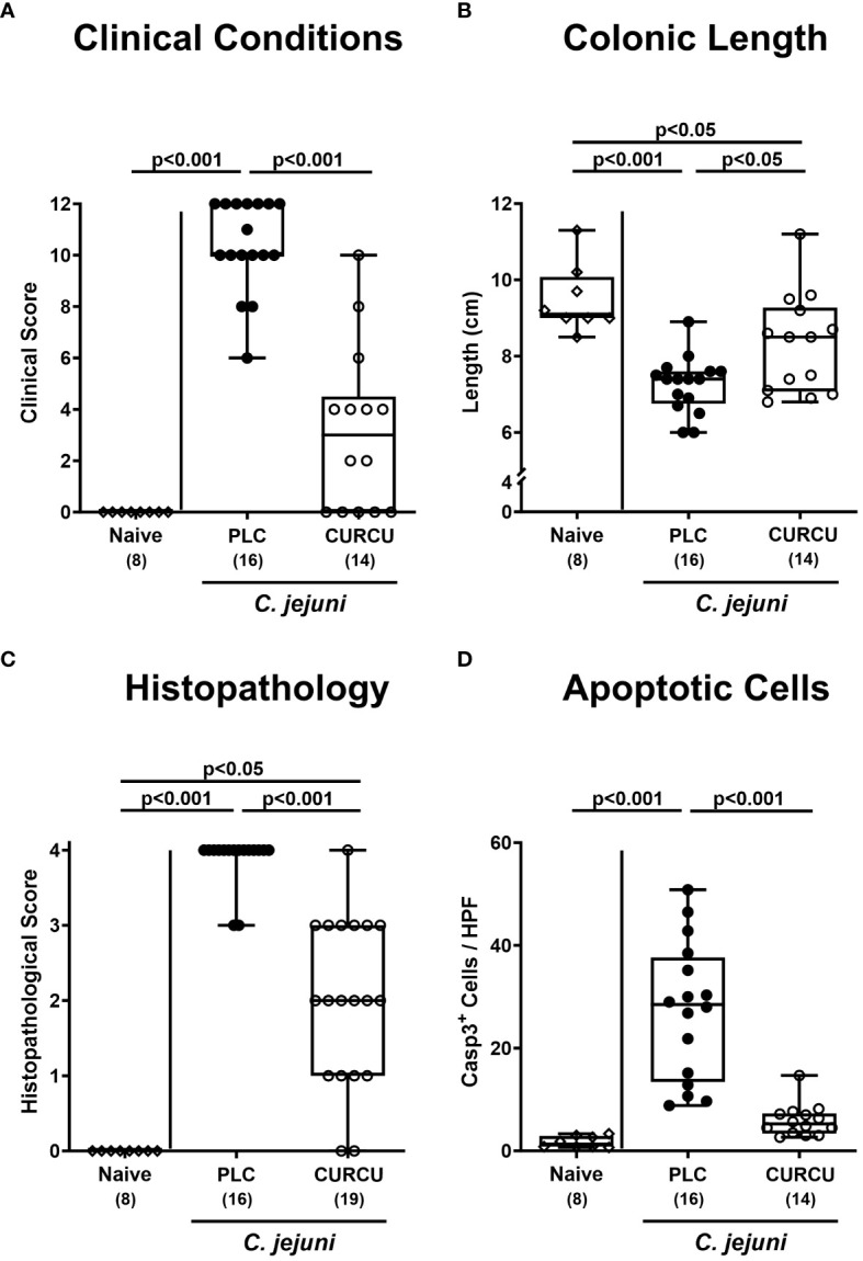
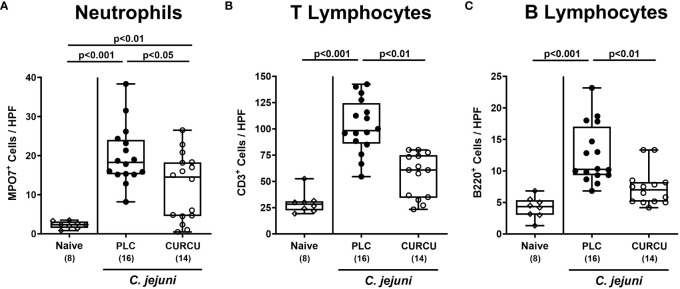
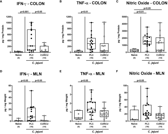
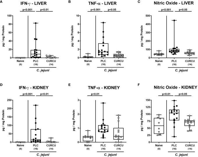
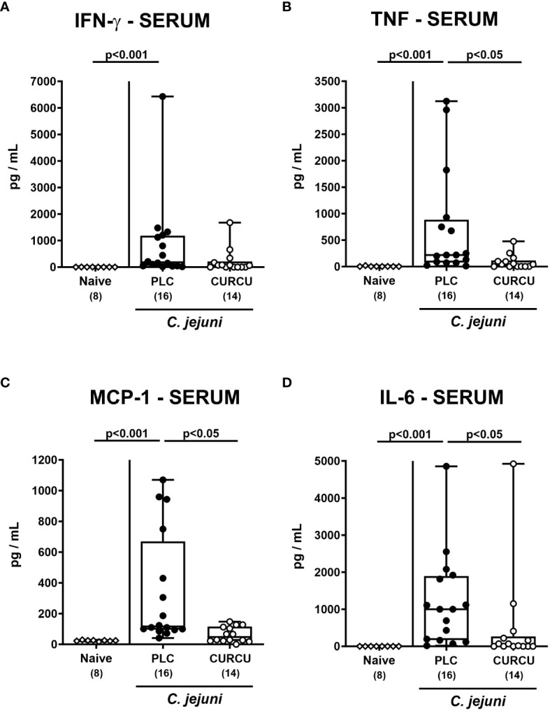
Results and discussion: Whereas placebo counterparts were suffering from severe enterocolitis characterized by wasting symptoms and bloody diarrhea on day 6 post-infection, curcumin pretreated mice, however, were clinically far less compromised and displayed less severe microscopic inflammatory sequelae such as histopathological changes and epithelial cell apoptosis in the colon. In addition, curcumin pretreatment could mitigate pro-inflammatory innate and adaptive immune responses in the intestinal tract and importantly, rescue colonic epithelial barrier integrity upon C. jejuni infection. Remarkably, the disease-mitigating effects of exogenous curcumin was also observed in organs beyond the infected intestines and strikingly, even systemically given basal hepatic, renal, and serum concentrations of pro-inflammatory mediators measured in curcumin pretreated mice on day 6 post-infection. In conclusion, the anti-Campylobacter and disease-mitigating including anti-inflammatory effects upon oral curcumin application observed here highlight the polyphenolic compound as a promising antibiotics-independent option for the prevention from severe acute campylobacteriosis and its potential post-infectious complications.

Keywords: Campylobacter jejuni; acute enterocolitis; campylobacteriosis model; curcumin; host-pathogen interaction; polyphenols; preclinical placebo-controlled intervention study; secondary abiotic IL-10 -/-mice.


Conflict of interest statement

The authors declare that the research was conducted in the absence of any commercial or financial relationships that could be construed as a potential conflict of interest. The author(s) declared that they were an editorial board member of Frontiers, at the time of submission. This had no impact on the peer review process and the final decision.

Figures

Figure 1
Figure 1
Fecal pathogen shedding over time following C. jejuni infection of mice with curcumin prophylaxis. Secondary abiotic IL-10-/- mice were pretreated with curcumin (CURCU; open circles) or placebo (PLC; closed circles) via the drinking water starting 7 days prior peroral infection with C. jejuni 81–176 strain on day (d) 0 and d1. Fecal C. jejuni numbers were determined daily post-infection by culture (in colony forming units per gram; CFU/g). Box plots (25th and 75th percentiles), whiskers (minimum and maximum values), medians (black bar in boxes), numbers of analyzed mice pooled from 3 independent experiments (in parentheses), and significance levels (p values) determined by the Mann-Whitney test are indicated.
Figure 2
Figure 2
Gastrointestinal pathogen counts following C. jejuni infection of mice with curcumin prophylaxis. Secondary abiotic IL-10-/- mice were pretreated with curcumin (CURCU; open circles) or placebo (PLC; closed circles) via the drinking water starting 7 days prior peroral infection with C. jejuni 81–176 strain on days 0 and 1. At necropsy (i.e., day 6 post-infection), luminal C. jejuni bacteria were quantified in the gastrointestinal tract (as indicated) by culture (in colony forming units per gram; CFU/g). Box plots (25th and 75th percentiles), whiskers (minimum and maximum values), medians (black bar in boxes), numbers of analyzed mice pooled from 3 independent experiments (in parentheses), and significance levels (p values) determined by the Mann-Whitney test are indicated.
Figure 3
Figure 3
Macroscopic and microscopic inflammatory signs following curcumin pretreatment of C. jejuni infected mice. Secondary abiotic IL-10-/- mice were pretreated with curcumin (CURCU; open circles) or placebo (PLC; closed circles) via the drinking water starting 7 days prior peroral infection with C. jejuni 81–176 strain on days 0 and 1. The macroscopic inflammatory signs including (A) the clinical conditions as quantified with a campylobacteriosis scoring system (see methods) and (B) the colonic lengths as measured with a ruler (in cm) were surveyed on day 6 post-infection. Furthermore, the microscopic inflammatory changes were quantitatively assessed in colonic paraffin sections with (C) histopathological scores (see methods) and (D) average numbers of apoptotic epithelial cells (positive for caspase3, Casp3) from 6 high power fields (HPF, 400-times magnification) per animal. Naive mice (open diamonds) served as non-infected controls without prophylaxis. Box plots (25th and 75th percentiles), whiskers (minimum and maximum values), medians (black bar in boxes), numbers of analyzed mice pooled from 3 independent experiments (in parentheses), and significance levels (p values) determined by the Kruskal-Wallis test with Dunn’s post-hoc test (A, C) or by the one sided ANOVA test with Tukey’s post-hoc test (B, D) are indicated.
Figure 4
Figure 4
Colonic immune cell responses following curcumin pretreatment of C. jejuni infected mice. Secondary abiotic IL-10-/- mice were pretreated with curcumin (CURCU; open circles) or placebo (PLC; closed circles) via the drinking water starting 7 days prior peroral infection with C. jejuni 81–176 strain on days 0 and 1. On day 6 post-infection, the average numbers of (A) neutrophils (MPO7+), (B) T lymphocytes (CD3+), and (C) B lymphocytes (B220+) were determined in the colonic mucosa and lamina propria from 6 high power fields (HPF, 400-times magnification) per animal in immunohistochemically stained paraffin sections. Naive mice (open diamonds) served as non-infected controls without prophylaxis. Box plots (25th and 75th percentiles), whiskers (minimum and maximum values), medians (black bar in boxes), numbers of analyzed mice pooled from 3 independent experiments (in parentheses), and significance levels (p values) determined by the Kruskal-Wallis test with Dunn’s post-hoc test are indicated.
Figure 5
Figure 5
Intestinal pro-inflammatory mediators following curcumin pretreatment of C. jejuni infected mice. Secondary abiotic IL-10-/- mice were pretreated with curcumin (CURCU; open circles) or placebo (PLC; closed circles) via the drinking water starting 7 days prior peroral infection with C. jejuni 81–176 strain on days 0 and 1. (A, D) IFN-γ, (B, E) TNF-α, and (C, F) nitric oxide concentrations were measured in supernatants of ex vivo biopsies derived from the colon (A–C) and mesenteric lymph nodes (MLN; D-F) on day 6 post-infection. Naive mice (open diamonds) served as non-infected controls without prophylaxis. Box plots (25th and 75th percentiles), whiskers (minimum and maximum values), medians (black bar in boxes), numbers of analyzed mice pooled from 3 independent experiments (in parentheses), and significance levels (p values) determined by the Kruskal-Wallis test with Dunn’s post-hoc test (A, B, D–F) or by the one-sided ANOVA test with Tukey’s post-hoc test (C) are indicated.
Figure 6
Figure 6
Colonic epithelial barrier function following curcumin pretreatment of C. jejuni infected mice. Secondary abiotic IL-10-/- mice were pretreated with curcumin (CURCU; open circles) or placebo (PLC; closed circles) via the drinking water starting 7 days prior peroral infection with C. jejuni 81–176 strain on days 0 and 1. On day 6 post-infection, the transmural electrical resistance of the distal colon was measured in Ussing chambers. Naive mice (open diamonds) served as non-infected controls without prophylaxis. Box plots (25th and 75th percentiles), whiskers (minimum and maximum values), medians (black bar in boxes), numbers of analyzed mice pooled from 3 independent experiments (in parentheses), and significance levels (p values) determined by the one-sided ANOVA test with Tukey’s post-hoc test are indicated.
Figure 7
Figure 7
Extra-intestinal pro-inflammatory mediators following curcumin pretreatment of C. jejuni infected mice. Secondary abiotic IL-10-/- mice were pretreated with curcumin (CURCU; open circles) or placebo (PLC; closed circles) via the drinking water starting 7 days prior peroral infection with C. jejuni 81–176 strain on days 0 and 1. (A, D) IFN-γ, (B, E) TNF-α, and (C, F) nitric oxide concentrations were measured in supernatants of ex vivo biopsies derived from the liver (A–C) and kidneys (D–F) on day 6 post-infection. Naive mice (open diamonds) served as non-infected controls without prophylaxis. Box plots (25th and 75th percentiles), whiskers (minimum and maximum values), medians (black bar in boxes), numbers of analyzed mice pooled from 3 independent experiments (in parentheses), and significance levels (p values) determined by the Kruskal-Wallis test with Dunn’s post-hoc test (A–C, E, F) or by the one sided ANOVA test with Tukey’s post-hoc test (D) are indicated.
Figure 8
Figure 8
Systemic pro-inflammatory mediators following curcumin pretreatment of C. jejuni infected mice. Secondary abiotic IL-10-/- mice were pretreated with curcumin (CURCU; open circles) or placebo (PLC; closed circles) via the drinking water starting 7 days prior peroral infection with C. jejuni 81–176 strain on days 0 and 1. (A) IFN-γ, (B) TNF-α, (C) MCP-1, and (D) IL-6 concentrations were measured in serum samples taken on day 6 post-infection. Naive mice (open diamonds) served as non-infected controls without prophylaxis. Box plots (25th and 75th percentiles), whiskers (minimum and maximum values), medians (black bar in boxes), numbers of analyzed mice pooled from 3 independent experiments (in parentheses), and significance levels (p values) determined by the Kruskal-Wallis test with Dunn’s post-hoc test are indicated.

Similar articles

Cited by

References

    1. WHO. World Health Organisation . Campylobacter (2020). Available online at: https://www.who.int/news-room/fact-sheets/detail/campylobacter.
    1. European Food Safety A. European Centre for Disease P, Control . The european union summary report on antimicrobial resistance in zoonotic and indicator bacteria from humans, animals and food in 2019–2020. EFSA J. (2022) 20:e07209. doi: 10.2903/j.efsa.2022.7209 - DOI - PMC - PubMed
    1. Wilson DJ, Gabriel E, Leatherbarrow AJ, Cheesbrough J, Gee S, Bolton E, et al. . Tracing the source of campylobacteriosis. PloS Genet. (2008) 4:e1000203. doi: 10.1371/journal.pgen.1000203 - DOI - PMC - PubMed
    1. Fitzgerald C. Campylobacter. Clin Lab Med. (2015) 35:289–98. doi: 10.1016/j.cll.2015.03.001 - DOI - PubMed
    1. Silva J, Leite D, Fernandes M, Mena C, Gibbs PA, Teixeira P. Campylobacter spp. As a foodborne pathogen: A review. Front Microbiol. (2011) 2:200. doi: 10.3389/fmicb.2011.00200 - DOI - PMC - PubMed