

Roles of the APETALA3-3 ortholog in the petal identity specification and morphological differentiation in Delphinium anthriscifolium flowers
- PMID: 38855416
- PMCID: PMC11161261
- DOI: 10.1093/hr/uhae097
Roles of the APETALA3-3 ortholog in the petal identity specification and morphological differentiation in Delphinium anthriscifolium flowers
Abstract
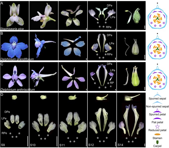
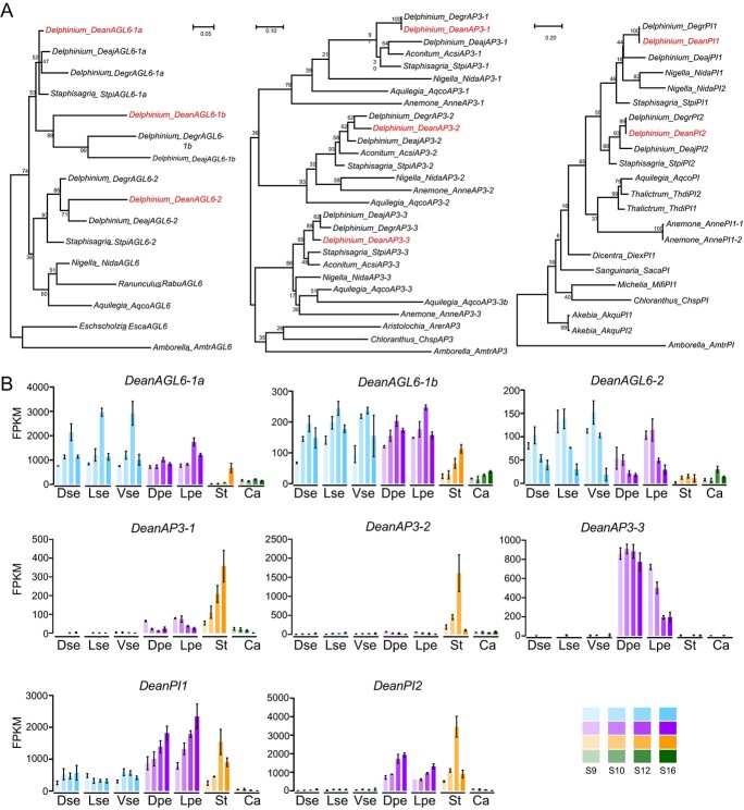
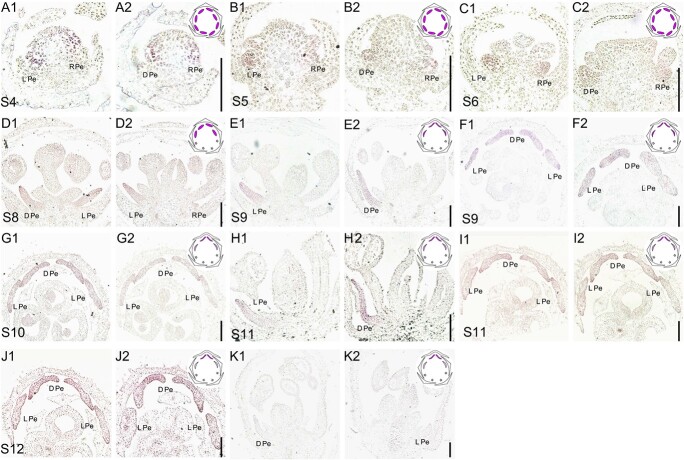
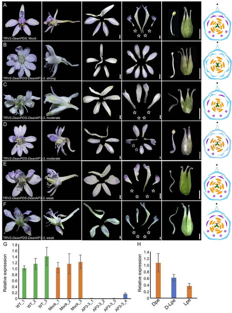
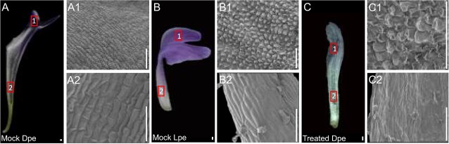
The genus Delphinium (Ranunculaceae) with its unique and highly complex floral structure is an ideal system to address some key questions in terms of morphological and evolutionary studies in flowers. In D. anthriscifolium, for example, the original eight petal primordia differentiate into three types at maturity (i.e., two dorsal spurred, two lateral flat, and four ventral reduced petals). The mechanisms underlying their identity determination and morphological differentiation remain unclear. Here, through a comprehensive approach combining digital gene expression (DGE) profiles, in situ hybridization, and virus-induced gene silencing (VIGS), we explore the role of the APETALLATA3-3 (AP3-3) ortholog in D. anthriscifolium. Our findings reveal that the DeanAP3-3 not only functions as a traditionally known petal identity gene but also plays a critical role in petal morphological differentiation. The DeanAP3-3 gene is expressed in all the petal primordia before their morphological differentiation at earlier stages, but shows a gradient expression level difference along the dorsventral floral axis, with higher expression level in the dorsal spurred petals, intermediate level in the lateral flat petals and lower level in the ventral reduced petals. VIGS experiments revealed that flowers with strong phenotypic changes showed a complete transformation of all the three types of petals into non-spurred sepals. However, in the flowers with moderate phenotypic changes, the transformation of spurred petals into flat petals is associated with moderate silencing of the DeanAP3-3 gene, suggesting a significant impact of expression level on petal morphological differentiation. This research also shed some insights into the role of changes in gene expression levels on morphological differentiation in plants.
© The Author(s) 2024. Published by Oxford University Press on behalf of Nanjing Agricultural University.
Conflict of interest statement
The authors declare no conflict of interest.
Figures
Similar articles
-
Delphinium as a model for development and evolution of complex zygomorphic flowers.Front Plant Sci. 2024 Aug 19;15:1453951. doi: 10.3389/fpls.2024.1453951. eCollection 2024. Front Plant Sci. 2024. PMID: 39224845 Free PMC article. Review.
-
The mechanism underlying asymmetric bending of lateral petals in Delphinium (Ranunculaceae).Curr Biol. 2024 Feb 26;34(4):755-768.e4. doi: 10.1016/j.cub.2024.01.004. Epub 2024 Jan 24. Curr Biol. 2024. PMID: 38272029
-
Disruption of the petal identity gene APETALA3-3 is highly correlated with loss of petals within the buttercup family (Ranunculaceae).Proc Natl Acad Sci U S A. 2013 Mar 26;110(13):5074-9. doi: 10.1073/pnas.1219690110. Epub 2013 Mar 11. Proc Natl Acad Sci U S A. 2013. PMID: 23479615 Free PMC article.
-
Delphinieae flowers originated from the rewiring of interactions between duplicated and diversified floral organ identity and symmetry genes.Plant Cell. 2023 Mar 15;35(3):994-1012. doi: 10.1093/plcell/koac368. Plant Cell. 2023. PMID: 36560915 Free PMC article.
-
Petal development and elaboration.J Exp Bot. 2022 Jun 2;73(11):3308-3318. doi: 10.1093/jxb/erac092. J Exp Bot. 2022. PMID: 35275176 Review.
Cited by
-
Delphinium as a model for development and evolution of complex zygomorphic flowers.Front Plant Sci. 2024 Aug 19;15:1453951. doi: 10.3389/fpls.2024.1453951. eCollection 2024. Front Plant Sci. 2024. PMID: 39224845 Free PMC article. Review.
-
Comparative case study of evolutionary insights and floral complexity in key early-diverging eudicot Ranunculales models.Front Plant Sci. 2024 Oct 30;15:1486301. doi: 10.3389/fpls.2024.1486301. eCollection 2024. Front Plant Sci. 2024. PMID: 39539296 Free PMC article. Review.
References
-
- Smyth DR. Evolution and genetic control of the floral ground plan. New Phytol. 2018;220:70–86 - PubMed
-
- Endress PK, Doyle JA. Reconstructing the ancestral angiosperm flower and its initial specializations. Am J Bot. 2009;96:22–66 - PubMed
-
- Keasar T. Patterns of flower complexity in plant communities. Annu Plant Rev. 2020;3:643–60
LinkOut - more resources
Full Text Sources
