. 2024 Jun 9:12:tkae035.
doi: 10.1093/burnst/tkae035. eCollection 2024.

Neuregulin-1, a member of the epidermal growth factor family, mitigates STING-mediated pyroptosis and necroptosis in ischaemic flaps

Affiliations

Neuregulin-1, a member of the epidermal growth factor family, mitigates STING-mediated pyroptosis and necroptosis in ischaemic flaps

Xuwei Zhu et al. Burns Trauma. .

Abstract

Background: Ensuring the survival of the distal end of a random flap during hypoperfusion (ischaemia) is difficult in clinical practice. Effective prevention of programmed cell death is a potential strategy for inhibiting ischaemic flap necrosis. The activation of stimulator of interferon genes (STING) pathway promotes inflammation and leads to cell death. The epidermal growth factor family member neuregulin-1 (NRG1) reduces cell death by activating the protein kinase B (AKT) signalling pathway. Moreover, AKT signalling negatively regulates STING activity. We aimed to verify the efficacy of NRG1 injection in protecting against flap necrosis. Additionally, we investigated whether NRG1 effectively enhances ischemic flap survival by inhibiting pyroptosis and necroptosis through STING suppression.

Methods: A random-pattern skin flap model was generated on the backs of C57BL/6 mice. The skin flap survival area was determined. The blood supply and vascular network of the flap was assessed by laser Doppler blood flow analysis. Cluster of differentiation 34 immunohistochemistry (IHC) and haematoxylin and eosin (H&E) staining of the flap sections revealed microvessels. Transcriptome sequencing analysis revealed the mechanism by which NRG1 promotes the survival of ischaemic flaps. The levels of angiogenesis, oxidative stress, necroptosis, pyroptosis and indicators associated with signalling pathways in flaps were examined by IHC, immunofluorescence and Western blotting. Packaging adeno-associated virus (AAV) was used to activate STING in flaps.

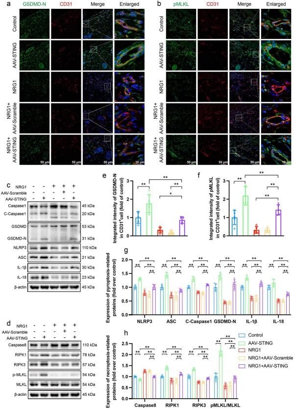
Results: NRG1 promoted the survival of ischaemic flaps. An increased subcutaneous vascular network and neovascularization were found in ischaemic flaps after the application of NRG1. Transcriptomic gene ontology enrichment analysis and protein level detection indicated that necroptosis, pyroptosis and STING activity were reduced in the NRG1 group. The phosphorylation of AKT and forkhead box O3a (FOXO3a) were increased after NRG1 treatment. The increased expression of STING in flaps induced by AAV reversed the therapeutic effect of NRG1. The ability of NRG1 to phosphorylate AKT-FOXO3a, inhibit STING and promote flap survival was abolished after the application of the AKT inhibitor MK2206.

Conclusions: NRG1 inhibits pyroptosis and necroptosis by activating the AKT-FOXO3a signalling pathway to suppress STING activation and promote ischaemic flap survival.

Keywords: Epidermal growth factor; Ischaemic flaps; Necroptosis; Neuregulin-1; Pyroptosis; STING.


Conflict of interest statement

None declared.

Figures

Figure 1
Figure 1
NRG1 increased the survival rate of ischaemic skin flaps. (a,b) Survial areas of control and NRG1 groups on days 1, 3 and 7 after surgery. (c,d) LDBF analysis on days 1 and 7. (e) Comparison of the percentage of survival area between two groups (n = 5). (f) Comparison of the LDBF analysis signals between two groups on day 7 (n = 5). (g) H&E staining of zone-II of the flaps in the two groups (scale bar = 50 μm, scale bar in the enlarged image = 10 μm). Yellow arrows indicate microvessels. (h) Immunohistochemical staining of CD34 (brown) in Zone-II of flaps in the two groups (scale bar = 50 μm, scale bar in the enlarged image = 10 μm). Yellow arrows indicate microvessels. (i) Immunohistochemical staining for SOD1 (brown) in zone-II of flaps in the two groups (scale bar = 50 μm, scale bar in the enlarged image = 10 μm). (j) Comparison of the vessel density (/mm 2) determined by H&E staining between two groups (n = 5). (k) Comparison of the number of CD34+ vessels (/mm 2) between two groups (n = 5). (l) Comparison of the integrated intensity of SOD1 between two groups (n = 5). Two-tailed, unpaired t tests were conducted and the data are presented as the means ± SD, **p < 0.01. NRG1 neuregulin-1, H&E haematoxylin and eosin, CD34 cluster of differentiation 34, SOD1 superoxide dismutase 1
Figure 2
Figure 2
NRG1 inhibited cell pyroptosis in ischaemic flaps. (a) GO analysis of the target genes showing the biological processes influenced by NRG1 treatment. (b) Western blot results of caspase1, GSDMD, ASC, NLRP3, IL-18 and IL-1β expression in control and NRG1 groups presented as typical images. β-Actin was utilized as a loading control. (c) Quantification of the protein levels of cleaved-caspase1, GSDMD-N, ASC, NLRP3, IL-18 and IL-1β (n = 5). (d) Immunofluorescence staining of caspase1 (green), CD31 (red) and DAPI (blue) (scale bar = 50 μm, scale bar in the enlarged image = 10 μm). (e) Immunofluorescence staining of GSDMD-N (red), CD31 (green) and DAPI (blue) (scale bar = 50 μm, scale bar in the enlarged image = 10 μm). (f,h) Comparison of the integrated intensities of caspase1 and GSDMD-N in CD31+ endothelial cells of the flap’s dermal layer between two groups (n = 5). (g) Schematic diagram of the flap tissue, with the top representing the epidermis layer, the middle representing the dermis layer and the bottom representing the subcutaneous layer. The section is positioned in the dermis layer. Two-tailed, unpaired t tests were performed and the data are presented as the means ± SD, *p < 0.05, **p < 0.01. NRG1 neuregulin1, GO gene ontology, CD31 cluster of differentiation 31, GSDMD gasdermin D, NLRP3 nucleotide-binding domain leucine-rich-containing family pyrin domain-containing-3, ASC adaptor apoptosis-associated speck-like protein containing a caspase-recruitment domain adaptor
Figure 3
Figure 3
NRG1 inhibited cell necroptosis in ischaemic flaps. (a) Western blot results of RIPK1, RIPK3, MLKL, pMLKL and caspase8 expression in control and NRG1 groups presented as typical images. β-Actin was utilized as a loading control. (b) Quantification of the protein levels of RIPK1, RIPK3, pMLKL/MLKL and caspase8 (n = 5). (c) Immunofluorescence staining of RIPK3 (green), CD31 (red) and DAPI (blue) (scale bar = 50 μm, scale bar in the enlarged image = 10 μm). (d) Immunofluorescence staining of pMLKL (green), CD31 (red) and DAPI (blue) (scale bar = 50 μm, scale bar in the enlarged image = 10 μm). (e,f) Comparison of the integrated intensities of RIPK3 and pMLKL in CD31+ endothelial cells of the flap’s dermal layer between two groups (n = 5). (g) schematic diagram of the flap tissue; the section positioned in the dermis layer. (h) Heatmap of genes upregulated or downregulated by NRG1 injection in mouse skin tissue. (i) Western blot results of pSTING, STING and cGAS expression in control and NRG1 groups presented as typical images. β-Actin was utilized as a loading control. (j) Quantification of the protein levels of pSTING, STING and cGAS (n = 5). (k) Immunofluorescence staining of STING (green) and DAPI staining (blue), scale bar = 10 μm. (l) Comparison of the integrated intensity of STING+ (green) cells between two groups (n = 5). Two-tailed, unpaired t tests were conducted and the data are presented as the means ± SD, *p < 0.05, **p < 0.01. NRG1 neuregulin-1, C control1, N neuregulin-1, DAPI 4,6-Diamino-2-phenyl indole, CD31 cluster of differentiation 31, RIPK receptor-interacting serine/threonine-protein kinase, MLKL mixed-lineage kinase domain-like, STING stimulator of interferon genes, cGAS cyclic GMP-AMP synthase
Figure 4
Figure 4
NRG1 inhibited STING activity in ischaemic flaps. (a,b,d) Immunofluorescence staining of STING (green) and DAPI staining (blue), scale bar = 10 μm. Comparison of the integrated intensity of STING+ (green) cells among control, AAV-STING, NRG1, NRG1 + AAV-scramble, and NRG1 + AAV-STING groups (n = 5). (c) Western blot results of pSTING, STING and cGAS expression in the five groups presented as typical images. β-Actin was utilized as a loading control. (e) Quantification of the protein levels of pSTING, STING and cGAS. (n = 5). Statistical analysis was performed using ANOVA with the least significant difference post hoc test or Dunnett’s T3 test. The data are presented as the means ± SD, *p < 0.05, **p < 0.01. NRG1 neuregulin-1, DAPI 4,6-diamino-2-phenyl indole, STING stimulator of interferon genes, cGAS cyclic GMP-AMP synthase, AAV adeno-associated virus
Figure 5
Figure 5
NRG1 promoted ischaemic flap survival by inhibiting STING activity. (a) Survival areas in control, AAV-STING, NRG1, NRG1 + AAV-scramble and NRG1 + AAV-STING groups on days 1, 3 and 7 after surgery. (b) LDBF analysis on days 1 and 7 after surgery. (c,d) H&E staining of skin tissues from zone-II of the flap in the five groups (scale bar = 50 μm, scale bar in the enlarged image = 10 μm). Yellow arrows indicating microvessels. (e) Comparison of the survival area among the five groups on day 7 (n = 5). (g) Comparison of the LDBF signals among the five groups on days 7 (n = 5). (g) Comparison of the mean vessel density (/mm2) among the five groups (n = 5). Statistical analysis was performed using ANOVA with least significant difference post hoc tests or Dunnett’s T3 test. The data are presented as the means ± SD, **p < 0.01. NRG1 neuregulin-1, H&E haematoxylin and eosin, AAV adeno-associated virus, STING stimulator of interferon genes
Figure 6
Figure 6
NRG1 decreased pyroptosis and necroptosis during ischaemic flap survival by inhibiting STING activity. (a) Immunofluorescence staining of GSDMD-N (green), CD31 (red) and DAPI staining (blue) (scale bar = 50 μm, scale bar in the enlarged image = 10 μm). (b) Immunofluorescence staining of pMLKL (green), CD31 (red) and DAPI (blue) (scale bar = 50 μm, scale bar in the enlarged image = 10 μm). (c) Western blot results of caspase1, GSDMD, ASC, NLRP3, IL-18 and IL-1β expression in control, AAV-STING, NRG1, NRG1 + AAV-scramble, and NRG1 + AAV-STING groups presented as typical images. β-Actin was utilized as a loading control. (d) Western blot results of caspase8, RIPK1, RIPK3, MLKL and pMLKL expression in five groups presented as typical images. β-Actin was used as a loading control. (e) Comparison of the integrated intensity of GSDMD-N among the five groups (n = 5). (f) Comparison of the integrated intensity of pMLKL among the five groups (n = 5). (g) Quantification of the protein levels of cleaved-caspase1, GSDMD-N, ASC, NLRP3, IL-18 and IL-1β (n = 5). (h) Quantification of the protein levels of caspase8, RIPK1, RIPK3 and pMLKL/MLKL (n = 5). Statistical analysis was performed using ANOVA with least significant difference post hoc tests or Dunnett’s T3 test. The data are presented as the means ± SD, *p < 0.05, **p < 0.01. NRG1 neuregulin-1, AAV adeno-associated virus, STING stimulator of interferon genes, CD31 cluster of differentiation 31, GSDMD gasdermin D, NLRP3 nucleotide-binding domain leucine-rich-containing family pyrin domain-containing-3, ASC adaptor apoptosis-associated speck-like protein containing a caspase-recruitment domain, RIPK receptor-interacting serine/threonine-protein kinase, MLKL mixed-lineage kinase domain-like
Figure 7
Figure 7
NRG1 inhibited STING activity via the AKT-FOXO3a signalling pathway. (a) KEGG enrichment plot displays pathways with statistically significant differences between the two groups related to AKT and NRG1. (b) Western blot results of AKT, pAKT, FOXO3a and pFOXO3a in control and NRG1 groups presented as typical images. β-Actin was utilized as a loading control. (c) Quantification of the protein levels of pAKT/AKT and pFOXO3a/FOXO3a (n = 5). (d) Survival areas of control, MK2206, NRG1 and NRG1 + MK2206 groups on days 1, 3 and 7 after surgery. (e) LDBF analysis of the four groups on days 1 and 7. (f) Comparison of the percentage of survival area on day 7 (n = 5). (g) Comparison of the LDBF signals among the four groups on day 7 (n = 5). (h) Immunofluorescence staining of FOXO3a (red) in skin tissue sections from dermal layer of the flap (scale bar = 10 μm). (i) Comparison of the FOXO3a+ cells in the cytoplasm among the four groups (n = 5). (j) Western blot results of AKT, pAKT, FOXO3a and pFOXO3a in the four groups presented as typical images. (k) Quantification of the protein levels of pAKT/AKT and pFOXO3a/FOXO3a (n = 5). (l) Western blot results of pSTING, STING and cGAS in the four groups presented as typical images. (m) Quantification of the protein levels of pSTING, STING and cGAS (n = 5). (n) Western blot results of GSDMD, caspase1, NLRP3, ASC, IL-1β and IL-18 in the four groups presented as typical images. (o) Quantification of the protein levels of GSDMD-N, cleaved-caspase1, NLRP3, ASC, IL-1β and IL-18 (n = 5). (p) Western blot results of caspase8, RIPK1, RIPK3, MLKL and pMLKL in the four groups presented as typical images. (q) Quantification of the protein levels of caspase8, RIPK1, RIPK3 and pMLKL/MLKL (n = 5). Statistical analysis was performed using ANOVA with least squares difference post hoc tests or Dunnett’s T3 test. Data are presented as means ± SD, ns stands for not significant, *p < 0.05, **p < 0.01. NRG1 neuregulin-1, KEGG Kyoto encyclopedia of genes and genomes, STING stimulator of interferon genes, cGAS cyclic GMP-AMP synthase, GSDMD gasdermin D, NLRP3 nucleotide-binding domain leucine-rich-containing family pyrin domain-containing-3, ASC adaptor apoptosis-associated speck-like protein containing a caspase-recruitment domain, RIPK receptor-interacting serine/threonine-protein kinase, MLKL mixed-lineage kinase domain-like, AKT protein kinase B, FOXO3a forkhead box O3a

Similar articles

Cited by

References

    1. Charafeddine AH, Drake R, McBride J. et al. Facelift: History and Anatomy. Clin Plast Surg 2019;46:505–13. - PubMed
    1. Summers BK, Siegle RJ. Facial cutaneous reconstructive surgery: general aesthetic principles. J Am Acad Dermatol 1993;29:669–81. - PubMed
    1. Chung S, Hazen A, Levine JP. et al. Vascularized acellular dermal matrix island flaps for the repair of abdominal muscle defects. Plast Reconstr Surg 2003;111:225–32. - PubMed
    1. Chen Z, Wu H, Yang J. et al. Activating Parkin-dependent mitophagy alleviates oxidative stress, apoptosis, and promotes random-pattern skin flaps survival. Communications Biology 2022;5:616. - PMC - PubMed
    1. Weinzierl A, Ampofo E, Menger MD. et al. Tissue-Protective Mechanisms of Bioactive Phytochemicals in Flap Surgery. Front Pharmacol 2022;13:864351. 10.3389/fphar.2022.864351. - DOI - PMC - PubMed