. 2024 May 31;13(5):720-735.
doi: 10.21037/tau-23-632. Epub 2024 May 24.

Interfascial planes as surgical landmarks for laparoscopic upper retroperitoneal surgery: a cadaveric and retrospectively clinical study

Affiliations

Interfascial planes as surgical landmarks for laparoscopic upper retroperitoneal surgery: a cadaveric and retrospectively clinical study

Jiayu Huang et al. Transl Androl Urol. .

Abstract

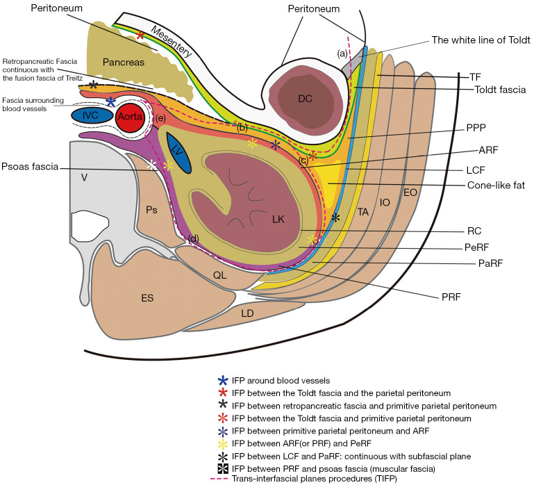
Background: Radiologists currently accept the concept of "interfascial plane (IFP)" to understand retroperitoneal anatomy, replacing Meyers' classic tricompartmental theory. Despite much research on retroperitoneal anatomy, its anatomical structure, embryonic origin and developmental process still require further exploration to guide the optimization of surgical process. This study aims to explore the anatomical basis of IFP related to laparoscopic upper retroperitoneal surgery (LURS) and to compare the clinical outcomes of trans-interfascial plane procedures for LURS (TIFP-LURS) with conventional LURS (Con-LURS).

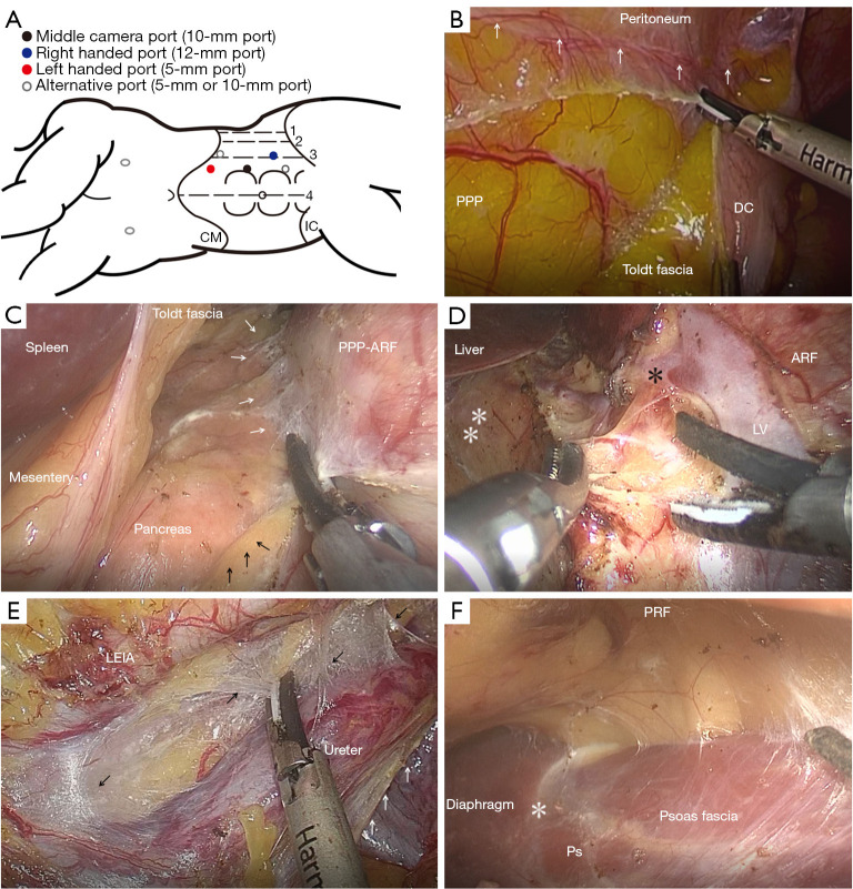
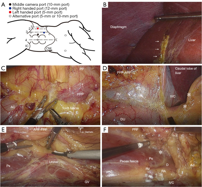
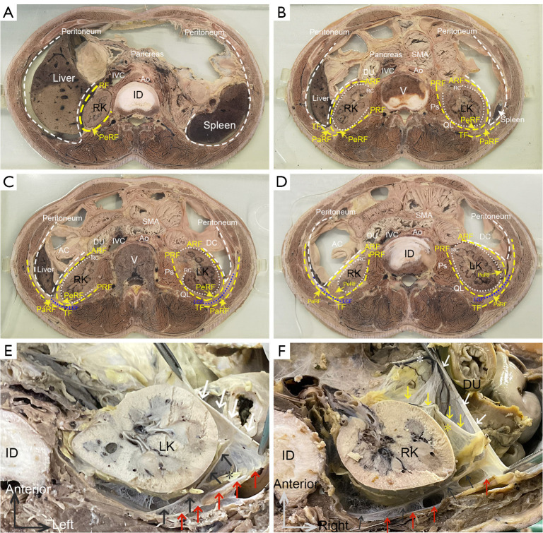
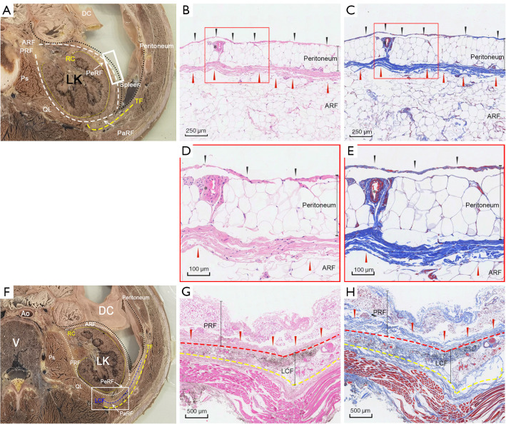
Methods: The study consisted of two parts: cadaveric and clinical study. The cadaveric study involved dissecting and observing the retroperitoneal fasciae and IFP in 32 cadavers using gross anatomical and histological methods. This retrospective clinical study compared the perioperative data and complications of 229 patients who underwent TIFP-LURS and 121 patients who underwent Con-LURS for upper retroperitoneal lesions at our center.

Results: The cadaveric study revealed that the retroperitoneal space was composed of multilaminar fasciae that formed potential bloodless spaces among them, that could be used as surgical landmarks and operating planes. The clinical study showed that TIFP-LURS had a significantly less estimated blood loss, lower intraoperative complication rate, lower postoperative complication rate, shorter hospital-stay and lower long-term postoperative complications rate than Con-LURS. Multivariate analysis indicated that the TIFP procedure was an independent protective factor for decreasing the risk of postoperative complications.

Conclusions: The IFP are potential avascular spaces that can be used during laparoscopic surgery, and TIFP-LURS is a novel surgical approach that can improve the safety and efficacy of laparoscopic surgery for upper retroperitoneal lesions.

Keywords: Retroperitoneal anatomy; interfascial plane (IFP); laparoscopic upper retroperitoneal surgery (LURS).


Conflict of interest statement

Conflicts of Interest: All authors have completed the ICMJE uniform disclosure form (available at https://tau.amegroups.com/article/view/10.21037/tau-23-632/coif). The authors have no conflicts of interest to declare.

Figures

Figure 1
Figure 1
Demonstration of trans-interfascial planes procedures for laparoscopic upper retroperitoneal surgery on the left side. (A) Surgical position and trocar placement. The patient was placed in the right lateral decubitus with the left flank up. Dashed line 1: middle axillary line; dashed line 2: anterior axillary line; dashed line 3: the parallel line 2 cm away from the lateral aspect of the rectus abdominis muscle; dashed line 4: anterior median line; the middle camera port was placed lateral to the rectus abdominis muscle and 2 cm superiorly up to the level of the umbilicus. The left-handed port was at the junction of the lateral aspect of the rectus muscle (alternative choice: dashed line 3) and the subcostal border. The right-handed port was placed on dashed line 3 and 2 cm inferiorly down to the level of the umbilicus. Alternative right-handed port was marked half the distance between the anterior superior iliac spine and the umbilicus. (B) Mobilization of the colon. Incise along the white line of Toldt (white arrows) medially and access the IFP between Toldt fascia and PPP. (C) Exposure of the retroperitoneum. Dissect along the IFP between Toldt fascia and PPP medially, and reflect DC to expose the urinary organs. Stay outside the paler yellow ARF and avoid entering the brighter yellow posterior mesentery (black arrows). Loose areolar tissues (white arrows) with minute vessels inside can be observed along the IFP between different fasciae, which can be dissected without hemorrhage. (D) Renal hilum dissection. Dissect along the IFP among retropancreatic fascia (white asterisks), ARF and fasciae surrounding vessels, and expose LV and adrenal vein (black asterisk). (E) Dissection along the ureter. Dissecting along loose areolar tissues (black arrows) in the IFP between fasciae surrounding LEIA and Gerota’s fascia surrounding the ureter (white arrows). (F) Exposure of the psoas muscle. Enter the IFP between PRF and the psoas fascia. Retract the kidney up but stay outside PRF. The medial arcuate ligament of diaphragm (white asterisk) is the anatomical landmark continuous with the fasciae of the psoas muscle. CM, costal margin; IC, iliac crest; IFP, interfascial plane; PPP, primitive parietal peritoneum; ARF, anterior renal fascia; DC, descending colon; LV, left renal vein; LEIA, left external iliac artery; Ps, psoas major; PRF, posterior renal fascia.
Figure 2
Figure 2
Demonstration of trans-interfascial planes procedures for laparoscopic upper retroperitoneal surgery on the right side. (A) Surgical position and trocar placement. The patient was placed in the left lateral decubitus with the right flank up. Dashed line 1: middle axillary line; dashed line 2: anterior axillary line; dashed line 3: the parallel line 2 cm away from the lateral aspect of the rectus abdominis muscle; dashed line 4: anterior median line; the middle camera port was placed lateral to the rectus abdominis muscle at the level of the umbilicus. The left-handed port was on dashed line 2 at the level of the umbilicus. The right-handed port was placed on dashed line 3 and 5 cm superiorly up to the level of the umbilicus. Alternative right-handed port was at the junction of the lateral aspect of the rectus muscle and the subcostal border. Alternative left-handed port was on dashed line 3 and 5 cm inferiorly down to the level of the umbilicus. (B) Retraction of the liver. Divide the hepatocolic ligament (white arrows indicated the cut edge) and retract the liver up. (C) Mobilization of the colon. Incise along the white line of Toldt (white arrows) medially and get the access to the IFP between Toldt fascia and PPP. Communicating vessels between PP and PPP were identified as the landmarks about correct surgical dissection plane between Toldt fascia and PPP. (D) Dissection at the renal hilum. Dissect along the IFP among the fusion fascia of Treitz (white asterisks, continuous with retropancreatic fascia), ARF and fasciae surrounding vessels, and expose IVC and RV. (E) Dissection along the ureter. Dissect along loose areolar tissues in the IFP between fasciae surrounding the GV and Gerota’s fasciae (ARF-PRF) surrounding ureter. (F) Exposure of the psoas muscle. Ligate and divide the RA and RV. Then retract the kidney up but stay outside PRF and enter the IFP between PRF and psoas fascia. Medial arcuate ligament (white asterisk) of diaphragm (letter D) was the anatomical landmark continuous with fasciae of the psoas muscle. CM, costal margin; IC, iliac crest; IFP, interfascial plane; PPP, primitive parietal peritoneum; PP, parietal peritoneum; ARF, anterior renal fascia; AC, ascending colon; DU, duodenum; IVC, inferior vena cava; RV, right renal vein; RA, right renal artery; GV, gonadal vein; Ps, psoas major; PRF, posterior renal fascia; Ad, adrenal gland.
Figure 3
Figure 3
Transverse-sectional diagrams of abdomen and gross anatomy of the retroperitoneum (at the L1 and L2 level). (A-D) Transverse-sectional diagrams of the abdomen from a male cadaver (70 years of age at death) observed from the caudal aspect. Multilaminar fasciae surrounding the kidney in the retroperitoneal space were observed in transverse sectional specimens, including RC, PeRF, RF, parietal peritoneum, PaRF, etc. There was a membranous structure with a clear demarcation between PeRF and PaRF. (A) Transverse-section at the intervertebral disc level between T12 and L1. (B) Transverse-section at the L1 vertebral body level. (C) Transverse-section at the L2 vertebral body level. (D) Transverse-section at the intervertebral disc level between L2 and L3. (E,F) Gross anatomy of the retroperitoneum. A transverse section of the retroperitoneum from a male cadaver (61 years of age at death) at the intervertebral disc level between L1 and L2. (E) The interfascial planes of perirenal space of left kidney observed from the caudal aspect. The fibrous connective tissue was observed between PRF and muscular fascia. White arrow: parietal peritoneum; Black arrow: PRF; Red arrow: muscular fascia of diaphragm; Black asterisk (*): PaRF. (F) The interfascial planes of perirenal space of right kidney observed from the cephalic aspect. White arrow: parietal peritoneum; Yellow arrow: ARF; Black arrow: PRF; Red arrow: muscular fascia of diaphragm; White asterisk (*): the blood supply of peritoneum within the areolar connective tissue. The vessels travelled and branched within the sub-peritoneal layer. Yellow asterisk (*): the blood supply of ARF, which was independent of peritoneum. The vessels ran in the internal space of ARF and communicated with vessels within PeRF. SMA, superior mesenteric artery; IVC, inferior vena cava; Ao, aorta; RF, renal fascia; RK, right kidney; PeRF, perirenal fat; ID, intervertebral disc; DU, duodenum; ARF, anterior renal fascia; PRF, posterior renal fascia; RC, renal capsule; PaRF, pararenal fat; TF, transversalis fascia; Ps, psoas major; LK, left kidney; V, vertebra; QL, quadratus lumborum; AC, ascending colon; DC, descending colon; LCF, lateral conal fascia.
Figure 4
Figure 4
Histology of the retroperitoneum. (A) A transverse section of the left retroperitoneum at the L1 vertebral body level. (B,C) Representative images showed H&E staining (B) and Masson’s trichrome staining (C) of white square section in (A). A fibrous tissue gap was observed between peritoneum and ARF (red arrowheads). Black arrowheads: abdominal aspect of the peritoneum; Black asterisk (*): the vessel ran in the internal space (sub-mesothelial tissue) of peritoneum. Bars, 250 µm. (D,E) High magnification of the red square sections in (B,C). Fascial tissue consisted of mesothelial (black arrowheads) and sub-mesothelial tissue while the fusion layer of different fasciae consisted of multilaminar parallel strands of collagen fibers (red arrowheads), which was pink in HE staining (D) and blue in Masson staining (E). Bars, 100 µm. (F) A transverse section of the left retroperitoneum at the L2 vertebral body level. (G,H) Representative images showed H&E staining (G) and Masson’s trichrome staining (H) of white square section in (F). Red dashed line indicated the fusion layer between PRF and LCF. Yellow dashed line indicated the fusion layer between LCF and muscular fascia. Red arrowheads: strands of collagen fibers in fusion layer; Bars, 500 µm. Ao, aorta; DC, descending colon; LK, left kidney; V, vertebra; Ps, psoas major; QL, quadratus lumborum; TF, transversalis fascia; RF, renal fascia; ARF, anterior renal fascia; PRF, posterior renal fascia; RC, renal capsule; LCF, lateral conal fascia; PeRF, perirenal fat; PaRF, pararenal fat.
Figure 5
Figure 5
Schematic diagram of retroperitoneal anatomy and trans-interfascial planes procedures in surgery. The transverse sectional diagram of the left part of the retroperitoneum was drawn based on the concept of “interfascial plane”. The interfascial plane is a potential space within a multilaminar membranous structure, indicated by asterisks (*) with different shapes and colors. We considered anterior renal fascia continuous with posterior renal fascia, which was composed of both mesothelial layer and sub-mesothelial loose connective tissue. We considered the combination of the primitive parietal peritoneum, cone-like fat, and anterior renal fascia as the traditional anterior layer of Gerota’s fascia, and the combination of the posterior renal fascia, lateral conal fascia and fasciae of the psoas muscle and quadratus lumborum muscle as the posterior layer of Gerota’s fascia (also known as Zuckerkandl’s fascia). We considered Toldt fascia sandwiched between the overlying mesothelial layer of the mesocolon and underlying mesothelial layer of the primitive parietal peritoneum. The anterior layer of Gerota’s fascia extended transversely in two directions: medially to fuse with the fascia overlying the adventitia layer of the aorta and IVC, laterally to fuse with Toldt fascia and the posterior layer, and then tapered at the area below the reflection of visceral and parietal peritoneum. The interfascial planes that sandwiched by different fasciae comprised areolar tissues with minute vessels inside. Therefore, surgical dissection plane is readily developed within interfascial planes. We designed the trans-interfascial planes procedures for laparoscopic upper retroperitoneal surgery (pink dashed line) using transperitoneal approach, which included: (a) incise along the white line of Toldt and get the access to IFP between Toldt fascia and primitive parietal peritoneum, which could mobilize and reflect the colon; (b) dissect along the IFP and medially get the access to the IFP between primitive parietal peritoneum and ARF, which actually stay outside ARF; (c) identify cone-like fat as an anatomical landmark and laterally dissect along the IFP between renal fascia (ARF and PRF) and LCF; (d) dissect along the IFP between PRF and LCF and then get the access to the IFP between PRF and fasciae of the psoas muscle and quadratus lumborum muscle, which divide the lateral and dorsal attachments of urinary system; (e) communicate the IFP between renal fascia (ARF and PRF) and fascia around blood vessels, which could control and dissect the renal hilum with great care. IFP, interfascial plane; IVC, inferior vena cava; DC, descending colon; LV, left renal vein; LK, left kidney; V, vertebra; Ps, psoas major; QL, quadratus lumborum; ES, erector spinae; LD, latissimus dorsi; TF, transversalis fascia; PPP, primitive parietal peritoneum; ARF, anterior renal fascia; PRF, posterior renal fascia; RC, renal capsule; LCF, lateral conal fascia; PeRF, perirenal fat; PaRF, pararenal fat; TA, transverse abdominis; IO, internal obliques; EO, external obliques.

Similar articles

References

    1. Rosevear HM, Montgomery JS, Roberts WW, et al. Characterization and management of postoperative hemorrhage following upper retroperitoneal laparoscopic surgery. J Urol 2006;176:1458-62. 10.1016/j.juro.2006.06.023 - DOI - PubMed
    1. Ochi A, Muro S, Adachi T, et al. Zoning inside the renal fascia: The anatomical relationship between the urinary system and perirenal fat. Int J Urol 2020;27:625-33. 10.1111/iju.14248 - DOI - PMC - PubMed
    1. Ishikawa K, Nakao S, Nakamuro M, et al. The retroperitoneal interfascial planes: current overview and future perspectives. Acute Med Surg 2016;3:219-29. 10.1002/ams2.188 - DOI - PMC - PubMed
    1. Meyers MA, Whalen JP, Peelle K, et al. Radiologic features of extraperitoneal effusions. An anatomic approach. Radiology 1972;104:249-57. 10.1148/104.2.249 - DOI - PubMed
    1. Chesbrough RM, Burkhard TK, Martinez AJ, et al. Gerota versus Zuckerkandl: the renal fascia revisited. Radiology 1989;173:845-6. 10.1148/radiology.173.3.2682777 - DOI - PubMed