. 2024 Apr 17;15(5):3112-3127.
doi: 10.1364/BOE.515781. eCollection 2024 May 1.

Deep learning based characterization of human organoids using optical coherence tomography

Affiliations

Deep learning based characterization of human organoids using optical coherence tomography

Bingjie Wang et al. Biomed Opt Express. .

Abstract

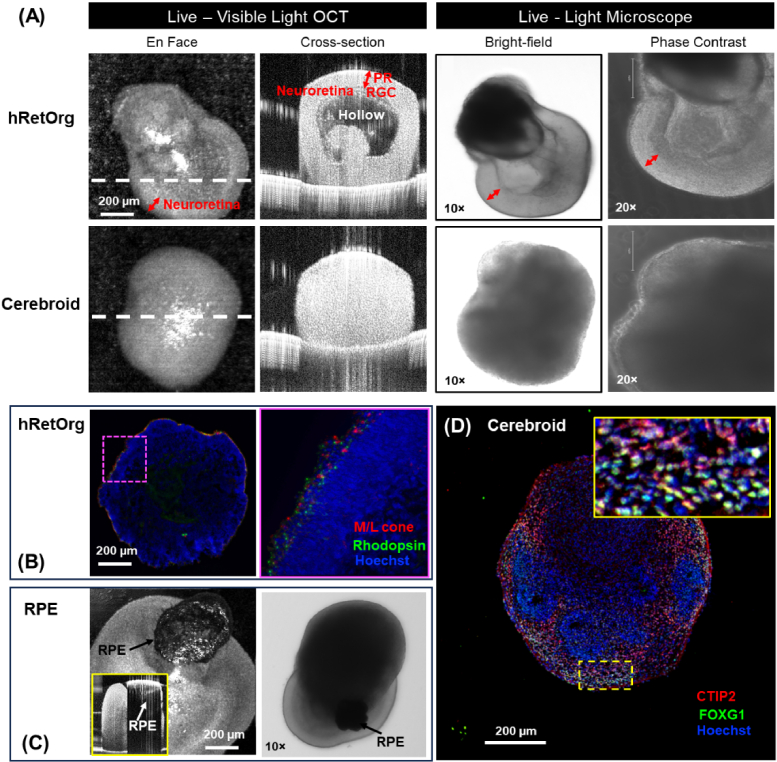
Organoids, derived from human induced pluripotent stem cells (hiPSCs), are intricate three-dimensional in vitro structures that mimic many key aspects of the complex morphology and functions of in vivo organs such as the retina and heart. Traditional histological methods, while crucial, often fall short in analyzing these dynamic structures due to their inherently static and destructive nature. In this study, we leveraged the capabilities of optical coherence tomography (OCT) for rapid, non-invasive imaging of both retinal, cerebral, and cardiac organoids. Complementing this, we developed a sophisticated deep learning approach to automatically segment the organoid tissues and their internal structures, such as hollows and chambers. Utilizing this advanced imaging and analysis platform, we quantitatively assessed critical parameters, including size, area, volume, and cardiac beating, offering a comprehensive live characterization and classification of the organoids. These findings provide profound insights into the differentiation and developmental processes of organoids, positioning quantitative OCT imaging as a potentially transformative tool for future organoid research.


Conflict of interest statement

The authors declare no conflicts of interest.

Figures

Fig. 1.
Fig. 1.
The architecture of the proposed OrgSegNet for deep learning segmentation of organoid tissue and internal structure in OCT B-scan images. A cardiac organoid is shown as a representative example.
Fig. 2.
Fig. 2.
Representative OCT B-scan images of human organoids, the ground truth of binary images, and subsequent segmentations using OrgSegNet. Tissue segmentation is in cyan and hollows in magenta. Arrows indicate the challenging conditions for segmentation including specular reflection (yellow and red), base membrane of culture plate (green), reflectance attenuation (magenta), and ambiguous inner structure (orange). The zoom-in overlay of hollow segmentations on panel C inset demonstrated representative cases causing a discrepancy between the ground truth (green color) and deep learning (red color) results where the OrgSegNet highlighted the regions that are challenging in manual delineation.
Fig. 3.
Fig. 3.
Representative images of hRetOrg and cerebroids at different time points of development. (A) Live hRetOrg and cerebroid at day (D) D31 in OCT and microscopy. Note the presence of neuroretina lamination and hollow structure in hRetOrg but not in the cerebroids, which were more evident in OCT B-scans. (B) hRetOrg at D117 immuno-stained for M/L cones and rods counterstained with Hoechst. Note the location of these photoreceptor cells at the outer edge of the hRetOrg. (C) OCT and microscopy images of a hRetOrg with RPE (arrows). Note the presence of a strong shadow on OCT scans corresponding to RPE (white arrow). (D) Cerebroid at D60 immuno-stained for the cerebral markers FOXG1 and CTIP2 counterstained with Hoechst. PR, photoreceptors; RGC, retinal ganglion cells; RPE, retinal pigment epithelium.
Fig. 4.
Fig. 4.
hRetOrg and cerebroid quantitative features extracted from OCT images and OrgSegNet platform. (A) – (C) Comparison between hRetOrg (red) and cerebroid size (green) (A), tissue volume (B) and hollow volume (C). ‘*’ indicated the significance of the hollow structure between hRetOrg and cerebroid. (D) – (E) Hollow volume (D), and ratio of hollow per total organoid volume (E) for each individual hRetOrg sample. (F) The area under the receiver operating characteristic (ROC) curve (AROC) performance (F) distinguishing hRetOrg from cerebroid based on hollow structure was 0.99, much higher than 0.69 based on tissue volume.
Fig. 5.
Fig. 5.
(A) The 3D and projected en face visualization of a cardiac organoid by OCT. (B) cross-sectional and depth plane visualization of chambers (marked with stars) in cardiac organoids, as well as their segmentations. Each isolated chamber was assigned a different color. (C) 3D visualization of the segmented chambers of cardiac organoid. (D) The association of the number of chambers with the percentage of chamber to total volume. An exponential fitting (C/T = 34.82×e-0.156N) was performed with an R-square of 0.82. (E) Projected en face image of an OCT beating scan for cardiac organoids. Note the occasional specular reflection due to the movement. (F) Appreciation of the cardiac beating by the axial position of detected organoid tissue surface. (G) Appreciation of the cardiac beating by examination of the axial position in specific locations of organoids (point c and 1-4 in E), in contrast to the static background board (point b1 and b2 in E).

Update of

  • doi: 10.1364/opticaopen.24790584.

References

    1. Alhaque S., Themis M., Rashidi H., “Three-dimensional cell culture: from evolution to revolution,” Phil. Trans. R. Soc. B 373(1750), 20170216 (2018).10.1098/rstb.2017.0216 - DOI - PMC - PubMed
    1. Akkerman N., Defize L. H., “Dawn of the organoid era: 3D tissue and organ cultures revolutionize the study of development, disease, and regeneration,” BioEssays 39(4), 1600244 (2017).10.1002/bies.201600244 - DOI - PubMed
    1. Shoji J. Y., Davis R. P., Mummery C. L., et al. , “Global meta-analysis of organoid and organ-on-chip research,” Adv. Healthcare Mater., 2301067 (2023). - PubMed
    1. Liu C., Oikonomopoulos A., Sayed N., et al. , “Modeling human diseases with induced pluripotent stem cells: from 2D to 3D and beyond,” Development 145(5), dev156166 (2018).10.1242/dev.156166 - DOI - PMC - PubMed
    1. Takahashi T., “Organoids for drug discovery and personalized medicine,” Annu. Rev. Pharmacol. Toxicol. 59(1), 447–462 (2019).10.1146/annurev-pharmtox-010818-021108 - DOI - PubMed

LinkOut - more resources