

FRD-Net: a full-resolution dilated convolution network for retinal vessel segmentation
- PMID: 38855685
- PMCID: PMC11161363
- DOI: 10.1364/BOE.522482
FRD-Net: a full-resolution dilated convolution network for retinal vessel segmentation
Abstract
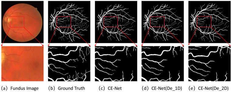
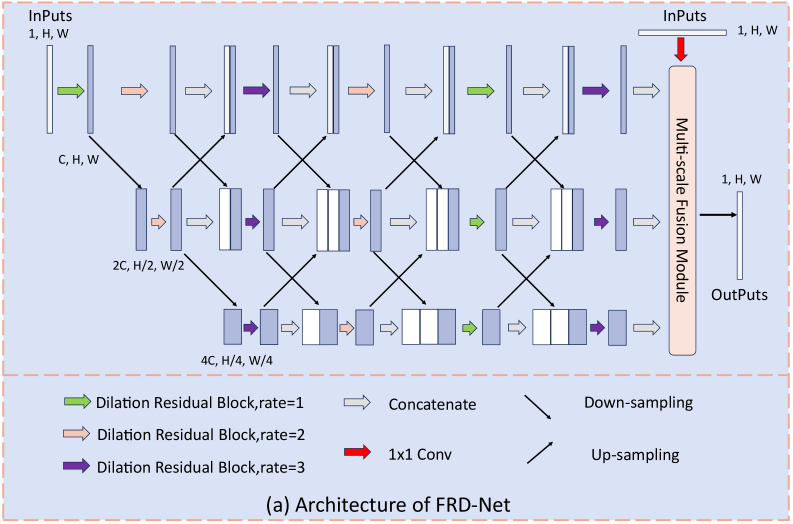
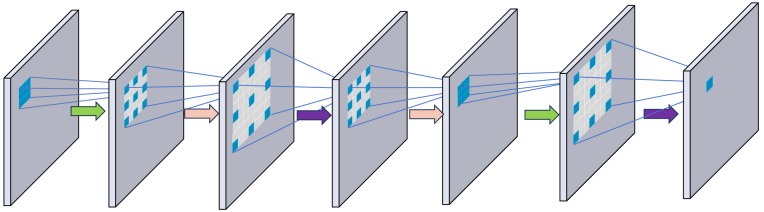
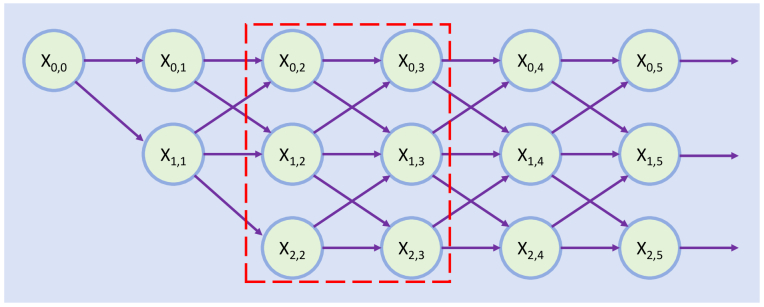
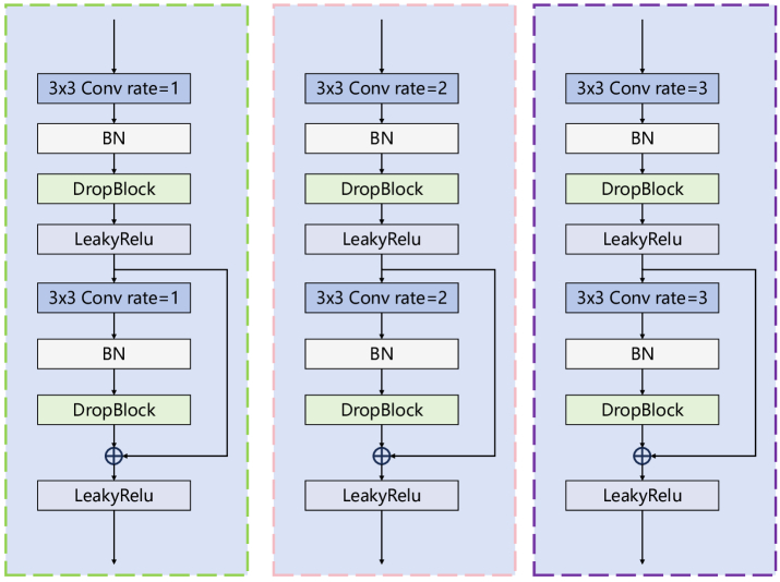
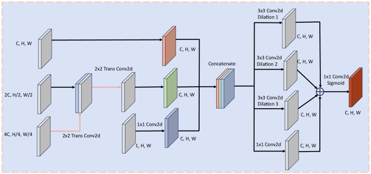
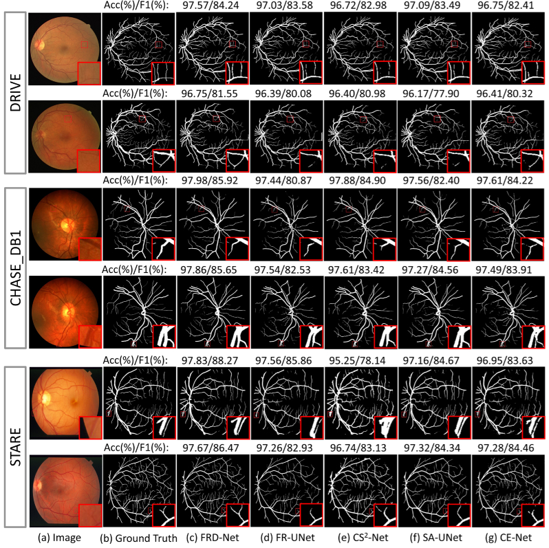
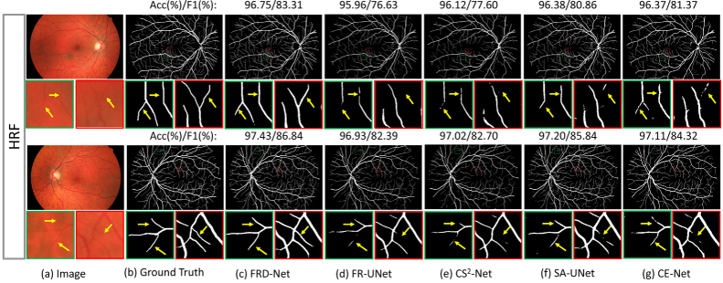
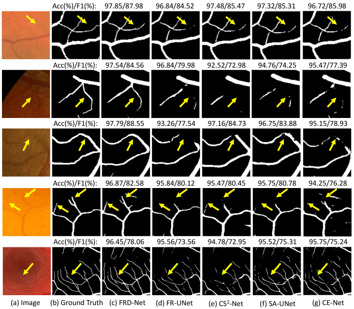
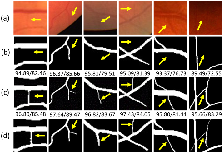
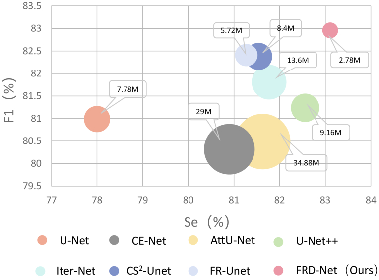
Accurate and automated retinal vessel segmentation is essential for performing diagnosis and surgical planning of retinal diseases. However, conventional U-shaped networks often suffer from segmentation errors when dealing with fine and low-contrast blood vessels due to the loss of continuous resolution in the encoding stage and the inability to recover the lost information in the decoding stage. To address this issue, this paper introduces an effective full-resolution retinal vessel segmentation network, namely FRD-Net, which consists of two core components: the backbone network and the multi-scale feature fusion module (MFFM). The backbone network achieves horizontal and vertical expansion through the interaction mechanism of multi-resolution dilated convolutions while preserving the complete image resolution. In the backbone network, the effective application of dilated convolutions with varying dilation rates, coupled with the utilization of dilated residual modules for integrating multi-scale feature maps from adjacent stages, facilitates continuous learning of multi-scale features to enhance high-level contextual information. Moreover, MFFM further enhances segmentation by fusing deeper multi-scale features with the original image, facilitating edge detail recovery for accurate vessel segmentation. In tests on multiple classical datasets,compared to state-of-the-art segmentation algorithms, FRD-Net achieves superior performance and generalization with fewer model parameters.
© 2024 Optica Publishing Group.
Conflict of interest statement
The authors declare that they have no known competing financial interests or personal relationships that could have appeared to influence the work reported in this paper.
Figures
Similar articles
-
DEAF-Net: Detail-Enhanced Attention Feature Fusion Network for Retinal Vessel Segmentation.J Imaging Inform Med. 2025 Feb;38(1):496-519. doi: 10.1007/s10278-024-01207-6. Epub 2024 Aug 5. J Imaging Inform Med. 2025. PMID: 39103564 Free PMC article.
-
TDCAU-Net: retinal vessel segmentation using transformer dilated convolutional attention-based U-Net method.Phys Med Biol. 2023 Dec 22;69(1). doi: 10.1088/1361-6560/ad1273. Phys Med Biol. 2023. PMID: 38052089
-
TP-Net: Two-Path Network for Retinal Vessel Segmentation.IEEE J Biomed Health Inform. 2023 Apr;27(4):1979-1990. doi: 10.1109/JBHI.2023.3237704. Epub 2023 Apr 4. IEEE J Biomed Health Inform. 2023. PMID: 37021912
-
Fully connected network with multi-scale dilation convolution module in evaluating atrial septal defect based on MRI segmentation.Comput Methods Programs Biomed. 2022 Mar;215:106608. doi: 10.1016/j.cmpb.2021.106608. Epub 2022 Jan 11. Comput Methods Programs Biomed. 2022. PMID: 35063713
-
HRD-Net: High resolution segmentation network with adaptive learning ability of retinal vessel features.Comput Biol Med. 2024 May;173:108295. doi: 10.1016/j.compbiomed.2024.108295. Epub 2024 Mar 19. Comput Biol Med. 2024. PMID: 38520920
References
-
- Xu W., Yang H., Zhang M., et al. , “Decnet: A dual-stream edge complementary network for retinal vessel segmentation,” in 2021 IEEE International Conference on Bioinformatics and Biomedicine (BIBM), (2021), pp. 1595–1600.
LinkOut - more resources
Full Text Sources