. 2024 Apr 17;15(5):3094-3111.
doi: 10.1364/BOE.511806. eCollection 2024 May 1.

Two-photon autofluorescence lifetime assay of rabbit photoreceptors and retinal pigment epithelium during light-dark visual cycles in rabbit retina

Affiliations

Two-photon autofluorescence lifetime assay of rabbit photoreceptors and retinal pigment epithelium during light-dark visual cycles in rabbit retina

Trung Duc Nguyen et al. Biomed Opt Express. .

Abstract

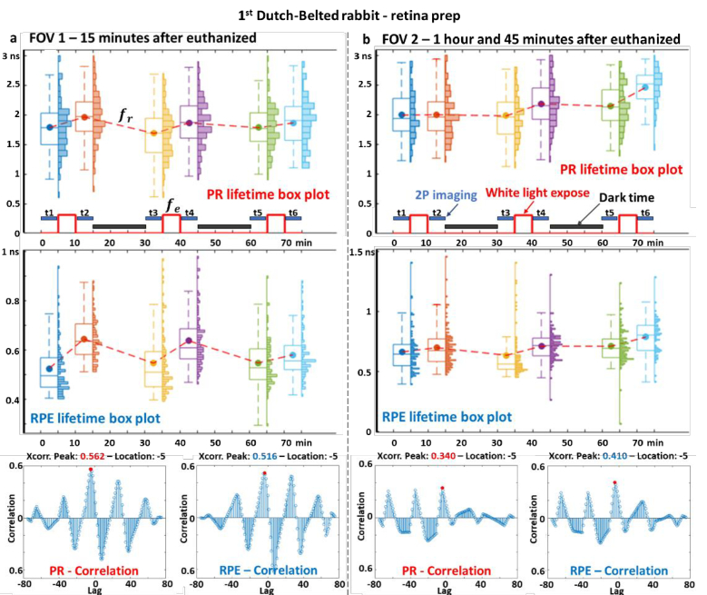
Two-photon excited fluorescence (TPEF) is a powerful technique that enables the examination of intrinsic retinal fluorophores involved in cellular metabolism and the visual cycle. Although previous intensity-based TPEF studies in non-human primates have successfully imaged several classes of retinal cells and elucidated aspects of both rod and cone photoreceptor function, fluorescence lifetime imaging (FLIM) of the retinal cells under light-dark visual cycle has yet to be fully exploited. Here we demonstrate a FLIM assay of photoreceptors and retinal pigment epithelium (RPE) that reveals key insights into retinal physiology and adaptation. We found that photoreceptor fluorescence lifetimes increase and decrease in sync with light and dark exposure, respectively. This is likely due to changes in all-trans-retinol and all-trans-retinal levels in the outer segments, mediated by phototransduction and visual cycle activity. During light exposure, RPE fluorescence lifetime was observed to increase steadily over time, as a result of all-trans-retinol accumulation during the visual cycle and decreasing metabolism caused by the lack of normal perfusion of the sample. Our system can measure the fluorescence lifetime of intrinsic retinal fluorophores on a cellular scale, revealing differences in lifetime between retinal cell classes under different conditions of light and dark exposure.


Conflict of interest statement

The authors declare no conflicts of interest.

Figures

Fig. 1.
Fig. 1.
(a) Schematic of 2P-FLIO system with raw data presentation, (b) Multiple retinal layers imaging and unmixing with endogenous fluorophores (c) Photoreceptor and retinal pigment epithelium lifetime images after unmixing. Abbreviations: PMT, photomultiplier tube; 1P, one-photon; 2P, two-photon; FLIO, fluorescence lifetime ophthalmoscopy; TCSPC, time-correlated single photon counting; SLO, scanning laser ophthalmoscopy; IP, image plane; P, pinhole; L, lens; PBS, polarizing beam splitter; EM, emission filter; M, mirror; DM, dichroic mirror; GM, galvo mirror; NZW, New Zealand White rabbit; DB, Dutch-Belted rabbit; GMM, Gaussian Mixture Models; PR, photoreceptor; RPE, retinal pigment epithelium. DM1 and DM2 were mounted on flip mounts (TRF90, Thorlabs) to select 2P-FLIO mode or 1P-FLIO mode. Scale bar is 50 µm.
Fig. 2.
Fig. 2.
(a) Lifetime phasors can be obtained from time-domain decay data, after conducting Fourier transform [33,34]. For a single species, lifetime decreases clockwise along the universal semicircle. (b) Lifetime phasor calibration using fluorescein (4 ns, excited by 780 nm femtosecond laser) for 2P-FLIO. (c) τ phasor plot of a convallaria sample. (d) False-colored FLIM image of the convallaria. Scale bar is 5 µm.
Fig. 3.
Fig. 3.
Data analysis procedure with Gaussian Mixture Models. (a) The 98 × 1 fluorescence decay curve I(t) at each pixel is separately transformed into τ phasors. (b) The 256 × 256 τ phasor sets (, ) are used as inputs for the GMM, after GMM unmixing, the corresponding clusters for photoreceptor and RPE cells are selected. (c) Photoreceptors segmentation with the watershed algorithm and RPE segmentation with the k-nearest neighbors (KNN) algorithm. (d) Summary of lifetime changes during light-dark visual cycles. Abbreviations: KNN, k-nearest neighbors algorithm; PR, photoreceptor; RPE, retinal pigment epithelium; BG, background. Scale bars are 50 µm.
Fig. 4.
Fig. 4.
Retina heating induced by near-infrared laser and white light exposure during light-dark visual cycles. (a) Near-infrared laser (blue box) and white light exposure scheme (red line). (b) Retina’s temperature changes during light-dark visual cycles, red circle denoted white light exposure.
Fig. 5.
Fig. 5.
1st Dutch-Belted rabbit - retina prep - (a) FOV 1–15 minutes after euthanized (b) FOV 2–1 hour and 45 minutes after euthanized. 1st row - Photoreceptor lifetime changes during light-dark visual cycles. 2nd row - RPE lifetime changes during light-dark visual cycles. 3rd row - Quantify photoreceptor and RPE lifetime changes during light-dark visual cycles with normalized cross-correlation in the time domain. Abbreviations: DB, Dutch-Belted rabbits, PR, photoreceptor; RPE, retinal pigment epithelium; Xcorr. Peak, time domain cross-correlation peak.
Fig. 6.
Fig. 6.
2nd Dutch-Belted rabbit - retina prep - (a) FOV 1–1 hour after euthanized (b) FOV 2–4 hours after euthanized. 1st row - Photoreceptor lifetime changes during light-dark visual cycles. 2nd row - RPE lifetime changes during light-dark visual cycles. 3rd row - Quantify photoreceptor and RPE lifetime changes during light-dark visual cycles with normalized cross-correlation in the time domain. Abbreviations: PR, photoreceptor; RPE, retinal pigment epithelium; Xcorr. Peak, time domain cross-correlation peak.
Fig. 7.
Fig. 7.
1st New Zealand White rabbit - retina prep - (a) FOV 1–30 minutes after euthanized (b) FOV 2–2 hours after euthanized. 1st row - Photoreceptor lifetime changes during light-dark visual cycles. 2nd row - RPE lifetime changes during light-dark visual cycles. 3rd row - Quantify photoreceptor and RPE lifetime changes during light-dark visual cycles with normalized cross-correlation in the time domain. Abbreviations: PR, photoreceptor; RPE, retinal pigment epithelium; Xcorr. Peak, time domain cross-correlation peak.
Fig. 8.
Fig. 8.
2nd New Zealand White rabbit - retina prep - (a) FOV 1–1 hour after euthanized (b) FOV 2–2 hours and 30 minutes after euthanized. 1st row - Photoreceptor lifetime changes during light-dark visual cycles. 2nd row - RPE lifetime changes during light-dark visual cycles. 2nd row - Quantify photoreceptor and RPE lifetime changes during light-dark visual cycles with normalized cross-correlation in the time domain. Abbreviations: PR, photoreceptor; RPE, retinal pigment epithelium; Xcorr. Peak, time domain cross-correlation peak.
Fig. 9.
Fig. 9.
Schematic representation of the pigment epithelium visual cycle for rod and cone photoreceptors and Müller cells. Abbreviations: ROS, rod outer segment; RIS, rod inner segment; COS, cone outer segment; CIS, cone inner segment, RPE, retinal pigment epithelium; 11-cis-RAL, 11-cis-retinal; 11-cis-ROL, 11-cis-retinol; 11-cis-RE, 11-cis- retinyl esters; AT-ROL, all-trans-retinol; AT-RAL, all-trans-retinal; AT-RE, all-trans-retinyl esters.

Similar articles

References

    1. Rando R. R., “The Biochemistry of the Visual Cycle,” Chem. Rev. 101(7), 1881–1896 (2001).10.1021/cr960141c - DOI - PubMed
    1. Strauss O., “The Retinal Pigment Epithelium in Visual Function,” Physiol. Rev. 85(3), 845–881 (2005).10.1152/physrev.00021.2004 - DOI - PubMed
    1. Rattner A., Smallwood P. M., Nathans J., et al. , “Identification and Characterization of All-trans-retinol Dehydrogenase from Photoreceptor Outer Segments, the Visual Cycle Enzyme That Reduces All-trans-retinal to All-trans-retinol,” J. Biol. Chem. 275(15), 11034–11043 (2000).10.1074/jbc.275.15.11034 - DOI - PubMed
    1. Rossi E. A., Granger C. E., Sharma R., et al. , “Imaging individual neurons in the retinal ganglion cell layer of the living eye,” Proc. Natl. Acad. Sci. 114(3), 586–591 (2017).10.1073/pnas.1613445114 - DOI - PMC - PubMed
    1. Sharma R., Schwarz C., Williams D. R., et al. , “In Vivo Two-Photon Fluorescence Kinetics of Primate Rods and Cones,” Invest. Ophthalmol. Vis. Sci. 57(2), 647 (2016).10.1167/iovs.15-17946 - DOI - PMC - PubMed

LinkOut - more resources