

Shear wave elastography for solid breast masses evaluation: Quantitative measurement of mean elasticity value and elasticity ratio
- PMID: 38855720
- PMCID: PMC11157203
- DOI: 10.1016/j.ejro.2024.100573
Shear wave elastography for solid breast masses evaluation: Quantitative measurement of mean elasticity value and elasticity ratio
Abstract
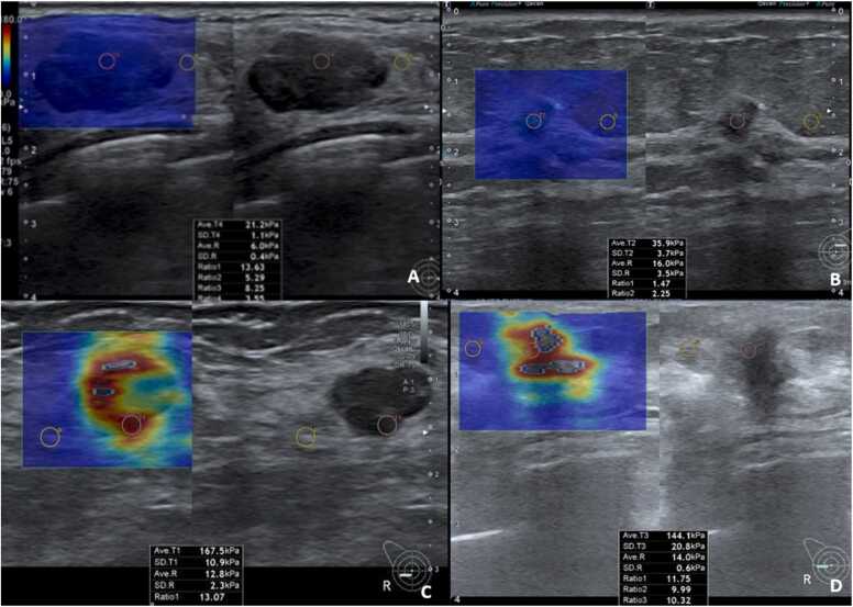
Purpose: Shear wave elastography (SWE), an ultrasonographic technique to measure the elasticity of mass lesions to evaluate breast mass. This study aimed to find out the cutoff values identifying breast malignancy using the mean elasticity (E-mean) and elasticity ratio (E-ratio) of breast masses.
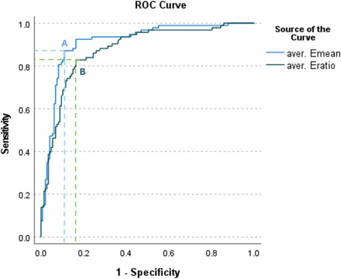
Methods: This retrospective study included women underwent SWE and US-guided biopsy of breast masses. During conventional US, the SWE mode was also performed, determining elasticity measurements, E-mean and E-ratio. Histopathological reports were obtained to identify mass status. The optimal and alternative cutoff values for E-mean and E-ratio to determine malignancy were assessed by receiver operating characteristic (ROC) curve analysis and Youden's index score.
Results: Among 147 benign and 93 malignant masses, the median of E-means were 26.20 (IQR 15.70-56.60) and 141.60 (IQR 119.80-154.60) kPa and the median E-ratios were 3.11 (IQR 1.83-5.23) and 9.24 (IQR 6.76-12.44), respectively. Using Youden's index, the optimal cutoff values for E-mean and E-ratio were 90.35 and 5.89, with sensitivity of 87.1 % and 82.8 %, specificity of 89.1 % and 83.7 %, positive predictive value (PPV) of 83.5 % and 76.2 %, negative predictive value (NPV) of 91.6 % and 88.5 %, positive likelihood ratio (LR+) of 8.00 and 5.07, and negative likelihood ratio (LR-) of 0.14 and 0.21, respectively.
Conclusion: This study revealed that SWE is useful in predicting malignancy. With the optimal cutoff values of E-mean and E-ratio at 90.35 kPa and 5.89, the sensitivity was nearly 90 % with E-mean and slightly over 80 % with E-ratio, respectively. These findings could be used in conjunction with conventional US.
Keywords: Breast mass; Elasticity ratio; Mean elasticity; Shear wave elastography; Ultrasound.
© 2024 The Authors. Published by Elsevier Ltd.
Conflict of interest statement
All authors declare that they have no conflicts of interest.
Figures
Similar articles
-
Diagnostic performance of qualitative and quantitative shear wave elastography in differentiating malignant from benign breast masses, and association with the histological prognostic factors.Quant Imaging Med Surg. 2019 Mar;9(3):386-398. doi: 10.21037/qims.2019.03.04. Quant Imaging Med Surg. 2019. PMID: 31032186 Free PMC article.
-
Diagnostic performance of quantitative shear wave elastography in the evaluation of solid breast masses: determination of the most discriminatory parameter.AJR Am J Roentgenol. 2014 Sep;203(3):W328-36. doi: 10.2214/AJR.13.11693. AJR Am J Roentgenol. 2014. PMID: 25148191
-
Comparative analysis of conventional ultrasound and shear wave elastography features in primary breast diffuse large B-cell lymphoma.World J Clin Cases. 2023 Nov 26;11(33):7994-8002. doi: 10.12998/wjcc.v11.i33.7994. World J Clin Cases. 2023. PMID: 38075578 Free PMC article.
-
A qualitative and quantitative assessment of simultaneous strain, shear wave, and point shear wave elastography to distinguish malignant and benign breast lesions.Acta Radiol. 2021 Sep;62(9):1155-1162. doi: 10.1177/0284185120961422. Epub 2020 Oct 18. Acta Radiol. 2021. PMID: 33070635
-
Diagnostic performances of shear-wave elastography and B-mode ultrasound to differentiate benign and malignant breast lesions: the emphasis on the cutoff value of qualitative and quantitative parameters.Clin Imaging. 2018 Jul-Aug;50:302-307. doi: 10.1016/j.clinimag.2018.05.007. Epub 2018 May 4. Clin Imaging. 2018. PMID: 29751202
References
-
- Mendelson E.B., Böhm-Vélez, M., Berg, W.A., et al., ACR BI-RADS® ultrasound, in: ACR BI-RADS® Atlas, Breast Imaging Reporting and Data System, American College of Radiology, Reston, VA, 2013, 2013.
-
- Youk J.H., Gweon H.M., Son E.J., Han K.H., Kim J.A. Diagnostic value of commercially available shear-wave elastography for breast cancers: integration into BI-RADS classification with subcategories of category 4. Eur. Radiol. 2013;23(10):2695–2704. - PubMed
-
- Berg W.A., Cosgrove D.O., Doré C.J., Schäfer F.K.W., Svensson W.E., Hooley R.J., et al. Shear-wave elastography improves the specificity of breast US: the BE1 multinational study of 939 masses. Radiology. 2012;262(2):435–449. - PubMed
-
- Liu B., Zheng Y., Huang G., Lin M., Shan Q., Lu Y., et al. Breast lesions: quantitative diagnosis using ultrasound shear wave elastography–a systematic review and meta–analysis. Ultrasound Med. Biol. 2016;42(4):835–847. - PubMed
LinkOut - more resources
Full Text Sources
