Review
. 2024 Nov 6;30(6):409-418.
doi: 10.4274/dir.2024.242740. Epub 2024 Jun 10.

Pivotal role of the synovioentheseal complex in the imaging of arthritis and rheumatic diseases

Affiliations
Review

Pivotal role of the synovioentheseal complex in the imaging of arthritis and rheumatic diseases

Adalet Elçin Yıldız et al. Diagn Interv Radiol. .

Abstract

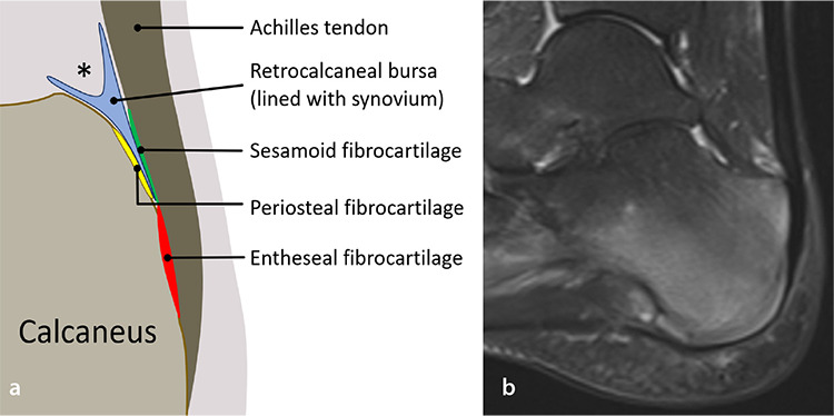
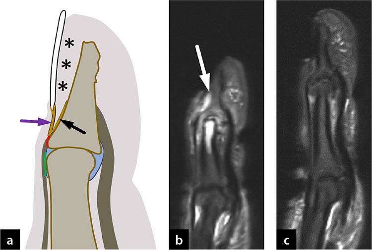
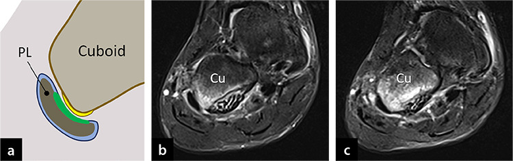
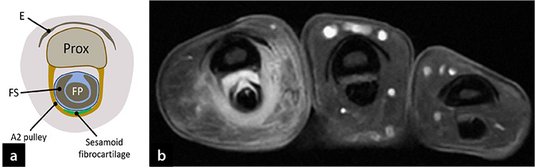
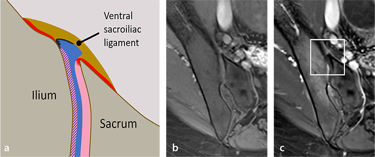
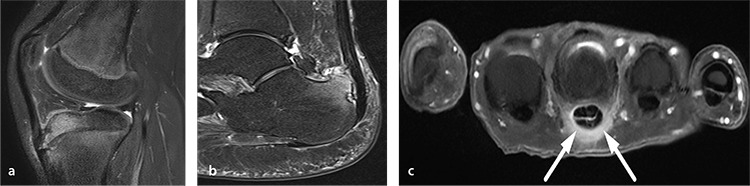
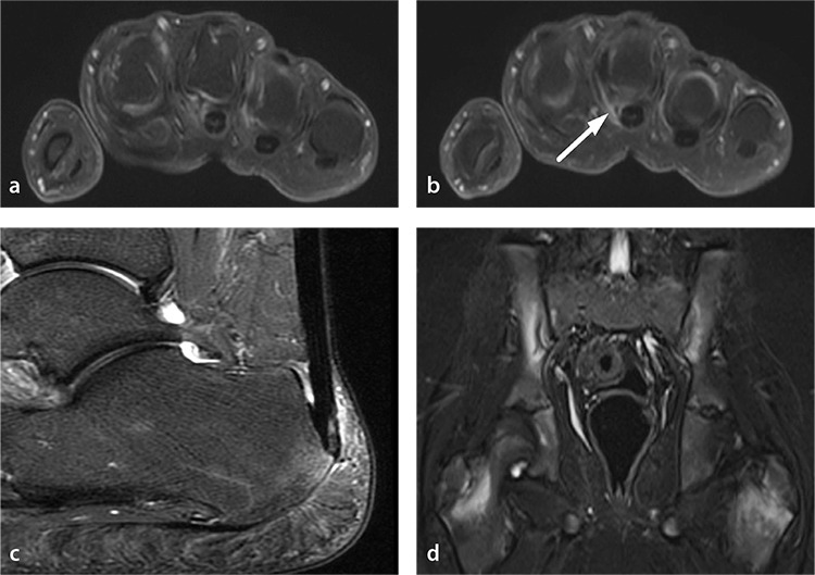
Imaging plays a key role in the diagnosis and management of rheumatic diseases. Although joints and periarticular tissue are commonly involved in rheumatic diseases, entheses further away from joints, such as in the Achilles tendon or plantar fascia insertion onto the calcaneus, as well as skin and subcutaneous tissue, are among other -sometimes overlooked- targets. The link of enthesitis, which describes inflammation at the insertions of ligaments, tendons, or joint capsules, with spondyloarthritis (SpA) was established just before the turn of the century as a characteristic feature based on imaging studies with histopathological correspondence. To highlight the association between enthesitis and synovitis in SpA, the anatomical unit of the “synovioentheseal complex” (SEC) and the concepts of “functional enthesis” and “articular enthesis,” apart from the better known “insertional enthesis,” were introduced to encompass other inflammatory lesions associated with SpA. Studies from the last two decades revealed the involvement of the SEC in rheumatic and non-rheumatic disorders with different pathogeneses. Although such involvement is sometimes distinctive, it does not necessarily point to a specific diagnosis at other times. Nevertheless, the potential of SEC inflammation in the differentiation of SpA from other forms of arthritis remains important. The purpose of this review was to provide essential information concerning the involvement of the SEC in the diagnosis of rheumatic diseases and arthritis, focusing on imaging characteristics.

Keywords: Arthritis; enthesitis; magnetic resonance imaging; synovioentheseal complex; ultrasonography.


Conflict of interest statement

Conflict of interest disclosure: The authors declared no conflicts of interest.

Figures

Figure 1
Figure 1
Synovioentheseal complex in a classic fibrocartilaginous enthesis in the ankle. (a) Schematic illustration of a midsagittal ankle section through the Achilles tendon shows the synovioentheseal complex including subunits of the entheseal organ: distal tendon, sesamoid (green), and entheseal (red) fibrocartilages of the tendon, periosteal fibrocartilage (yellow) of the calcaneus, retrocalcaneal bursa lined with synovium (blue), and the surrounding fat pad (asterisk). (b) Sagittal fat-saturated T2W magnetic resonance image shows Achilles tendon enthesitis as well as insertional plantar fasciitis, which also is a type of enthesitis, in a 16-year-old girl with enthesitis-related arthritis. Note retrocalcaneal bursitis and extensive bone marrow edema related to enthesitis.
Figure 2
Figure 2
Synovioentheseal complex in the classic fibrocartilaginous enthesis of extensor tendon with its nail root extensions. (a) Schematic illustration of a midsagittal distal finger section shows sesamoid fibrocartilage (green) and entheseal fibrocartilage (red) of the extensor tendon with its extensions to the nail root (superficial lamina, purple arrow) and the dorsal distal phalangeal periosteum (deep lamina, black arrow). Asterisks denote the nail bed. (b, c) Sagittal fat-saturated T2W magnetic resonance images of the second (b) and third (c) fingers in a 58-year-old woman with psoriatic arthritis reveal extensor tendon enthesitis of the second finger characterized with peritendinous, nail root, and bed inflammation (b, arrow), extensive periarticular osteitis of the middle and distal phalanges and synovitis of the second distal interphalangeal joint in addition to uniform joint space narrowing and mild periarticular bone proliferation. Extensor tendon enthesis, nail root, and bed of the third finger (c) are normal.
Figure 3
Figure 3
Synovioentheseal complex in a functional enthesis at a tendon–bone friction site. (a) Schematic illustration of a midfoot coronal section shows the peroneus longus tendon (PL) enveloped in its synovial tendon sheath. The sesamoid fibrocartilage (green) at the peroneus longus tendon facing the cuboid and the corresponding periosteal fibrocartilage (yellow) at this site constitute a functional enthesis. (b, c) Consecutive coronal fat-saturated T1W post-contrast magnetic resonance images through midfoot show functional enthesitis at the cuboid pulley in a 16-year-old girl with enthesitis-related arthritis. Cu, cuboid.
Figure 4
Figure 4
Synovioentheseal complex in a functional enthesis at the finger pulley-flexor tendon gliding site. (a) Schematic illustration of a transverse section through the mid-level of a proximal phalanx shows the extensor mechanism (E), proximal phalanx (Prox), superficial (FS), and deep (FP) flexor tendons, which are enveloped in a synovial sheath (blue). Phalangeal insertions of pulleys are classic entheses. The sesamoid fibrocartilage at the A2 pulley (green) facing the flexor tendon group (which is a friction site) is a functional enthesis. (b) Transverse fat-saturated T1W post-contrast magnetic resonance image shows functional enthesitis of A2 pulley with accompanying subcutaneous inflammation, dactylitis, in a 34-year-old woman with psoriatic arthritis.
Figure 5
Figure 5
Synovioentheseal complex (SEC) in the articular enthesis at the sacroiliac joint. (a) Schematic illustration of an oblique axial section through the distal third of the sacroiliac joint shows entheseal fibrocartilages (red) of the ventral sacroiliac ligament (VSIL) at the sacral and iliac bone insertions and at the transition zone where hyaline cartilage of the sacral side (pink) blend with the VSIL. The articular cartilage on the iliac side is thinner than on the sacral side and consists of a mixture of hyaline and fibrous cartilages (shown shaded in pink and purple diagonal stripes, respectively), and at the joint periphery it merges with the periosteum and is covered by the joint synovium (deep blue). (b, c) Transverse oblique fat-saturated T1W pre- (b) and post-contrast (c) magnetic resonance images (MRI) show linear fibrocartilaginous enhancement at the iliac joint surface with subchondral osteitis and enhancement of the VSIL compatible with active sacroiliitis (the white rectangular box in c roughly corresponds to the area drawn in a). Although the large field-of-view MRI does not distinctively depict each subunit of the SEC, it nevertheless shows the involvement of this complex.
Figure 6
Figure 6
Synovioentheseal complex inflammation in chronic overuse, degenerative, and traumatic injuries. (a) Sagittal fat-saturated proton density-weighted magnetic resonance image (MRI) of a 12-year-old boy with Osgood-Schlatter disease, which is an overuse injury, reveals distal patellar tendon enthesopathy characterized by intra- and peritendinous hyperintensity, subcortical bone marrow edema at the entheseal insertion, and deep infrapatellar bursitis. (b) Sagittal fat-saturated T2W MRI of a 54-year-old woman with Haglund syndrome shows Achilles tendon enthesopathy characterized by intra- and peritendinous hyperintensity, subcortical bone marrow edema, and erosion at the entheseal insertion, and retrocalcaneal bursitis with Haglund deformity of the calcaneus. (c) Transverse fat-saturated T1W post-contrast MR image of a 44-year-old man with a 1-month history of pain following an episode of lifting luggage with a single finger shows traumatic A1 pulley enthesopathy (arrows) with mild flexor tenosynovitis.
Figure 7
Figure 7
Synovioentheseal complex inflammation in rheumatic diseases other than spondyloarthritis. (a, b) Transverse fat-saturated T1W post-contrast magnetic resonance images (MRI) of a 58-year-old woman with seronegative rheumatoid arthritis show mild second through fourth metacarpophalangeal joint synovitis along with A1 pulley enthesitis of the third finger (b, arrow). (c) Sagittal fat-saturated T2W MR image of a 54-year-old woman with Sjögren syndrome shows Achilles tendon enthesitis characterized by intra- and peritendinous hyperintensity, subcortical bone marrow edema at the entheseal insertion, and retrocalcaneal bursitis. (d) Coronal short tau inversion recovery MR image of a 12-year-old boy with chronic non-bacterial osteomyelitis shows bilateral active on chronic sacroiliitis prominent on iliac sides with characteristic periphyseal osteitis at proximal femurs.

Similar articles

References

    1. Centers for Disease Control and Prevention. Disability and Health Related Conditions. Accessed February 28, 2024.
    1. Shiraishi M, Fukuda T, Igarashi T, Tokashiki T, Kayama R, Ojiri H. Differentiating rheumatoid and psoriatic arthritis of the hand: multimodality imaging characteristics. Radiographics. 2020;40(5):1339–1354. doi: 10.1148/rg.2020200029. - DOI - PubMed
    1. Jacobson JA, Girish G, Jiang Y, Sabb BJ. Radiographic evaluation of arthritis: degenerative joint disease and variations. Radiology. 2008;248(3):737–747. doi: 10.1148/radiol.2483062112. - DOI - PubMed
    1. Niepel GA, Kostka D, Kopecký S, Manca S. Enthesopathy. Acta Rheumatologica et Balneologica Pistiniana. 1966;1:1–64.
    1. McGonagle D, Gibbon W, O'Connor P, Green M, Pease C, Emery P. Characteristic magnetic resonance imaging entheseal changes of knee synovitis in spondylarthropathy. Arthritis Rheum. 1998;41(4):694–700. doi: 10.1002/1529-0131(199804)41:4<694::AID-ART17>3.0.CO;2-#. - DOI - PubMed

LinkOut - more resources