

Exploring the utility of retinal optical coherence tomography as a biomarker for idiopathic intracranial hypertension: a systematic review
- PMID: 38856724
- PMCID: PMC11319609
- DOI: 10.1007/s00415-024-12481-3
Exploring the utility of retinal optical coherence tomography as a biomarker for idiopathic intracranial hypertension: a systematic review
Abstract
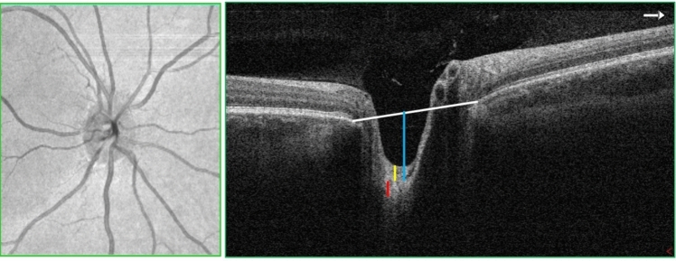
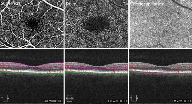
This study aimed to examine the existing literature that investigated the effectiveness of optical coherence tomography (OCT) and optical coherence tomography angiography (OCT-A) as a biomarker for idiopathic intracranial hypertension (IIH). Our search was conducted on January 17th, 2024, and included the databases, Medline, Scopus, Embase, Cochrane, Latin American and Caribbean Health Sciences Literature (LILACS), International Standard Randomized Controlled Trial Number (ISRCTN) registry, and the International Clinical Trials Registry Platform (ICTRP). Our final review included 84 articles. In 74 studies, OCT was utilized as the primary ocular imaging method, while OCT-A was employed in two studies including eight studies that utilized both modalities. Overall, the results indicated that IIH patients exhibited significant increases in retinal nerve fiber layer (RNFL) thickness, total retinal and macular thickness, optic nerve head volume, and height, optic disc diameter and area, rim area, and thickness compared to controls. A significant correlation was observed between cerebrospinal fluid (CSF) pressure and OCT parameters including RNFL thickness, total retinal thickness, macular thickness, optic nerve head volume, and optic nerve head height. Interventions aimed at lowering CSF pressure were associated with a substantial improvement in these parameters. Nevertheless, studies comparing peripapillary vessel density using OCT-A between IIH patients and controls yielded conflicting results. Our systematic review supports OCT as a powerful tool to accurately monitor retinal axonal and optic nerve head changes in patients with IIH. Future research is required to determine the utility of OCT-A in IIH.
Keywords: Idiopathic intracranial hypertension; Optic nerve head; Optical coherence tomography; Optical coherence tomography angiography; Peripapillary vessel density; Retinal nerve fiber layer.
© 2024. The Author(s).
Conflict of interest statement
No potential conflict of interest was reported by the authors.
Figures


Similar articles
-
Ganglion Cell Layer Thickness as a Biomarker for Amyotrophic Lateral Sclerosis Functional Outcome: An OCT study.Rom J Ophthalmol. 2025 Apr-Jun;69(2):200-207. doi: 10.22336/rjo.2025.32. Rom J Ophthalmol. 2025. PMID: 40698100 Free PMC article.
-
The Relationship between Lumbar Puncture Opening Pressure and Retinal Nerve Fiber Layer Thickness in the Diagnosis of Idiopathic Intracranial Hypertension: Is a Lumbar Puncture Always Necessary?Neurologist. 2024 Mar 1;29(2):91-95. doi: 10.1097/NRL.0000000000000528. Neurologist. 2024. PMID: 37839079
-
Optical coherence tomography (OCT) for detection of macular oedema in patients with diabetic retinopathy.Cochrane Database Syst Rev. 2015 Jan 7;1(1):CD008081. doi: 10.1002/14651858.CD008081.pub3. Cochrane Database Syst Rev. 2015. PMID: 25564068 Free PMC article.
-
Optic nerve head and fibre layer imaging for diagnosing glaucoma.Cochrane Database Syst Rev. 2015 Nov 30;2015(11):CD008803. doi: 10.1002/14651858.CD008803.pub2. Cochrane Database Syst Rev. 2015. PMID: 26618332 Free PMC article.
-
Evaluating the impact of anti-CGRP monoclonal antibodies on retinal features in migraine patients: a retrospective optical coherence tomography study.Ther Adv Neurol Disord. 2025 Jun 24;18:17562864251347277. doi: 10.1177/17562864251347277. eCollection 2025. Ther Adv Neurol Disord. 2025. PMID: 40568185 Free PMC article.
Cited by
-
Optical coherence tomography surpasses fundus imaging and intracranial pressure measurement in monitoring idiopathic intracranial hypertension.Sci Rep. 2025 Apr 28;15(1):14859. doi: 10.1038/s41598-025-96831-9. Sci Rep. 2025. PMID: 40295571 Free PMC article.
-
Longitudinal visual outcomes in idiopathic intracranial hypertension: the role of early prognostic indicators and risk stratification in disease management.J Neurol. 2025 Jan 15;272(2):108. doi: 10.1007/s00415-024-12859-3. J Neurol. 2025. PMID: 39812855
References
-
- Markey KA, Mollan SP, Jensen RH, Sinclair AJ (2016) Understanding idiopathic intracranial hypertension: mechanisms, management, and future directions. Lancet Neurol 15:78–91 - PubMed
-
- Kesler A, Gadoth N (2001) Epidemiology of idiopathic intracranial hypertension in Israel. J Neuroophthalmol 21:12–14 - PubMed
-
- Raoof N, Sharrack B, Pepper IM, Hickman SJ (2011) The incidence and prevalence of idiopathic intracranial hypertension in Sheffield, UK. Eur J Neurol 18:1266–1268 - PubMed
-
- Durcan FJ, Corbett JJ, Wall M (1988) The incidence of pseudotumor cerebri. Population studies in Iowa and Louisiana. Arch Neurol 45:875–877 - PubMed
Publication types
MeSH terms
Substances
LinkOut - more resources
Full Text Sources
