

MRI-based virtual pathology of the prostate
- PMID: 38856839
- PMCID: PMC12320515
- DOI: 10.1007/s10334-024-01163-w
MRI-based virtual pathology of the prostate
Abstract
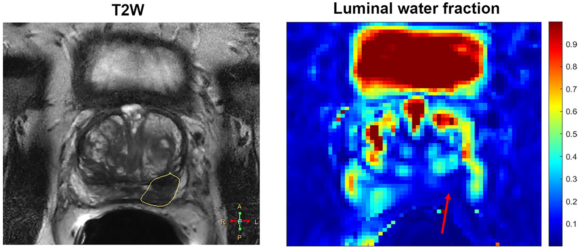
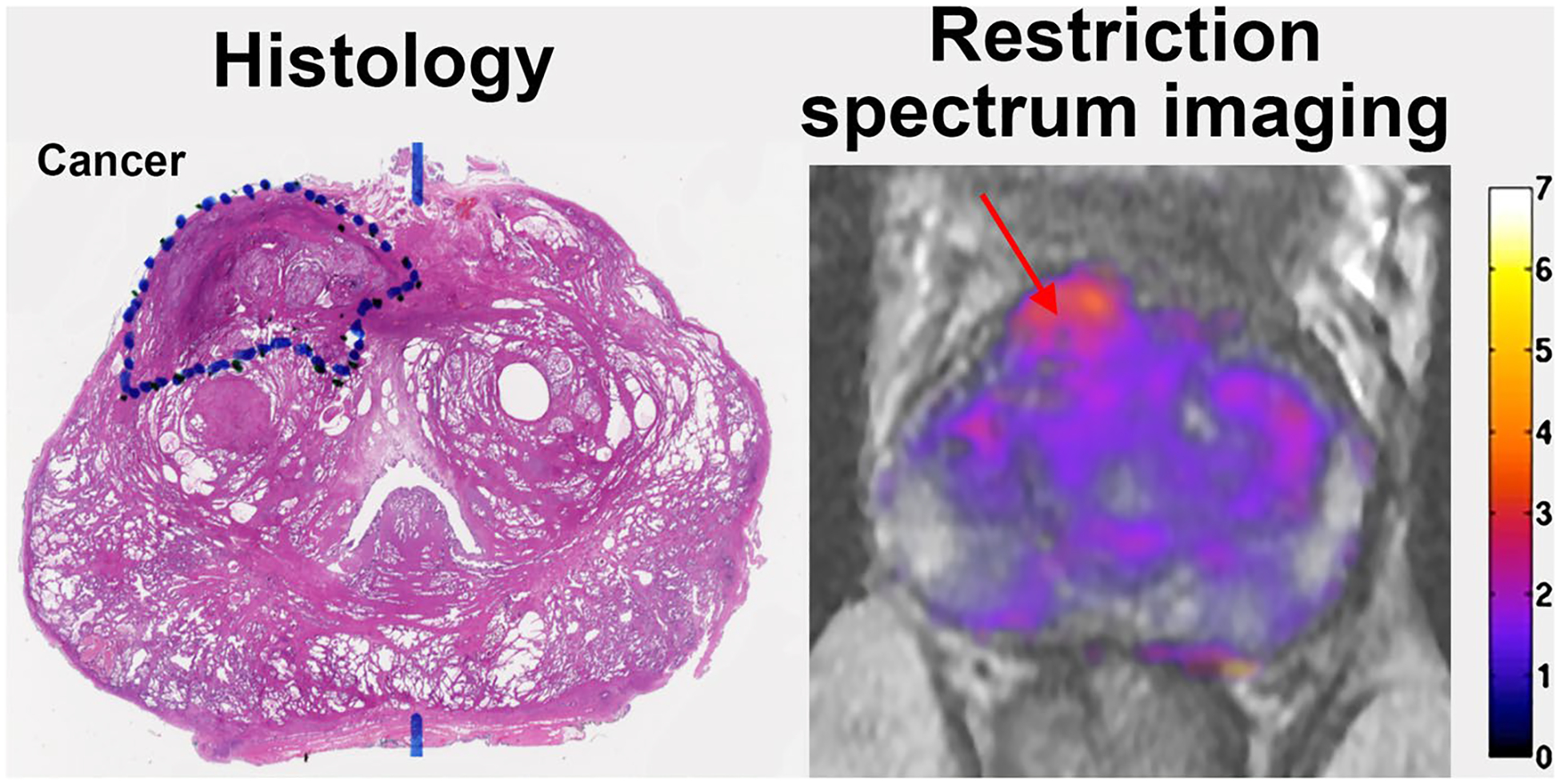
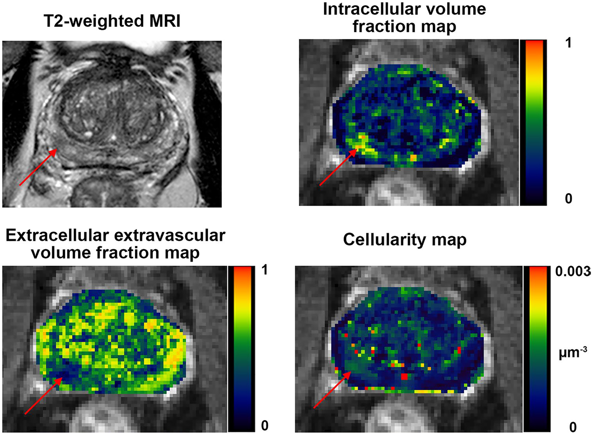
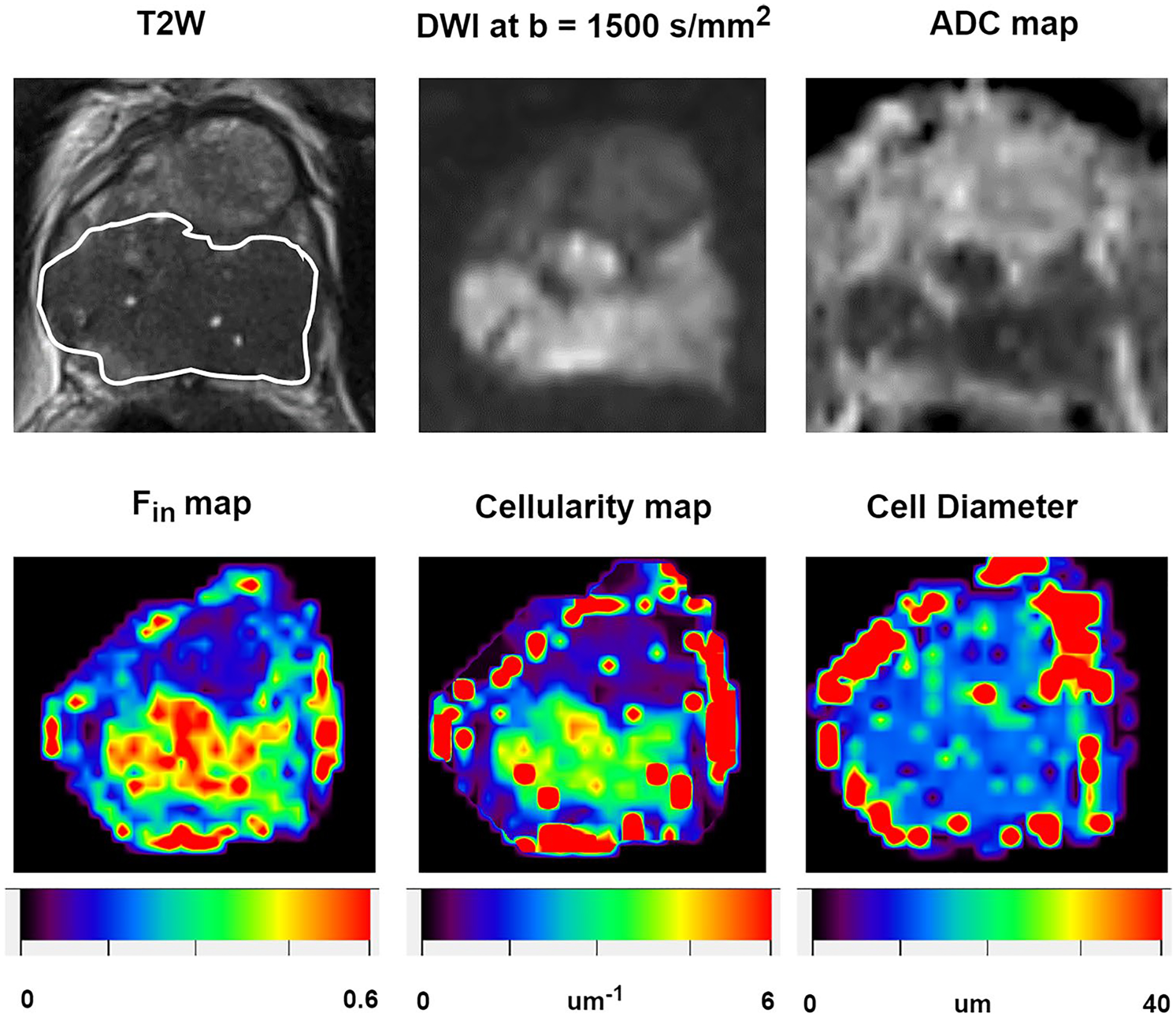
Prostate cancer poses significant diagnostic challenges, with conventional methods like prostate-specific antigen (PSA) screening and transrectal ultrasound (TRUS)-guided biopsies often leading to overdiagnosis or miss clinically significant cancers. Multiparametric MRI (mpMRI) has emerged as a more reliable tool. However, it is limited by high inter-observer variability and radiologists missing up to 30% of clinically significant cancers. This article summarizes a few of these recent advancements in quantitative MRI techniques that look at the "Virtual Pathology" of the prostate with an aim to enhance prostate cancer detection and characterization. These techniques include T2 relaxation-based techniques such as luminal water imaging, diffusion based such as vascular, extracellular, and restricted diffusion for cytometry in tumors (VERDICT) and restriction spectrum imaging or combined relaxation-diffusion techniques such as hybrid multi-dimensional MRI (HM-MRI), time-dependent diffusion imaging, and diffusion-relaxation correlation spectrum imaging. These methods provide detailed insights into underlying prostate microstructure and tissue composition and have shown improved diagnostic accuracy over conventional MRI. These innovative MRI methods hold potential for augmenting mpMRI, reducing variability in diagnosis, and paving the way for MRI as a 'virtual histology' tool in prostate cancer diagnosis. However, they require further validation in larger multi-center clinical settings and rigorous in-depth radiological-pathology correlation are needed for broader implementation.
Keywords: Prostate cancer; Quantitative MRI; Tissue composition; Virtual pathology.
© 2024. The Author(s), under exclusive licence to European Society for Magnetic Resonance in Medicine and Biology (ESMRMB).
Conflict of interest statement
Figures


Similar articles
-
The diagnostic accuracy and cost-effectiveness of magnetic resonance spectroscopy and enhanced magnetic resonance imaging techniques in aiding the localisation of prostate abnormalities for biopsy: a systematic review and economic evaluation.Health Technol Assess. 2013 May;17(20):vii-xix, 1-281. doi: 10.3310/hta17200. Health Technol Assess. 2013. PMID: 23697373 Free PMC article.
-
Prospective Validation of an Automated Hybrid Multidimensional MRI Tool for Prostate Cancer Detection Using Targeted Biopsy: Comparison with PI-RADS-based Assessment.Radiol Imaging Cancer. 2025 Jan;7(1):e240156. doi: 10.1148/rycan.240156. Radiol Imaging Cancer. 2025. PMID: 39836080 Free PMC article. Clinical Trial.
-
What Is the Negative Predictive Value of Multiparametric Magnetic Resonance Imaging in Excluding Prostate Cancer at Biopsy? A Systematic Review and Meta-analysis from the European Association of Urology Prostate Cancer Guidelines Panel.Eur Urol. 2017 Aug;72(2):250-266. doi: 10.1016/j.eururo.2017.02.026. Epub 2017 Mar 21. Eur Urol. 2017. PMID: 28336078
-
MRI software and cognitive fusion biopsies in people with suspected prostate cancer: a systematic review, network meta-analysis and cost-effectiveness analysis.Health Technol Assess. 2024 Oct;28(61):1-310. doi: 10.3310/PLFG4210. Health Technol Assess. 2024. PMID: 39367754 Free PMC article.
-
Comparing Three Different Techniques for Magnetic Resonance Imaging-targeted Prostate Biopsies: A Systematic Review of In-bore versus Magnetic Resonance Imaging-transrectal Ultrasound fusion versus Cognitive Registration. Is There a Preferred Technique?Eur Urol. 2017 Apr;71(4):517-531. doi: 10.1016/j.eururo.2016.07.041. Epub 2016 Aug 25. Eur Urol. 2017. PMID: 27568655
Cited by
-
Quantitative Multi-Parametric MRI of the Prostate Reveals Racial Differences.Cancers (Basel). 2024 Oct 16;16(20):3499. doi: 10.3390/cancers16203499. Cancers (Basel). 2024. PMID: 39456593 Free PMC article.
-
Quantitative Prostate MRI, From the AJR Special Series on Quantitative Imaging.AJR Am J Roentgenol. 2025 Aug;225(2):e2431715. doi: 10.2214/AJR.24.31715. Epub 2024 Oct 2. AJR Am J Roentgenol. 2025. PMID: 39356481 Free PMC article. Review.
References
-
- Siegel RL, Miller KD, Wagle NS, Jemal A (2023) Cancer statistics. CA Cancer J Clin 73(1):17–48 - PubMed
-
- Sung H, Ferlay J, Siegel RL, Laversanne M, Soerjomataram I, Jemal A, Bray F (2021) Global cancer statistics 2020: GLOBOCAN estimates of incidence and mortality worldwide for 36 cancers in 185 countries. CA Cancer J Clin 71(3):209–249 - PubMed
-
- Schouten MG, van der Leest M, Pokorny M, Hoogenboom M, Barentsz JO, Thompson LC, Futterer JJ (2017) Why and where do we miss significant prostate cancer with multi-parametric magnetic resonance imaging followed by magnetic resonance-guided and transrectal ultrasound-guided biopsy in biopsy-naive men? Eur Urol 71(6):896–903 - PubMed
-
- Force UPST (2018) Screening for prostate cancer: US Preventive Services Task Force recommendation statement. JAMA 319(18):1901–1913 - PubMed
-
- Kasivisvanathan V, Stabile A, Neves JB, Giganti F, Valerio M, Shanmugabavan Y, Clement KD, Sarkar D, Philippou Y, Thurtle D, Deeks J, Emberton M, Takwoingi Y, Moore CM (2019) Magnetic resonance imaging-targeted biopsy versus systematic biopsy in the detection of prostate cancer: a systematic review and meta-analysis. Eur Urol 76(3):284–303 - PubMed
Publication types
MeSH terms
Grants and funding
LinkOut - more resources
Full Text Sources
Medical
Research Materials
Miscellaneous
