. 2024 Jun 10;10(1):30.
doi: 10.1186/s40729-024-00548-9.

Round and flat zygomatic implants: effectiveness after a 3‑year follow‑up non‑interventional study

Affiliations

Round and flat zygomatic implants: effectiveness after a 3‑year follow‑up non‑interventional study

Carlos Aparicio et al. Int J Implant Dent. .

Abstract

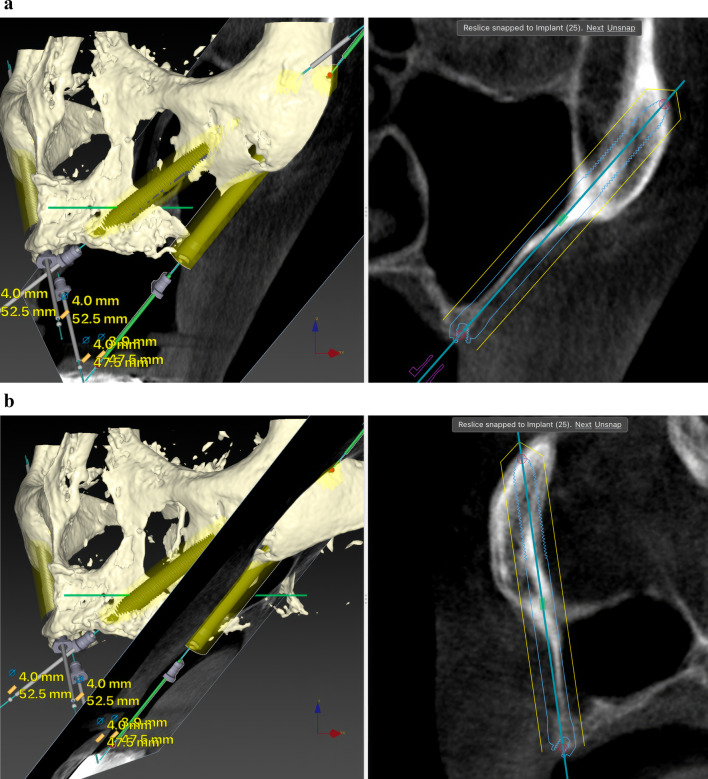
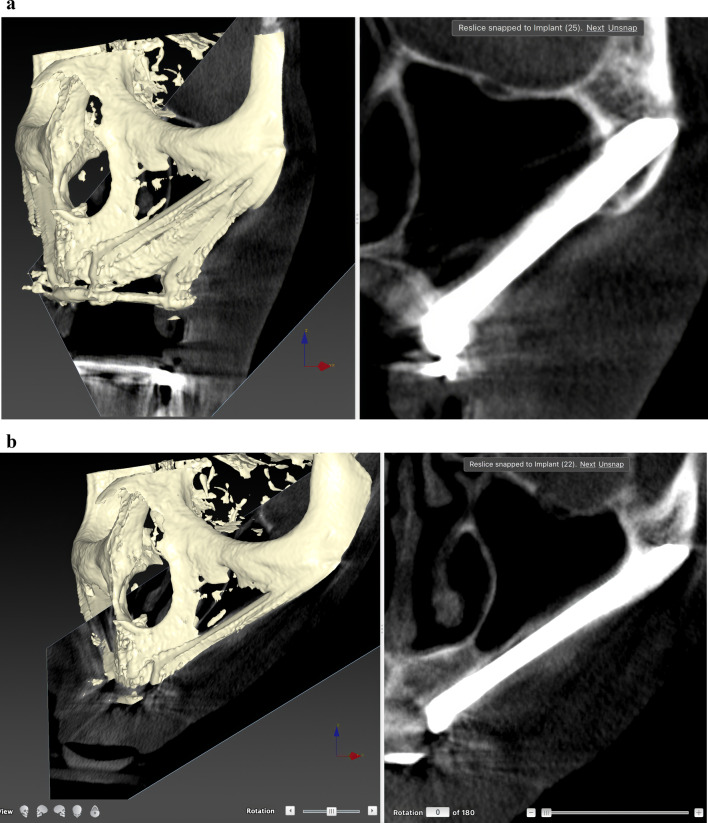
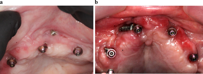
Purpose: This non-interventional study investigates variations in the type and frequency of late complications linked to novel zygomatic implant designs, installed adhering to the Zygoma Anatomy-Guided Approach (ZAGA) concept, over an extended follow-up period of at least 3 years.

Methods: Consecutive patients presenting indications for treatment with ZIs were treated according to ZAGA recommendations. Implants were immediately loaded. The ORIS success criteria for prosthetic offset, stability, sinus changes and soft-tissue status were used to evaluate the outcome.

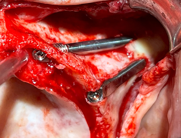
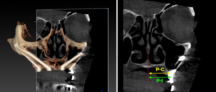
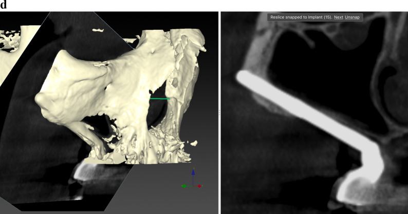
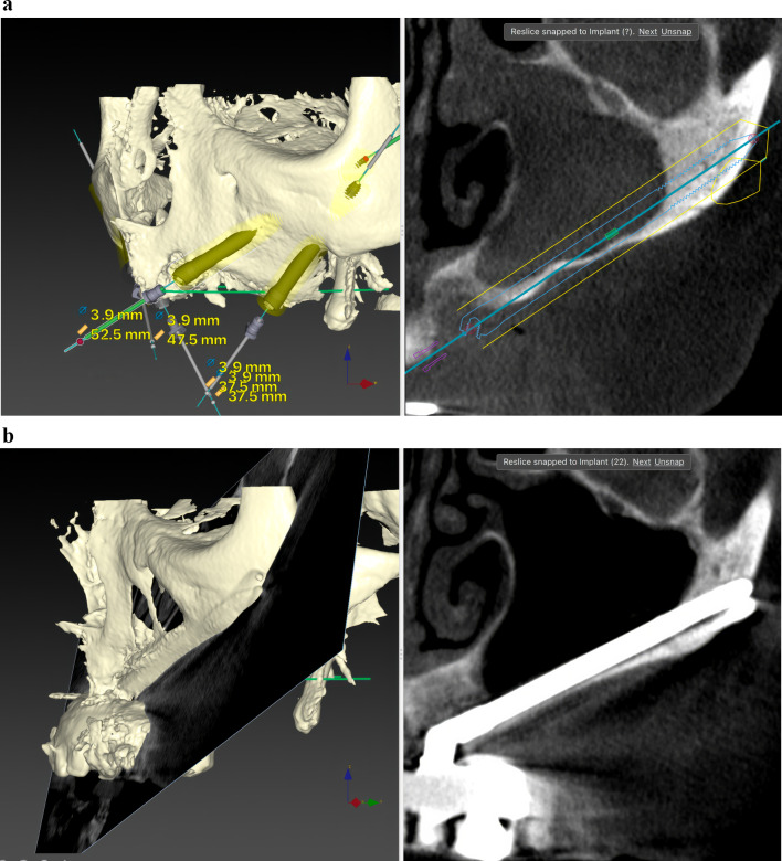
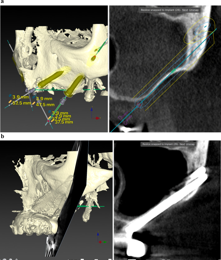
Results: Twenty patients were treated. Ten patients received two ZIs and regular implants; one received three ZIs plus regular implants, and nine received four ZIs. Fifty-nine ZIs were placed: thirty-six (61%) Straumann ZAGA-Flat implants and twenty-three (39%) Straumann ZAGA-Round implants. Four patients (20%) presented earlier sinus floor discontinuities. Fifteen patients (75%) had prior sinus opacities. Nineteen patients were followed for between 38 and 53 months (mean 46.5 months). One patient dropped out after 20 months. When comparing pre-surgical CBCT with post-surgical CBCT, 84.7% of the sites presented identical or less sinus opacity; nine locations (15%) showed decreased, and another nine increased (15%) post-surgical sinus opacity. Fifty-three ZIs (89.8%) maintained stable soft tissue. Six ZIs had recessions with no signs of infection. ZIs and prosthesis survival rate was 100%.

Conclusions: The study highlights the effectiveness of ZAGA-based zygomatic implant rehabilitations using Round and Flat designs. Despite patient number constraints, minimal changes in the frequency of late complications from the 1-year follow-up were observed. 100% implant and prosthesis survival rate over a mean follow-up of 46.5 months is reported.

Keywords: ORIS criteria; ZAGA; ZAGA implants; ZAGA-flat; ZAGA-round; Zygomatic implant late complications; Zygomatic implants.


Conflict of interest statement

Carlos Aparicio is the founding president of Zygoma ZAGA Centers and occasionally provides CE activities for NobelBiocare, Straumann Group, SouthernImplants and Versah Osseodensification Company. Waldemar Polido occasionally performs CE activities for Straumann Group and Geistlich Biomaterials. Bilal Al-Nawas occasionally performs CE activities and obtains research grants for StraumannGroup, BEGO, Camlog, Dentsply and Geistlich Biomaterials. All other authors declare that they have no conflicts of interest.

Figures

Fig. 1
Fig. 1
The clinical photograph shows two different Straumann ZAGA implants. In the anterior position, we can visualize the ZAGA Round model with a round section and macro-threads in its collar. In the premolar/molar position, we see the ZAGA Flat model. It has a circumferential arch section designed not to compress the soft tissue
Fig. 2
Fig. 2
a Oblique CBCT slice, taken using DTX Studio Implant software, showing the planning and anatomy at the level of the second premolar/upper left first molar of the ZAGA Flat implant in Fig. 1. b The DTX Studio Implant software allows the rotation of the oblique plane attached to the implant. Thus, we can observe that the planning for the placement of the ZAGA Flat implant in Fig. 1 includes the partial use of the anterior wall of the temporal fossa. By increasing the number of cortices crossed by the implant, we will increase its primary stability
Fig. 3
Fig. 3
The oblique CBCT slice, taken using DTX Studio Implant software, shows the planning and anatomy at the level of the left lateral incisor of the ZAGA Round implant in Fig. 1. Although the patient moved during the acquisition, we declined to perform another CBCT
Fig. 4
Fig. 4
3D stereolithographic model obtained with manual segmentation used to familiarize ourselves with the patient’s anatomy and planning. Models provided by ZAGA Centers S.L., Barcelona, Spain
Fig. 5
Fig. 5
a Oblique CBCT slice taken in the control of the Straumann ZAGA Flat implant placed in the left posterior position in Fig. 1. The image, taken using DTX Studio Implant software, is to be compared with the one in Fig. 2a before the placement of the ZI. This will enable us to understand the condition of the implant and the possible variations in the adjacent structures. b Oblique CBCT slice taken in the control of the Straumann ZAGA Round implant placed in the left anterior position in Fig. 1. The image, taken using DTX Studio Implant software, is to be compared with the one in Fig. 3 before the placement of the ZI. This will enable us to understand the condition of the implant and the possible variations in the adjacent structures
Fig. 6
Fig. 6
Immediate prosthesis placed on the implants in Fig. 1
Fig. 7
Fig. 7
Using the DTX Studio Implant, the left 3D image is cut in an orthoradial plane to the emergence of the ZAGA Flat implant in Fig. 1. On the right side of the illustration, we visualize that the red dashed line divides the palate in two. In yellow, the P–C segment indicates the distance from the center of the palate (P) to the center of the ridge (C). In green, the P-I segment indicates the distance from the center of the palate to the center of the implant platform (I). The subtraction of both segments will indicate the measurement and type of offset, if any
Fig. 8
Fig. 8
Status of soft tissues around Straumann abutments 50 months after surgery for zygomatic implant placement
Fig. 9
Fig. 9
The occlusal photograph shows the final position of the implants with their abutments of the case illustrated in Fig. 1 and subsequent ones. The two Straumann ZAGA Round implants, placed in the anterior position, typically appear on the palatal side of the ridge (green circles). The platforms of the Straumann ZAGA Flat implants in posterior positions are placed in the middle of the ridge (yellow circle)
Fig. 10
Fig. 10
a Oblique CBCT slice taken on November 11th 2020. DTX Studio Implant software was used to plan the implant trajectory using the ZAGA Channel approach. b December 11th 2020. One week after surgery, planned in a, the patient presents with acute symptoms of rhinosinusitis. The oblique CBCT slice using DTX Studio Implant software shows complete opacity of the right maxillary sinus. c March 3rd 2021. Three months after surgery. After medical treatment, the clinician’s symptoms disappear, and when we compare the image of this figure with the one in Fig. 11a, we may verify that the M-LM is negative. d The DTX Studio Implant software allows us to superimpose a virtual implant on the real one and analyze it. The new CBCT was taken on December 22, 2023, 3 years after the surgery. When we compare it with the CBCT before surgery, we verify that the M-LM is negative
Fig. 10
Fig. 10
a Oblique CBCT slice taken on November 11th 2020. DTX Studio Implant software was used to plan the implant trajectory using the ZAGA Channel approach. b December 11th 2020. One week after surgery, planned in a, the patient presents with acute symptoms of rhinosinusitis. The oblique CBCT slice using DTX Studio Implant software shows complete opacity of the right maxillary sinus. c March 3rd 2021. Three months after surgery. After medical treatment, the clinician’s symptoms disappear, and when we compare the image of this figure with the one in Fig. 11a, we may verify that the M-LM is negative. d The DTX Studio Implant software allows us to superimpose a virtual implant on the real one and analyze it. The new CBCT was taken on December 22, 2023, 3 years after the surgery. When we compare it with the CBCT before surgery, we verify that the M-LM is negative
Fig. 11
Fig. 11
a This heavy smoker patient (P6) presented, as a consequence of implant failure and previous augmentation surgeries, multiple bony discontinuities in the sinus and nasal floor on radiological examination. As observed in the oblique CBCT slice taken in the direction of the left anterior implant, both the maxillary sinuses and the ostium are opaque. The patient had no clinical symptoms. b The same patient (P6) in a presented this CBCT image 18 months after surgery performed according to the recommendations of the ZAGA protocol and using a Straumann ZAGA Round implant. The authors need to determine the reasons for the improved patency at both the sinuses and the ostium. Note also the spontaneous healing of the initial osseous defect at the level of the left incisors
Fig. 12
Fig. 12
a The oblique CBCT slice shows the left posterior sector with a ZAGA channel osteotomy planning. As in the anterior sector, the maxillary sinus and the ostium are opaque. b The same patient (P6) in a presented this CBCT image showing sinus patency 18 months after a zygoma surgery performed according to the recommendations of the ZAGA protocol and using a Straumann ZAGA Flat implant
Fig. 13
Fig. 13
a The clinical photograph shows a soft tissue defect in the window pattern that appeared in this patient 2 years after surgery. Soft tissue color is normal. b To close the window in a, surgery was performed by mobilizing soft tissues and partially removing the threads from the collar of the implant. The result was a wider defect. After 3 years, it remains stable

Similar articles

Cited by

References

    1. Aparicio C, Branemark PI, Keller EE, Olivé J. Reconstruction of the premaxilla with autogenous lliac bone in combination with osseointegrated implants. Int J Oral Maxillofac Implants. 1993;8:1–15.
    1. Jensen OT, Brownd C, Blacker J. Nasofacial prostheses supported by osseointegrated implants. Int J Oral Maxillofac Implants. 1992;7(2):203–211. - PubMed
    1. Chrcanovic BR, Albrektsson T, Wennerberg A. Survival and complications of zygomatic implants: an updated systematic review. J Oral Maxillofac Surg. 2016;74(10):1949–1964. doi: 10.1016/j.joms.2016.06.166. - DOI - PubMed
    1. Sáez-Alcaide LM, Cortés-Bretón-Brinkmann J, Sánchez-Labrador L, Pérez-González F, Forteza-López A, Molinero-Mourelle P, et al. Patient-reported outcomes in patients with severe maxillary bone atrophy restored with zygomatic implant-supported complete dental prostheses: a systematic review. Acta Odontol Scand. 2022;80(5):363–373. doi: 10.1080/00016357.2021.2018494. - DOI - PubMed
    1. Solà Pérez A, Pastorino D, Aparicio C, Pegueroles Neyra M, Khan RS, Wright S, et al. Success rates of zygomatic implants for the rehabilitation of severely atrophic maxilla: a systematic review. Dent J. 2022;10(8):151. doi: 10.3390/dj10080151. - DOI - PMC - PubMed

Substances

LinkOut - more resources