. 2024 Jun 10;81(1):254.
doi: 10.1007/s00018-024-05253-9.

Pyruvate kinase M2 sustains cardiac mitochondrial quality surveillance in septic cardiomyopathy by regulating prohibitin 2 abundance via S91 phosphorylation

Affiliations

Pyruvate kinase M2 sustains cardiac mitochondrial quality surveillance in septic cardiomyopathy by regulating prohibitin 2 abundance via S91 phosphorylation

Yingzhen Du et al. Cell Mol Life Sci. .

Abstract

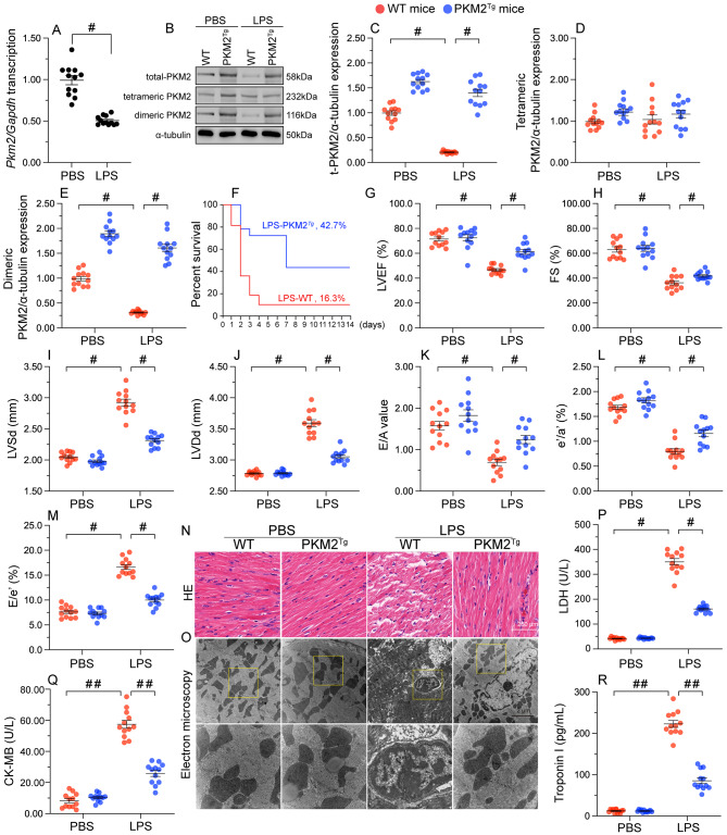
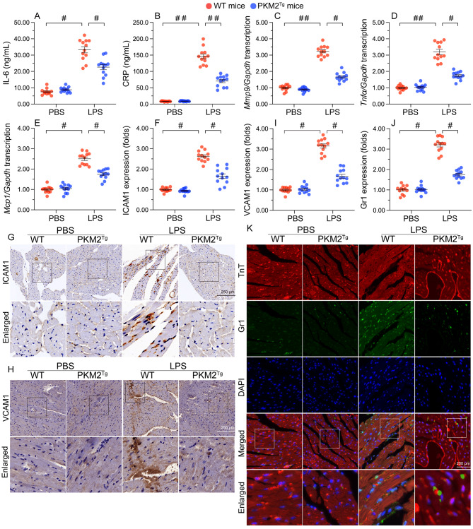
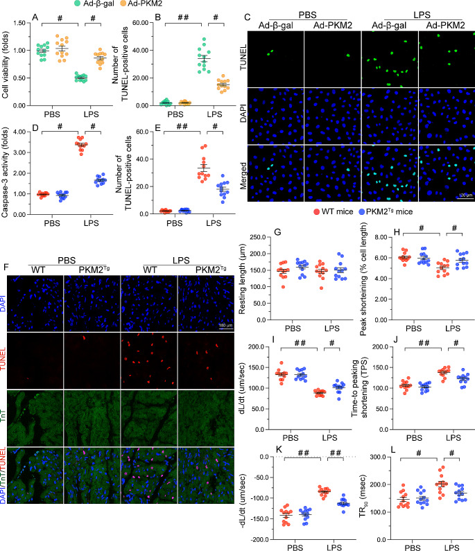
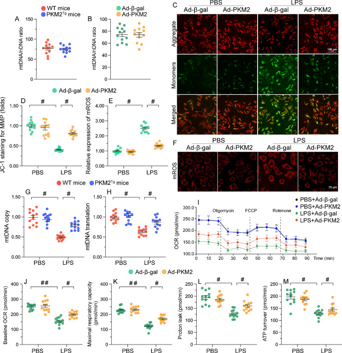
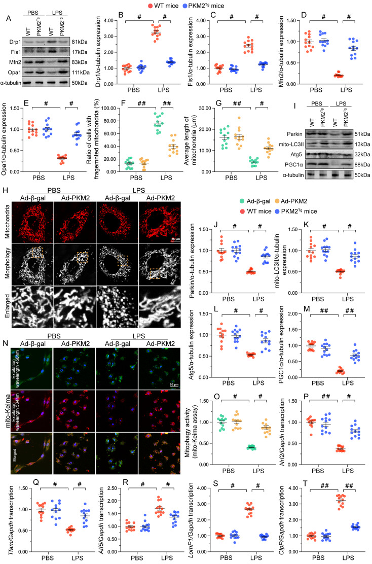
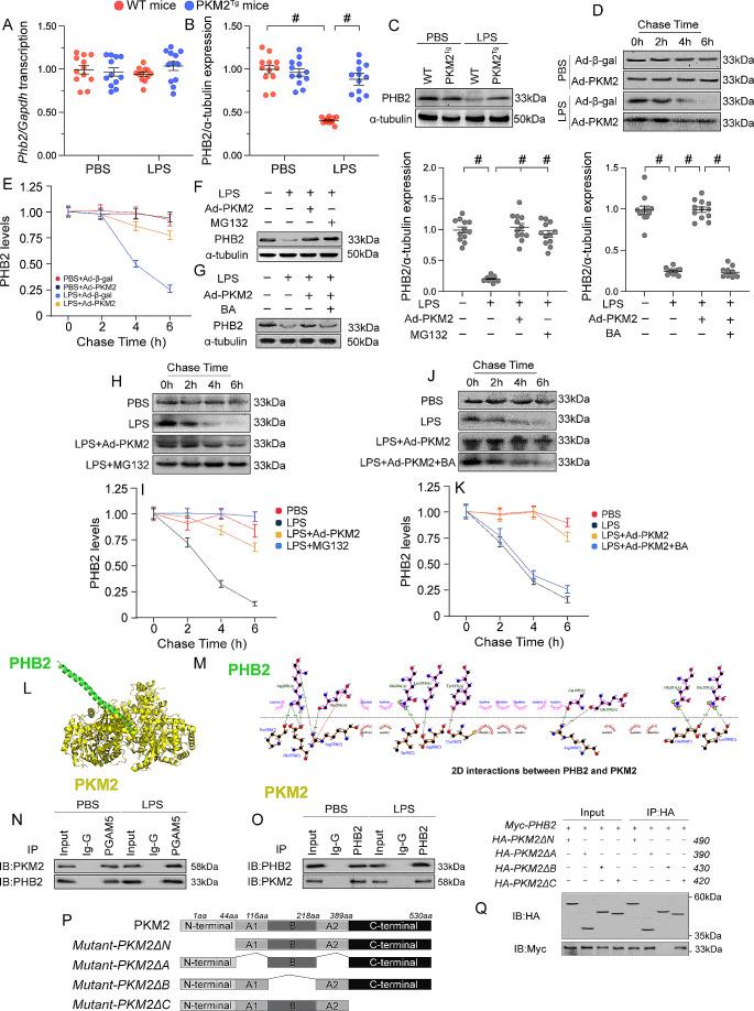
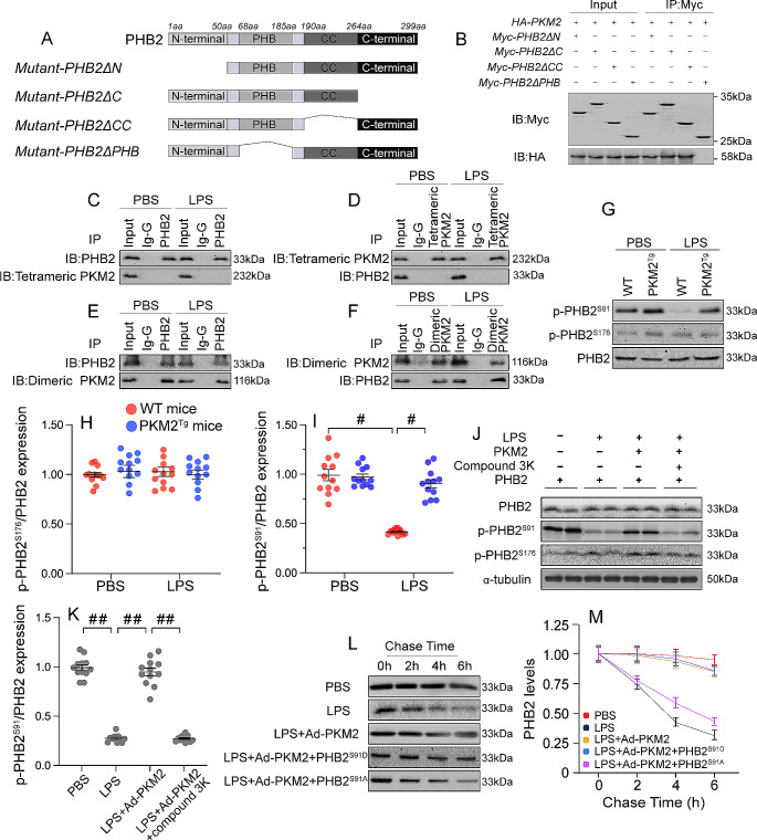
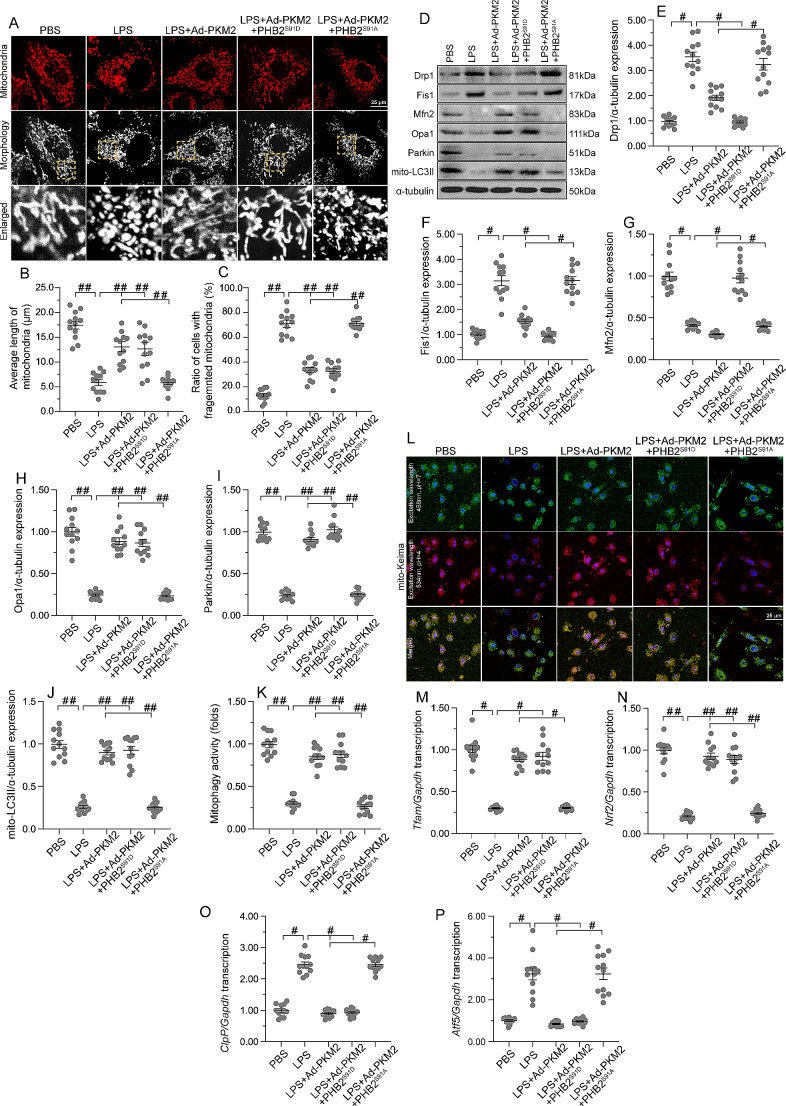
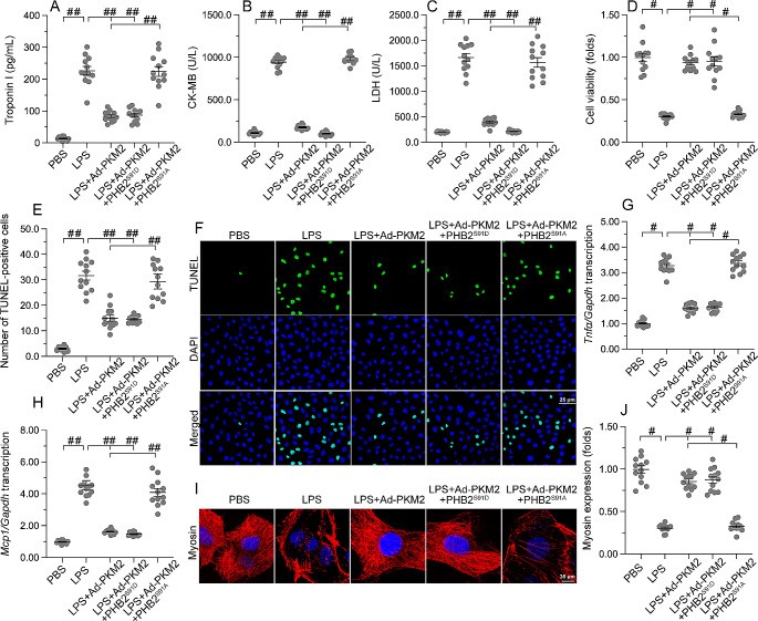
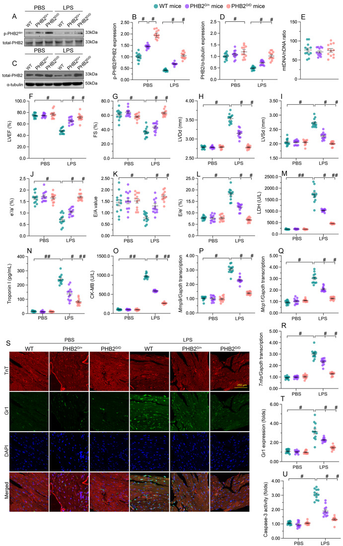
The endogenous mitochondrial quality control (MQC) system serves to protect mitochondria against cellular stressors. Although mitochondrial dysfunction contributes to cardiac damage during many pathological conditions, the regulatory signals influencing MQC disruption during septic cardiomyopathy (SC) remain unclear. This study aimed to investigate the involvement of pyruvate kinase M2 (PKM2) and prohibitin 2 (PHB2) interaction followed by MQC impairment in the pathogenesis of SC. We utilized LPS-induced SC models in PKM2 transgenic (PKM2TG) mice, PHB2S91D-knockin mice, and PKM2-overexpressing HL-1 cardiomyocytes. After LPS-induced SC, cardiac PKM2 expression was significantly downregulated in wild-type mice, whereas PKM2 overexpression in vivo sustained heart function, suppressed myocardial inflammation, and attenuated cardiomyocyte death. PKM2 overexpression relieved sepsis-related mitochondrial damage via MQC normalization, evidenced by balanced mitochondrial fission/fusion, activated mitophagy, restored mitochondrial biogenesis, and inhibited mitochondrial unfolded protein response. Docking simulations, co-IP, and domain deletion mutant protein transfection experiments showed that PKM2 phosphorylates PHB2 at Ser91, preventing LPS-mediated PHB2 degradation. Additionally, the A domain of PKM2 and the PHB domain of PHB2 are required for PKM2-PHB2 binding and PHB2 phosphorylation. After LPS exposure, expression of a phosphorylation-defective PHB2S91A mutant negated the protective effects of PKM2 overexpression. Moreover, knockin mice expressing a phosphorylation-mimetic PHB2S91D mutant showed improved heart function, reduced inflammation, and preserved mitochondrial function following sepsis induction. Abundant PKM2 expression is a prerequisite to sustain PKM2-PHB2 interaction which is a key element for preservation of PHB2 phosphorylation and MQC, presenting novel interventive targets for the treatment of septic cardiomyopathy.

Keywords: MQC; Mitochondria; PHB2; PKM2; Septic cardiomyopathy.


Conflict of interest statement

The authors declared no conflict of interest.

Figures

Fig. 1
Fig. 1
PKM2 downregulation contributes to myocardial injury in SC. PKM2 transgenic (PKM2Tg) mice and wild-type (WT) mice aged 8–10 weeks were injected intraperitoneally with 10 mg/kg LPS to induce SC. In vivo measurements were performed after 24 h later, and mice administered an equal volume of phosphate buffer saline served as controls. (A) Analysis of transcriptional levels of cardiac Pkm2 in WT mice by qPCR. (B-E) Western blot analysis of total PKM2, dimeric PKM2, and tetrameric PKM2 protein expression in mouse heart tissues. (F) Survival analysis of control and PKM-overexpressing mice after SC induction. (G-M) Assessment of cardiac function in WT or PKM2Tg mice by echocardiography. LVDd, left ventricular diastolic dimension; LVDs, left ventricular systolic dimension; IVS, interventricular septum thickness; E/A, ratio of early to late transmitral flow velocities; FS, ratio of left ventricular fractional shortening. (N) HE staining of myocardial tissue. (O) Representative images of electron microscopy of heart tissues. Yellow arrows indicate disorganized muscle fibers and deformed mitochondria with edematous cristae. (P-R) ELISA-based analysis of LDH, TnI, and CK-MB levels in mice sera. Values are presented as mean ± SEM. For in vivo data, n = 6 mice per group. For in vitro data, n = 4 independent experiments. #p < 0.05, and ##p < 0.01
Fig. 2
Fig. 2
PKM2 overexpression reduces LPS-mediated myocardial inflammation. PKM2 transgenic (PKM2Tg) mice and wild-type (WT) mice aged 8–10 weeks were injected intraperitoneally with 10 mg/kg LPS to induce SC. In vivo measurements were performed after 24 h later, and mice administered an equal volume of phosphate buffer saline served as controls. (A, B) ELISA-based measurement of IL-6 and CRP levels in mouse serum. (C-E) Transcriptional analysis of cardiac Mmp9, Mcp1, and Tnfα expression in WT or PKM2Tg mice by qPCR. (F, G) Quantification and representative images of immunohistochemical staining of ICAM1 in heart tissues. (H, I) Quantification and representative images of immunohistochemical staining of VCAM1 in heart tissues. (J, K) Immunofluorescent staining of Gr-1-positive neutrophils in heart tissues. Values are presented as mean ± SEM. For in vivo data, n = 6 mice per group. For in vitro data, n = 4 independent experiments. #p < 0.05, and ##p < 0.01
Fig. 3
Fig. 3
PKM2 overexpression preserves cardiomyocyte viability after septic insult. PKM2 transgenic (PKM2Tg) mice and wild-type (WT) mice aged 8–10 weeks were injected intraperitoneally with 10 mg/kg LPS to induce SC. In vivo and ex-vivo measurements were performed after 24 h later, and mice administered an equal volume of phosphate buffer saline served as controls. Immortalized mouse cardiac muscle HL-1 cells were treated with 10 µg/mL of LPS for 24 h to simulate SC in vitro. Cells treated with an equal volume of phosphate buffer saline were used as controls. Before LPS treatment, cardiomyocytes were transduced with adenovirus encoding PKM2 (Ad-PKM2). β-gal-overexpressing (Ad-β-gal) cells were used as controls. (A) Cell viability was measured in vitro via CCK-8 assay. (B, C) TUNEL staining was applied to analyze the number of apoptotic HL-1 cells in vitro in the presence of LPS. (D) ELISA-based measurement of cardiac caspase-3 activity following SC induction in vivo. (E, F) Results of TUNEL assays conducted in heart tissues following SC induction in vivo. (G-L) Analysis of contraction and relaxation parameters in field-stimulated single adult cardiomyocytes isolated from WT and PKM2Tg mice. Values are presented as mean ± SEM. For in vivo data, n = 6 mice per group. For in vitro data, n = 4 independent experiments. #p < 0.05, and ##p < 0.01
Fig. 4
Fig. 4
PKM2 protects mitochondrial function in cardiomyocytes exposed to LPS. PKM2 transgenic (PKM2Tg) mice and wild-type (WT) mice aged 8–10 weeks were injected intraperitoneally with 10 mg/kg LPS to induce SC. In vivo measurements were performed after 24 h later, and mice administered an equal volume of phosphate buffer saline served as controls. Immortalized mouse cardiac muscle HL-1 cells were treated with 10 µg/mL of LPS for 24 h to simulate SC in vitro. Cells treated with an equal volume of phosphate buffer saline were used as controls. Before LPS treatment, cardiomyocytes were transduced with Adenovirus encoding PKM2 (Ad-PKM2). β-gal-overexpressing (Ad-β-gal) cells were used as controls. (A) The CO1 gene of mtDNA and the NDUFV1 gene of nDNA were amplified using qPCR to assess the relative ratio of mtDNA/nDNA in heart tissues obtained from WT or PKM2Tg mice under normal physiological conditions. (B) HL-1 cells were transfected with Ad-PKM2 or Ad-β-gal. Subsequently, the CO1 gene of mtDNA and the NDUFV1 gene of nDNA were amplified using qPCR to evaluate the relative mtDNA/nDNA ratio in vitro. (C, D) Analysis of the effect of LPS exposure on mitochondrial membrane potential in JC-1-loaded HL-1 cells overexpressing adenovirus PKM2 (Ad-PKM2) or β-gal (Ad-β-gal; control). (E, F) Detection of mitochondrial ROS production in HL-1 cells loaded with MitoSOX Red. (G, H) Estimation of mitochondrial DNA (mtDNA) copy number and transcription via qPCR. (I-M) Mitochondrial oxygen consumption rate (OCR) was determined by the Seahorse XF realtime ATP rate assay using an XF-24 Extracellular Flux Analyzer. ATP turnover, baseline OCR, maximal respiration capacity and proton leak were measured in HL-1 cells treated with Ad-β-gal or Ad-PKM2. Values are presented as mean ± SEM. For in vivo data, n = 6 mice per group. For in vitro data, n = 4 independent experiments. #p < 0.05, and ##p < 0.01
Fig. 5
Fig. 5
PKM2 overexpression maintains mitochondrial quality control. PKM2 transgenic (PKM2Tg) mice and wild-type (WT) mice aged 8–10 weeks were injected intraperitoneally with 10 mg/kg LPS to induce SC. In vivo measurements were performed after 24 h later, and mice administered an equal volume of phosphate buffer saline served as controls. Immortalized mouse cardiac muscle HL-1 cells were treated with 10 µg/mL of LPS for 24 h to simulate SC in vitro. Cells treated with an equal volume of phosphate buffer saline were used as controls. Before LPS treatment, cardiomyocytes were transduced with Adenovirus encoding PKM2 (Ad-PKM2). β-gal-overexpressing (Ad-β-gal) cells were used as controls. (A-E). Western blot analysis of cardiac Drp1, Fis1, Mfn2, and Opa1 expression in vivo. (F-H) Results of immunofluorescence analysis of mitochondrial morphology in HL-1 cells. Rate of cells with fragmented mitochondria (F), average length of mitochondria (G), and representative mitochondrial immunofluorescence images (H) are shown. (I-M) Representative immunoblots and quantification of changes in Parkin, Atg5, Beclin1, and PGC1α expression in cardiac tissues. (N, O). Representative images and quantitative results of mt-Keima assays assessing mitophagy in HL-1 cells. (P-T) Transcriptional analysis of cardiac Nrf2, Tfam, Atf6, LonP1, and mtHsp70 expression by qPCR in vivo. Values are presented as mean ± SEM. For in vivo data, n = 6 mice per group. For in vitro data, n = 4 independent experiments. #p < 0.05, and ##p < 0.01
Fig. 6
Fig. 6
PKM2 binds to and prevents PHB2 degradation. PKM2 transgenic (PKM2Tg) mice and wild-type (WT) mice aged 8–10 weeks were injected intraperitoneally with 10 mg/kg LPS to induce SC. In vivo measurements were performed after 24 h later, and mice administered an equal volume of phosphate buffer saline served as controls. Immortalized mouse cardiac muscle HL-1 cells were treated with 10 µg/mL of LPS for 24 h to simulate SC in vitro. Cells treated with an equal volume of phosphate buffer saline were used as controls. Before LPS treatment, cardiomyocytes were transduced with Adenovirus encoding PKM2 (Ad-PKM2). β-gal-overexpressing (Ad-β-gal) cells were used as controls. (A) Analysis of the effect of LPS exposure on cardiac Phb2 mRNA expression levels in WT and PKM2Tg mice. (B, C) Western blots analysis of cardiac PHB2 protein levels in WT and PKM2Tg mice. (D, E) Analysis of the half-life of PHB2 protein in HL-1 cells (pulse-chase assay). (F-G) Western blot analysis of PHB2 expression in HL-1 cells exposed to LPS in the presence or absence of MG132, a proteasome inhibitor, or betulinic acid (BA), a proteasome activator. (H, I) Pulse-chase analysis of PHB2 degradation rate in cultured in HL-1 cells treated with LPS and MG132. (J, K) Pulse-chase analysis of PHB2 degradation rate in cultured in HL-1 cells treated with LPS and BA. (L-M) Mapping of PHB2/PKM2 interacting regions by docking analysis. (N, O) Co-IP analysis of PKM2/PHB2 binding in HL-1 cells transfected with different domain deletion PKM2 mutants. (P) Mapping of PKM2 regions and deletion mutants. (Q) Western blot analysis of PHB2 expression in HL-1 cells transfected with domain deletion PKM2 mutants. Values are presented as mean ± SEM. For in vivo data, n = 6 mice per group. For in vitro data, n = 4 independent experiments. #p < 0.05
Fig. 7
Fig. 7
PKM2 induces PHB2 phosphorylation. PKM2 transgenic (PKM2Tg) mice and wild-type (WT) mice aged 8–10 weeks were injected intraperitoneally with 10 mg/kg LPS to induce SC. In vivo measurements were performed after 24 h later, and mice administered an equal volume of phosphate buffer saline served as controls. Immortalized mouse cardiac muscle HL-1 cells were treated with 10 µg/mL of LPS for 24 h to simulate SC in vitro. Cells treated with an equal volume of phosphate buffer saline were used as controls. Before LPS treatment, cardiomyocytes were transduced with Adenovirus encoding PKM2 (Ad-PKM2). β-gal-overexpressing (Ad-β-gal) cells were used as controls. (A) Mapping of PHB2 regions and deletion mutants. (B) Representative immunoblot from immunoprecipitation analysis of HL-1 cells transfected with PHB2 deletion mutants. (C-F) Co-IP analysis of the interaction between dimeric and tetrameric PKM2 forms and PHB2 in HL-1 cells. (G-I) Western blot analysis of p-PHB2S176 and p-PHB2Ser91 expression in cardiac tissues. (J, K) Representative results of an in vitro kinase assay evaluating binding of phospho-PHB2 variants to PKM2 in the presence of LPS or compound 3k, a PKM2 inhibitor. (L, M) Pulse-chase analysis of PHB2 protein half-life in HL-1 cells transfected with PHB2S91D and PHB2S91A mutant constructs. Values are presented as mean ± SEM. For in vivo data, n = 6 mice per group. For in vitro data, n = 4 independent experiments. #p < 0.05, and ##p < 0.01
Fig. 8
Fig. 8
PHB2 dephosphorylation abolishes PKM2-mediated mitochondrial protection. HL-1 cardiomyocytes were transfected with PKM2 overexpression adenovirus (Ad-PKM2), a phosphorylation-defective PHB2S91A mutant, or a phosphorylation-mimetic PHB2 S91D mutant before LPS treatment. Adenovirus loaded β-gal (Ad-β-gal) cells were used as controls. (A-C) Immunofluorescence analysis of mitochondrial morphology in HL-1 cells. Representative images of mitochondria immunofluorescence (A), average mitochondrial length (B), and proportion of cardiomyocytes with fragmented mitochondria (C) are shown. (D-J) Western blot analysis of Drp1, Fis1, Mfn2, Opa1, Parkin, and Atg5 in HL-1 cells. (K, L) Mitophagy analysis results (mt-Keima assay). (M-P). Transcriptional analysis of Tfam, Nrf2, mtHsp70, and Atf6 expression by qPCR. Values are presented as mean ± SEM. For in vivo data, n = 6 mice per group. For in vitro data, n = 4 independent experiments. #p < 0.05, and ##p < 0.01
Fig. 9
Fig. 9
PKM2-mediated cardiomyocyte protection against septic insult requires PHB2 phosphorylation. HL-1 cardiomyocytes were transfected with PKM2 overexpression Adenovirus (Ad-PKM2), a phosphorylation-defective PHB2S91A mutant, or a phosphorylation-mimetic PHB2 S91D mutant before LPS treatment. Adenovirus loaded β-gal (Ad-β-gal) cells were used as controls. (A, C) ELISA-based analysis of TnI, CK-MB, and LDH levels in culture media of HL-1 cells transfected with phosphorylation-defective (PHB2S91A) and phosphorylation-mimetic (PHB2S91D) mutant constructs. (D) Cell viability analysis via CCK-8 assay in vitro. (E, F) Apoptosis analysis by TUNEL staining in cultured HL-1 cells. (G, H) Transcriptional analysis of Tnfα and Mcp1 expression. (I, J) Representative images of myosin immunofluorescence. Myosin expression levels were normalized to those of the control group. Values are presented as mean ± SEM. For in vivo data, n = 6 mice per group. For in vitro data, n = 4 independent experiments. #p < 0.05, and ##p < 0.01
Fig. 10
Fig. 10
Knockin mice expressing phospho-mimetic Phb2S91 are less vulnerable to SC. Heterozygous Phb2S91D/+ mice, homozygous Phb2S91D/D mice, and wild-type (WT) mice aged 8–10 weeks were injected intraperitoneally with 10 mg/kg LPS to induce SC. Mice administered an equal volume of phosphate buffer saline served as controls. (A, B) Western blot analysis of cardiac p-PHB2S91 in WT, heterozygous Phb2S91D/+, and homozygous Phb2S91D/D mice treated with PBS or LPS. Total-PHB2 was used as the loading control. (C, D) Western blot analysis of total-PHB2 in WT, heterozygous Phb2S91D/+, and homozygous Phb2S91D/D mice treated with PBS or LPS. α-tubulin was used as the loading control. (E) The CO1 gene of mtDNA and the NDUFV1 gene of nDNA were amplified using qPCR to assess the relative ratio of mtDNA/nDNA in heart tissues obtained from WT, heterozygous Phb2S91D/+, and homozygous Phb2S91D/D mice under normal physiological conditions. (F-L) Echocardiographic evaluation. LVDd, left ventricular diastolic dimension; LVDs, left ventricular systolic dimension; IVS, interventricular septum thickness; E/A, ratio of early to late transmitral flow velocities; FS, ratio of left ventricular fractional shortening. (M-O) ELISA-based analysis of serum TnI, CK-MB, and LDH concentrations. (P-R) Transcriptional analysis of cardiac Mmp9, Mcp1, and Tnfα expression by qPCR. (S) Representative images of TnT and Gr-1 immunohistochemistry in cardiac sections. (T) Quantification of Gr-1-positive neutrophils in mouse heart tissues. (U) ELISA-based measurement of caspase-3 activity in heart tissues. Values are presented as mean ± SEM. For in vivo data, n = 6 mice per group. For in vitro data, n = 4 independent experiments. #p < 0.05, and ##p < 0.01

Similar articles

Cited by

References

    1. Montero S, Abrams D, Ammirati E, Huang F, Donker DW, Hekimian G, García-García C, Bayes-Genis A, Combes A, Schmidt M. Fulminant myocarditis in adults: a narrative review. J Geriatr Cardiol. 2022;19:137–151. - PMC - PubMed
    1. Zhao YW, Yan KX, Sun MZ, Wang YH, Chen YD, Hu SY. Inflammation-based different association between anatomical severity of coronary artery disease and lung cancer. J Geriatr Cardiol. 2022;19:575–582. - PMC - PubMed
    1. Frustaci A, Russo MA, Chimenti C. Randomized study on the efficacy of immunosuppressive therapy in patients with virus-negative inflammatory cardiomyopathy: the TIMIC study. Eur Heart J. 2009;30:1995–2002. doi: 10.1093/eurheartj/ehp249. - DOI - PubMed
    1. Huang BT, Yang L, Yang BS, Huang FY, Xiao QF, Pu XB, Peng Y, Chen M. Relationship of body fat and left ventricular hypertrophy with the risk of all-cause death in patients with coronary artery disease. J Geriatr Cardiol. 2022;19:218–226. - PMC - PubMed
    1. Haileselassie B, Mukherjee R, Joshi AU, Napier BA, Massis LM, Ostberg NP, Queliconi BB, Monack D, Bernstein D, Mochly-Rosen D. Drp1/Fis1 interaction mediates mitochondrial dysfunction in septic cardiomyopathy. J Mol Cell Cardiol. 2019;130:160–169. doi: 10.1016/j.yjmcc.2019.04.006. - DOI - PMC - PubMed

MeSH terms

LinkOut - more resources