

The Fetal Spleen in Low-Risk Pregnancies and prior to Preterm Birth: Observational Study of the Role of Anatomical and Functional Magnetic Resonance Imaging
- PMID: 38857593
- PMCID: PMC11446336
- DOI: 10.1159/000539607
The Fetal Spleen in Low-Risk Pregnancies and prior to Preterm Birth: Observational Study of the Role of Anatomical and Functional Magnetic Resonance Imaging
Abstract
Introduction: Spontaneous preterm birth complicates ∼7% of pregnancies and causes morbidity and mortality. Although infection is a common etiology, our understanding of the fetal immune system in vivo is limited. This study aimed to utilize T2-weighted imaging and T2* relaxometry (which is a proxy of tissue oxygenation) of the fetal spleen in uncomplicated pregnancies and in fetuses that were subsequently delivered spontaneously prior to 32 weeks.
Methods: Women underwent imaging including T2-weighted fetal body images and multi-eco gradient echo single-shot echo planar sequences on a Phillips Achieva 3T system. Previously described postprocessing techniques were applied to obtain T2- and T2*-weighted imaging of the fetal spleen and T2-weighted fetal body volumes.
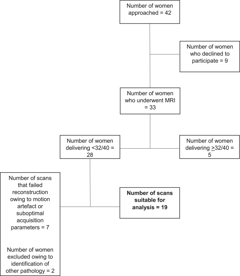
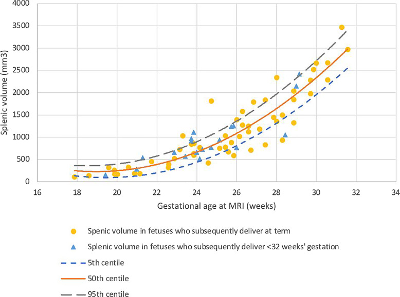
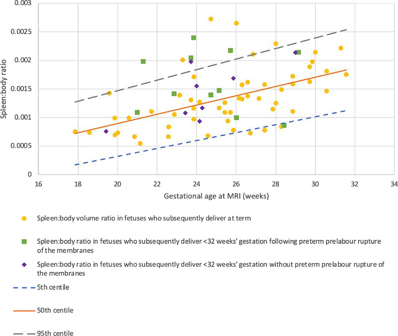
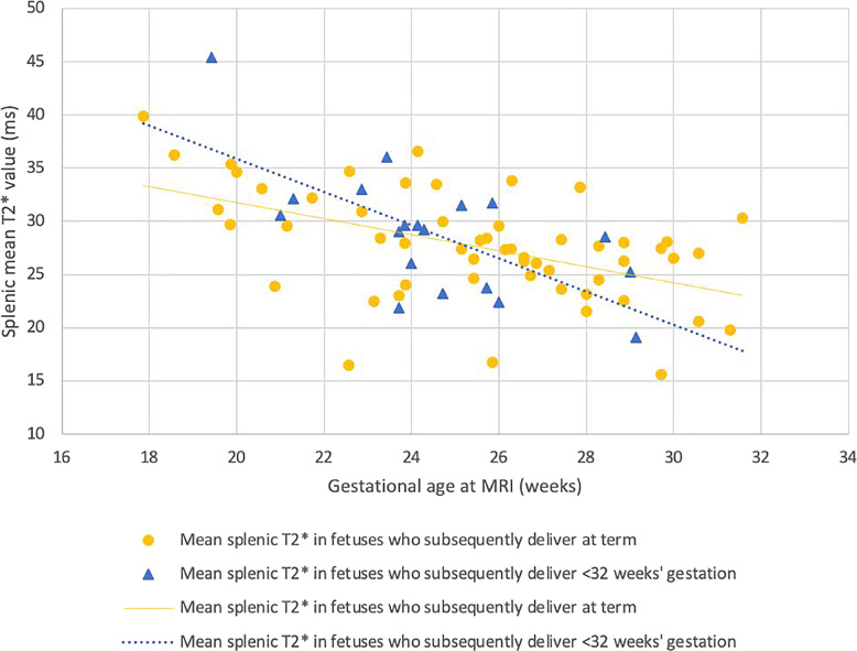
Results: Among 55 women with uncomplicated pregnancies, an increase in fetal splenic volume, splenic:body volume, and a decrease in splenic T2* signal intensity was demonstrated across gestation. Compared to controls, fetuses who were subsequently delivered prior to 32 weeks' gestation (n = 19) had a larger spleen when controlled for the overall size of the fetus (p = 0.027), but T2* was consistent (p = 0.76).
Conclusion: These findings provide evidence of a replicable method of studying the fetal immune system and give novel results on the impact of impending preterm birth on the spleen. While T2* decreases prior to preterm birth in other organs, preservation demonstrated here suggests preferential sparing of the spleen.
Keywords: Chorioamnionitis; Fetal immunity; Infection; Magnetic resonance imaging; Preterm delivery; Preterm rupture of membranes.
© 2024 The Author(s). Published by S. Karger AG, Basel.
Conflict of interest statement
The authors have no conflicts of interest to declare.
Figures


Similar articles
-
Antenatal thymus volumes in fetuses that delivered <32 weeks' gestation: An MRI pilot study.Acta Obstet Gynecol Scand. 2021 Jun;100(6):1040-1050. doi: 10.1111/aogs.13983. Epub 2020 Sep 24. Acta Obstet Gynecol Scand. 2021. PMID: 32865812 Free PMC article.
-
Functional MRI assessment of the lungs in fetuses that deliver very Preterm: An MRI pilot study.Eur J Obstet Gynecol Reprod Biol. 2024 Feb;293:106-114. doi: 10.1016/j.ejogrb.2023.12.015. Epub 2023 Dec 20. Eur J Obstet Gynecol Reprod Biol. 2024. PMID: 38141484 Free PMC article.
-
Assessment of the thymus in fetuses prior to spontaneous preterm birth using functional MRI.Early Hum Dev. 2025 Feb;201:106188. doi: 10.1016/j.earlhumdev.2024.106188. Epub 2025 Jan 13. Early Hum Dev. 2025. PMID: 39813902
-
Magnetic resonance imaging for prenatal estimation of birthweight in pregnancy: review of available data, techniques, and future perspectives.Am J Obstet Gynecol. 2019 May;220(5):428-439. doi: 10.1016/j.ajog.2018.12.031. Epub 2018 Dec 22. Am J Obstet Gynecol. 2019. PMID: 30582928 Review.
-
Magnetic resonance imaging of the fetal brain.Hong Kong Med J. 2016 Jun;22(3):270-8. doi: 10.12809/hkmj154678. Epub 2016 Apr 22. Hong Kong Med J. 2016. PMID: 27101791 Review.
References
-
- World Health Organisation . Born too soon: decade of action on preterm birth. Available from: https://www.who.int/publications/i/item/9789240073890 (accessed May 16th, 2023).
-
- Russell P. Inflammatory lesions of the human placenta: clinical significance of acute chorioamnionitis. Am J Diagn Gynecol Obstet. 1979;1.
-
- Golub R, Cumano A. Embryonic hematopoiesis. Blood Cells Mol Dis. 2013;51(4):226–31. - PubMed
Publication types
MeSH terms
Grants and funding
LinkOut - more resources
Full Text Sources
Medical
